
Rabbit Anti-Melan A antibody
MAR1_HUMAN; Melanoma antigen recognized by T-cells 1; MLANA; MART1; Antigen LB39-AA; Antigen SK29-AA; Protein Melan-A;
View History [Clear]
Details
Product Name Melan A Chinese Name 黑色素瘤相关抗原/黑色素-A抗体 Alias MAR1_HUMAN; Melanoma antigen recognized by T-cells 1; MLANA; MART1; Antigen LB39-AA; Antigen SK29-AA; Protein Melan-A; literatures Research Area Tumour Cell biology immunology TumourCell biologyMaker Immunogen Species Rabbit Clonality Polyclonal React Species Human, Applications ELISA=1:5000-10000 IHC-P=1:100-500 IHC-F=1:100-500 IF=1:100-500 (Paraffin sections need antigen repair)
not yet tested in other applications.
optimal dilutions/concentrations should be determined by the end user.Theoretical molecular weight 13kDa Cellular localization cytoplasmic The cell membrane Form Liquid Concentration 1mg/ml immunogen KLH conjugated synthetic peptide derived from human Melan-A: 51-118/118 Lsotype IgG Purification affinity purified by Protein A Buffer Solution 0.01M TBS(pH7.4) with 1% BSA, 0.03% Proclin300 and 50% Glycerol. Storage Shipped at 4℃. Store at -20 °C for one year. Avoid repeated freeze/thaw cycles. Attention This product as supplied is intended for research use only, not for use in human, therapeutic or diagnostic applications. PubMed PubMed Product Detail Melan A, a product of the MART-1 gene, is a melanocyte differentiation marker recognized by autologous cytotoxic T lymphocytes. Other melanoma-associated markers recognized by autologous cytotoxic T cells are reported to include MAGE-1, MAGE-3, tyrosinase, gp100, gp75, BAGE-1 and GAGE-1. The analysis of these different molecules and their expression in individual melanomas may be of help in the study of their particular molecular roles in melanocyte differentiation and tumorigenesis.
Function:
Involved in melanosome biogenesis by ensuring the stability of GPR143. Plays a vital role in the expression, stability, trafficking, and processing of melanocyte protein PMEL, which is critical to the formation of stage II melanosomes.
Subunit:
Interacts with PMEL. Interacts with GPR143.
Subcellular Location:
Endoplasmic reticulum membrane; Single-pass type III membrane protein. Golgi apparatus. Golgi apparatus, trans-Golgi network membrane. Melanosome. Note=Also found in small vesicles and tubules dispersed over the entire cytoplasm. A small fraction of the protein is inserted into the membrane in an inverted orientation. Inversion of membrane topology results in the relocalization of the protein from a predominant Golgi/post-Golgi area to the endoplasmic reticulum. Melanoma cells expressing the protein with an inverted membrane topology are more effectively recognized by specific cytolytic T-lymphocytes than those expressing the protein in its native membrane orientation.
Tissue Specificity:
Expression is restricted to melanoma and melanocyte cell lines and retina.
Post-translational modifications:
Acylated.
SWISS:
Q16655
Gene ID:
2315
Database links:Entrez Gene: 2315 Human
Omim: 605513 Human
SwissProt: Q16655 Human
Unigene: 154069 Human
信号中间体(Signaling Intermediates)
MelanA/MART-1是分化Tumour抗原类别中具有代表性的成员,它表达于正常的黑素细胞和大多数原发及转移的黑色素瘤中,故称:黑色素瘤相关抗原。
Product Picture 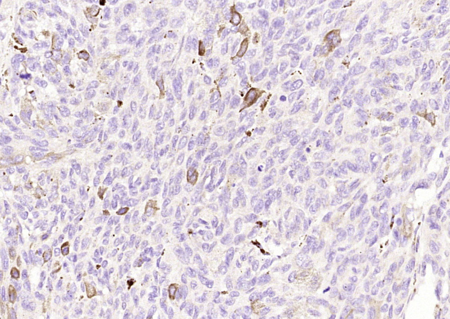 Paraformaldehyde-fixed, paraffin embedded (Human melanoma); Antigen retrieval by boiling in sodium citrate buffer (pH6.0) for 15min; Block endogenous peroxidase by 3% hydrogen peroxide for 20 minutes; Blocking buffer (normal goat serum) at 37°C for 30min; Antibody incubation with (Melan) Polyclonal Antibody, Unconjugated (SL0051R) at 1:200 overnight at 4°C, followed by operating according to SP Kit(Rabbit) (sp-0023) instructionsand DAB staining.Paraformaldehyde-fixed, paraffin embedded (Human melanoma); Antigen retrieval by boiling in sodium citrate buffer (pH6.0) for 15min; Block endogenous peroxidase by 3% hydrogen peroxide for 20 minutes; Blocking buffer (normal goat serum) at 37°C for 30min; Antibody incubation with (Melan) Polyclonal Antibody, Unconjugated (SL0051R) at 1:200 overnight at 4°C, followed by operating according to SP Kit(Rabbit) (sp-0023) instructionsand DAB staining.
Cartpieces
Totalgoods,subtotals:¥Checkout
Partial purchase records(bought amounts latest0)
No one bought this product
User Comment(Total0User Comment Num)
- No comment


 +86 571 56623320


