
Rabbit Anti-Caspase-9 antibody
Caspase-9 subunit p35; Apaf-3; APAF 3; APAF3; Apoptosis related cysteine peptidase; Apoptotic protease activating factor 3; Apoptotic protease MCH 6; Apoptotic protease MCH6; CASP 9; CASP9; Caspase 9; Caspase 9 apoptosis related cysteine protease; Caspase
View History [Clear]
Details
Product Name Caspase-9 Chinese Name 活化半胱胺酸蛋白酶蛋白-9抗体 Alias Caspase-9 subunit p35; Apaf-3; APAF 3; APAF3; Apoptosis related cysteine peptidase; Apoptotic protease activating factor 3; Apoptotic protease MCH 6; Apoptotic protease MCH6; CASP 9; CASP9; Caspase 9; Caspase 9 apoptosis related cysteine protease; Caspase 9 precursor; Caspase 9c; Caspase9; Caspase9 subunit p10; ICE LAP6; ICE like apoptotic protease 6; RNCASP9; MCH 6; MCH6; OTTHUMP00000044594; CASP9_HUMAN. literatures Research Area Tumour Cell biology Neurobiology Signal transduction Apoptosis Immunogen Species Rabbit Clonality Polyclonal React Species Human, Mouse, Rat, Applications WB=1:500-2000 ELISA=1:5000-10000 IHC-P=1:100-500 IHC-F=1:100-500 Flow-Cyt=1μg/Test ICC=1:100 IF=1:100-500 (Paraffin sections need antigen repair)
not yet tested in other applications.
optimal dilutions/concentrations should be determined by the end user.Theoretical molecular weight 35/50kDa Cellular localization The nucleus cytoplasmic Form Liquid Concentration 1mg/ml immunogen KLH conjugated synthetic peptide derived from human Caspase-9 subunit p35: 271-314/416 Lsotype IgG Purification affinity purified by Protein A Buffer Solution 0.01M TBS(pH7.4) with 1% BSA, 0.03% Proclin300 and 50% Glycerol. Storage Shipped at 4℃. Store at -20 °C for one year. Avoid repeated freeze/thaw cycles. Attention This product as supplied is intended for research use only, not for use in human, therapeutic or diagnostic applications. PubMed PubMed Product Detail Caspase 9 (also known as ICE like apoptotic protease 6 (ICE LAP6), apoptotic protease Mch6, and apoptotic protease activating factor 3 (Apaf3)) is a member of the peptidase family C14 that contains a CARD domain. This caspase is active as a heterotetramer and has been reported to have two isoforms. ProCaspase 9 has been reported to be approximately 47 kD. This caspase is present in the cytosol and, upon activation, translocates to the mitochondria. Caspase 9 is involved in the caspase activation cascade responsible for apoptosis execution and cleaves/activates Caspase 3 and Caspase 6. Caspase 9 is inhibited by the dominant negative isoform, BclXL, cIAP1, cIAP2, XIAP, and Livin. This caspase becomes activated when recruited to Apaf1/cytochrome c complex, and following cleavage by Apaf1, granzyme B, Caspase 3, possibly Caspase 8 and Caspase 10 into large p37 and small p10 subunits. Caspase 9 intereacts with BIRC7 and has been shown to cleave PARP and vimentin.
Function:
Involved in the activation cascade of caspases responsible for apoptosis execution. Binding of caspase-9 to Apaf-1 leads to activation of the protease which then cleaves and activates caspase-3. Proteolytically cleaves poly(ADP-ribose) polymerase (PARP).
Isoform 2 lacks activity is an dominant-negative inhibitor of caspase-9.
Subunit:
Heterotetramer that consists of two anti-parallel arranged heterodimers, each one formed by a 35 kDa (p35) and a 10 kDa (p10) subunit. Caspase-9 and APAF1 bind to each other via their respective NH2-terminal CED-3 homologous domains in the presence of cytochrome C and ATP. Interacts (inactive form) with EFHD2. Interacts with HAX1. Interacts with BIRC2/c-IAP1, XIAP/BIRC4, BIRC5/survivin, BIRC6/bruce and BIRC7/livin.
Tissue Specificity:
Ubiquitous, with highest expression in the heart, moderate expression in liver, skeletal muscle, and pancreas. Low levels in all other tissues. Within the heart, specifically expressed in myocytes.
Post-translational modifications:
Cleavages at Asp-315 by granzyme B and at Asp-330 by caspase-3 generate the two active subunits. Caspase-8 and -10 can also be involved in these processing events.
Phosphorylated at Thr-125 by MAPK1/ERK2. Phosphorylation at Thr-125 is sufficient to block caspase-9 processing and subsequent caspase-3 activation.
Similarity:
Belongs to the peptidase C14A family.
Contains 1 CARD domain.
SWISS:
P55211
Gene ID:
842
Database links:Entrez Gene: 842 Human
Omim: 602234 Human
SwissProt: P55211 Human
Unigene: 329502 Human
Caspase-9半胱氨酸蛋白酶家族成员之一,又称ICE-Lap6(ICE Like apoptotease 6)参与Apoptosis过程和cell factor的加工过程,在许多胚胎和成人组织中都有分布。此抗体主要用于Tumour研究Product Picture 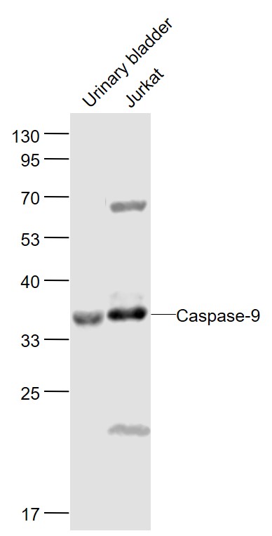 Sample:
Urinary bladder(Mouse) Lysate at 40 ug
Jurkat(Human) Cell Lysate at 30 ug
Primary: Anti-Caspase-9 (SL20773R) at 1/1000 dilution
Secondary: IRDye800CW Goat Anti-Rabbit IgG at 1/20000 dilution
Predicted band size: 46-51'35'37 kD
Observed band size: 35 kD
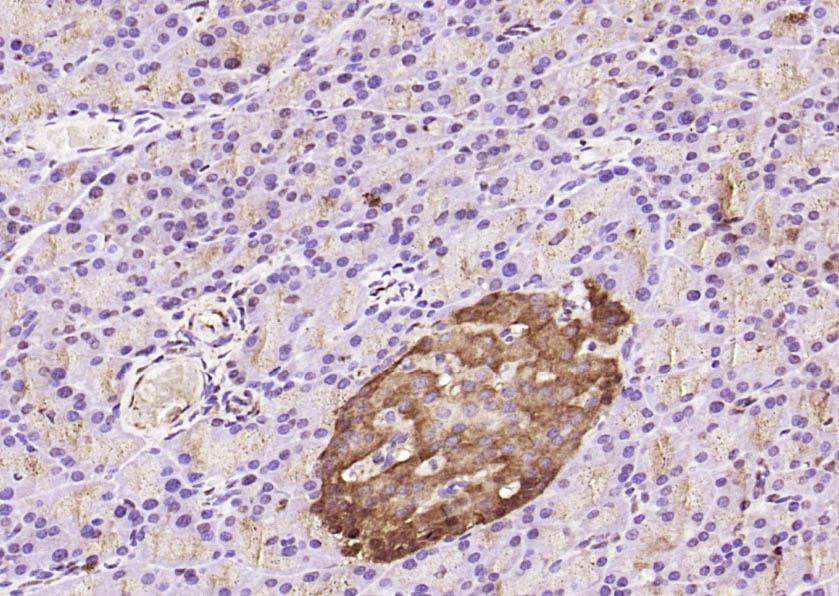 Paraformaldehyde-fixed, paraffin embedded (rat pancreas); Antigen retrieval by boiling in sodium citrate buffer (pH6.0) for 15min; Block endogenous peroxidase by 3% hydrogen peroxide for 20 minutes; Blocking buffer (normal goat serum) at 37°C for 30min; Antibody incubation with (Caspase-9) Polyclonal Antibody, Unconjugated (SL0050R) at 1:200 overnight at 4°C, followed by operating according to SP Kit(Rabbit) (sp-0023) instructionsand DAB staining.
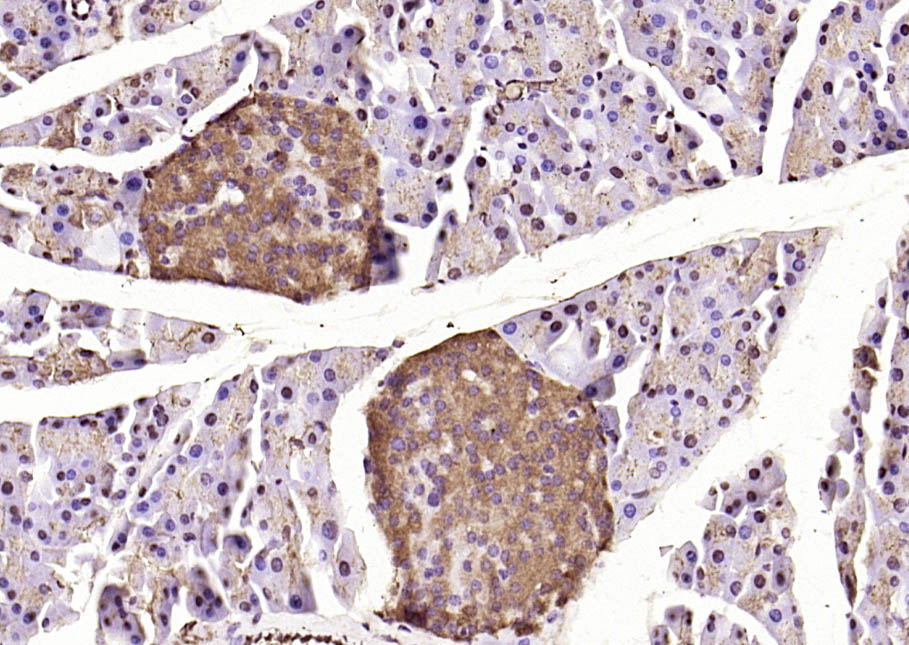 Paraformaldehyde-fixed, paraffin embedded (mouse pancreas); Antigen retrieval by boiling in sodium citrate buffer (pH6.0) for 15min; Block endogenous peroxidase by 3% hydrogen peroxide for 20 minutes; Blocking buffer (normal goat serum) at 37°C for 30min; Antibody incubation with (Caspase-9) Polyclonal Antibody, Unconjugated (SL0050R) at 1:200 overnight at 4°C, followed by operating according to SP Kit(Rabbit) (sp-0023) instructionsand DAB staining.
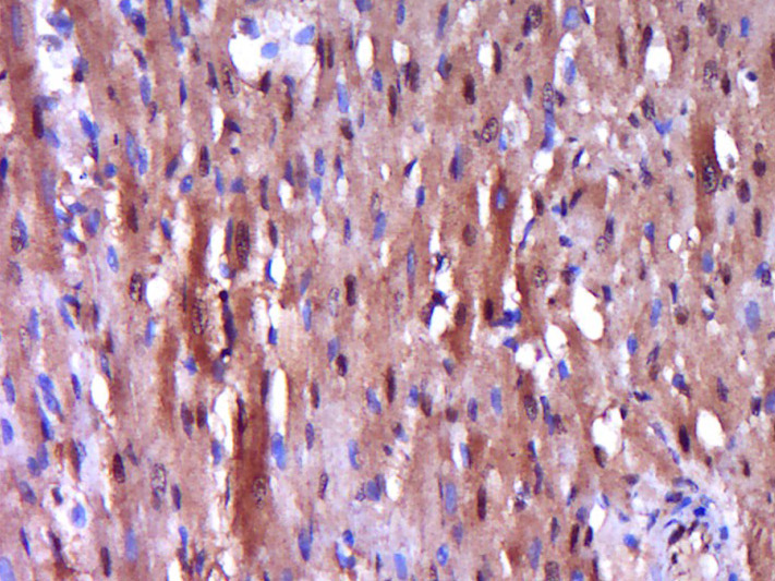 Paraformaldehyde-fixed, paraffin embedded (rat heart); Antigen retrieval by boiling in sodium citrate buffer (pH6.0) for 15min; Block endogenous peroxidase by 3% hydrogen peroxide for 20 minutes; Blocking buffer (normal goat serum) at 37°C for 30min; Antibody incubation with (Caspase-9) Polyclonal Antibody, Unconjugated (SL0050R) at 1:400 overnight at 4°C, followed by operating according to SP Kit(Rabbit) (sp-0023) instructionsand DAB staining.
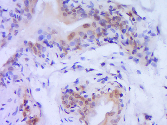 Paraformaldehyde-fixed, paraffin embedded (Rat bladder); Antigen retrieval by boiling in sodium citrate buffer (pH6.0) for 15min; Block endogenous peroxidase by 3% hydrogen peroxide for 20 minutes; Blocking buffer (normal goat serum) at 37°C for 30min; Antibody incubation with (Caspase-9) Polyclonal Antibody, Unconjugated (SL0050R) at 1:800 overnight at 4°C, followed by operating according to SP Kit(Rabbit) (sp-0023) instructionsand DAB staining.
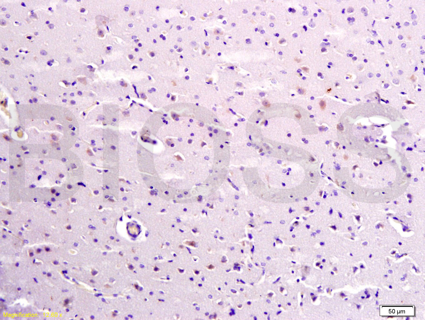 Tissue/cell: human brain tissue; 4% Paraformaldehyde-fixed and paraffin-embedded;
Antigen retrieval: citrate buffer ( 0.01M, pH 6.0 ), Boiling bathing for 15min; Block endogenous peroxidase by 3% Hydrogen peroxide for 30min; Blocking buffer (normal goat serum,C-0005) at 37℃ for 20 min;
Incubation: Anti-Caspase-9 Polyclonal Antibody, Unconjugated(SL0050R) 1:300, overnight at 4°C, followed by conjugation to the secondary antibody(SP-0023) and DAB(C-0010) staining
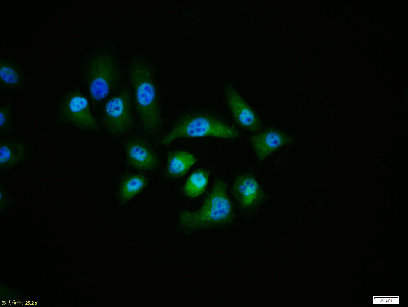 HepG2 cell; 4% Paraformaldehyde-fixed; Triton X-100 at room temperature for 20 min; Blocking buffer (normal goat serum, C-0005) at 37°C for 20 min; Antibody incubation with (Caspase-9) polyclonal Antibody, Unconjugated (SL0050R) 1:100, 90 minutes at 37°C; followed by a conjugated Goat Anti-Rabbit IgG antibody at 37°C for 90 minutes, DAPI (blue, C02-04002) was used to stain the cell nuclei.
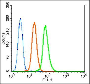 Blank control: K562 (blue).
Primary Antibody:Rabbit Anti-caspase-9 antibody (SL0050R,Green); Dilution: 1μg in 100 μL 1X PBS containing 0.5% BSA;
Isotype Control Antibody: Rabbit IgG(orange) ,used under the same conditions;
Secondary Antibody: Goat anti-rabbit IgG-FITC(white blue), Dilution: 1:200 in 1 X PBS containing 0.5% BSA.
Protocol
The cells were fixed with 80% methanol (5 min) and and then permeabilized with 0.01M PBS-Tween for 20 min . Primary antibody (SL0050R, 1μg /1x10^6 cells) were incubated for 30 min at room temperature, followed by 1 X PBS containing 0.5% BSA + 10% goat serum (30min) to block non-specific protein-protein interactions. Then the Goat Anti-rabbit IgG/FITC antibody was added into the blocking buffer mentioned above to react with the primary antibody at 1/200 dilution for 30 min at room temperature. Acquisition of 20,000 events was performed.
Cartpieces
Totalgoods,subtotals:¥Checkout
Partial purchase records(bought amounts latest0)
No one bought this product
User Comment(Total0User Comment Num)
- No comment


 +86 571 56623320


