
Rabbit Anti-IDE antibody
BC2; Insulin degrading enzyme; FLJ35968; insulin protease; insulinase; insulysin; Abeta-degrading protease; FLJ35968; Ide; IDE_HUMAN; Insulin-degrading enzyme; OTTHUMP00000020097.
View History [Clear]
Details
Product Name IDE Chinese Name 胰岛素降解酶抗体 Alias BC2; Insulin degrading enzyme; FLJ35968; insulin protease; insulinase; insulysin; Abeta-degrading protease; FLJ35968; Ide; IDE_HUMAN; Insulin-degrading enzyme; OTTHUMP00000020097. Research Area Cardiovascular Cell biology immunology Neurobiology Signal transduction Growth factors and hormones Synthesis and Degradation Diabetes Immunogen Species Rabbit Clonality Polyclonal React Species Mouse, Rat, (predicted: Human, Chicken, Pig, Cow, ) Applications WB=1:500-2000 ELISA=1:5000-10000 IHC-P=1:100-500 IHC-F=1:100-500 IF=1:100-500 (Paraffin sections need antigen repair)
not yet tested in other applications.
optimal dilutions/concentrations should be determined by the end user.Theoretical molecular weight 54/117kDa Cellular localization cytoplasmic The cell membrane Secretory protein Form Liquid Concentration 1mg/ml immunogen KLH conjugated synthetic peptide derived from human IDE: 491-590/1019 Lsotype IgG Purification affinity purified by Protein A Buffer Solution 0.01M TBS(pH7.4) with 1% BSA, 0.03% Proclin300 and 50% Glycerol. Storage Shipped at 4℃. Store at -20 °C for one year. Avoid repeated freeze/thaw cycles. Attention This product as supplied is intended for research use only, not for use in human, therapeutic or diagnostic applications. PubMed PubMed Product Detail Insulysin was identified nearly a century ago as an enzyme responsible for the degradation of insulin in cells, although the precise interactions between insulin and insulysin remain elusive. Human insulysin was cloned in 1988, and shown to be a 118 kDa protein that exists primarily as a homodimer, and perhaps also complexed with other molecules. The sequence is well conserved between humans, rats and mice, and the antibody recognizes these species. Insulysin is a metalloproteinase of the clan ME, family M16, which contains an active site HxxEH, a reversal of the canonical HExxH zinc binding motif. Considered a zinc metalloproteinase, the activity of insulysin can be blocked with EDTA or 1-10 phenanthroline. In addition to the active metalloproteinase domain, insulysin contains a second metalloproteinase site which is considered catalytically inactive, and is thought to assist in substrate binding. Insulysin is most closely related to the bacterial proteinase pitrilysin, (the human orthologue of which appears to be MPRP1) and the mammalian proteinsae nardilysin. Generally thought to be a cytoplasmic protein, insulysin has been isolated from many different tissues and cell lines, and can degrade intact insulin, insulin B chain, glucagon, denatured hemoglobin, alpha amyloid protein, TGF alpha and amylin. Recent work implicates insulysin in clearing beta amyloid plaques from the brain, and has generated much interest in Alzheimer’s disease research. The pH optimum for insulysin is basic, pH 8.5, which also distinguishes it from other metalloproteinases.
Function:
Plays a role in the cellular breakdown of insulin, IAPP, glucagon, bradykinin, kallidin and other peptides, and thereby plays a role in intercellular peptide signaling. Degrades amyloid formed by APP and IAPP. May play a role in the degradation and clearance of naturally secreted amyloid beta-protein by neurons and microglia.
Subunit:
Homodimer. Can form higher oligomers. Interacts (via N-terminus) with varicella-zoster virus (VZV) envelope glycoprotein E (via N-terminus); the membrane-associated isoform may function as an entry receptor for this virus.
Subcellular Location:
Cytoplasm. Cell membrane. Secreted. Note=Present at the cell surface of neuron cells. The membrane-associated isoform is approximately 5 kDa larger than the known cytosolic isoform.
Post-translational modifications:
The N-terminus is blocked.
Similarity:
Belongs to the peptidase M16 family.
SWISS:
P14735
Gene ID:
3416
Database links:Entrez Gene: 3416 Human
Entrez Gene: 15925 Mouse
Omim: 146680 Human
SwissProt: P14735 Human
SwissProt: Q9JHR7 Mouse
Unigene: 500546 Human
Unigene: 28366 Mouse
Unigene: 45029 Rat
Product Picture 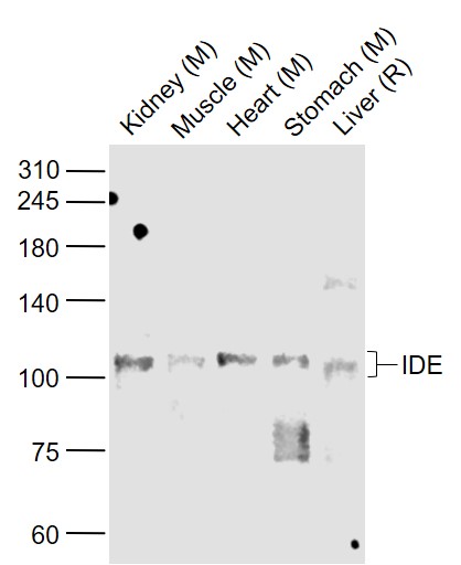 Sample:
Lane 1: Kidney (Mouse) Lysate at 40 ug
Lane 2: Muscle (Mouse) Lysate at 40 ug
Lane 3: Heart (Mouse) Lysate at 40 ug
Lane 4: Stomach (Mouse) Lysate at 40 ug
Lane 5: Liver (Rat) Lysate at 40 ug
Primary: Anti-IDE (SL0018R) at 1/1000 dilution
Secondary: IRDye800CW Goat Anti-Rabbit IgG at 1/20000 dilution
Predicted band size: 110 kD
Observed band size: 105 kD
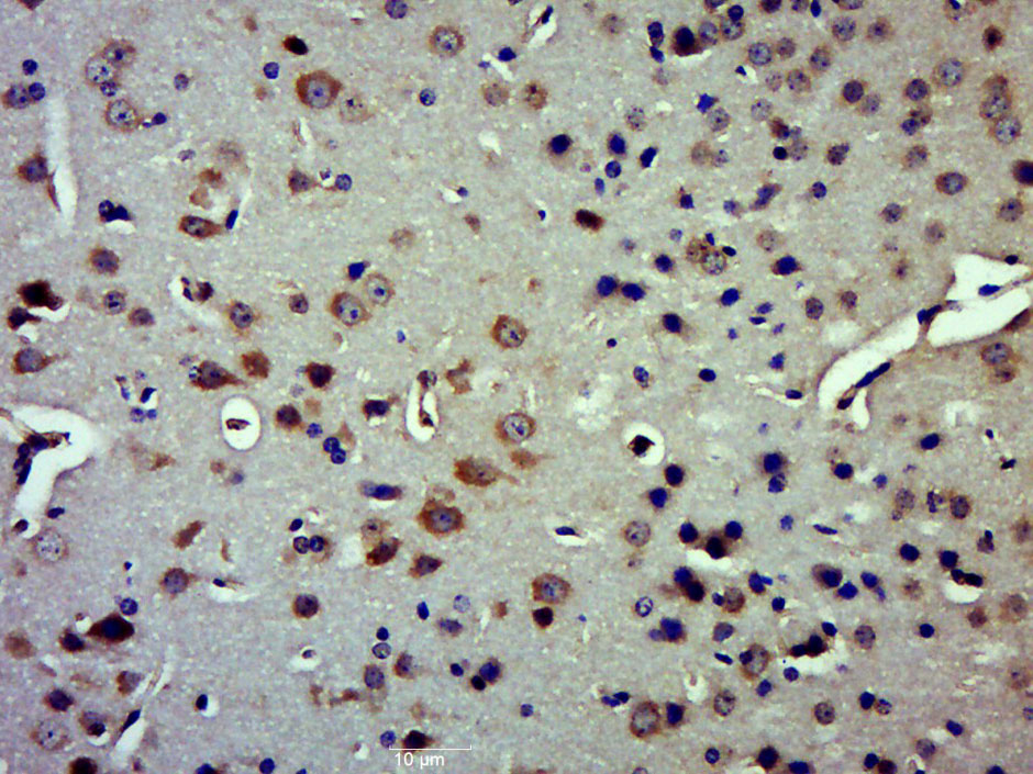 Paraformaldehyde-fixed, paraffin embedded (Mouse brain); Antigen retrieval by boiling in sodium citrate buffer (pH6.0) for 15min; Block endogenous peroxidase by 3% hydrogen peroxide for 20 minutes; Blocking buffer (normal goat serum) at 37°C for 30min; Antibody incubation with (IDE) Polyclonal Antibody, Unconjugated (SL0018R) at 1:500 overnight at 4°C, followed by a conjugated secondary (sp-0023) for 20 minutes and DAB staining.
Cartpieces
Totalgoods,subtotals:¥Checkout
Partial purchase records(bought amounts latest0)
No one bought this product
User Comment(Total0User Comment Num)
- No comment


 +86 571 56623320


