
Rabbit Anti-CD1c antibody
CD1c antigen; CD1C antigen c polypeptide; CD1c molecule; CD1C_HUMAN; Cortical thymocyte antigen CD1C; Differentiation antigen CD1 alpha 3 antibody R7; T cell surface glycoprotein CD1c; T-cell surface glycoprotein CD1c.
View History [Clear]
Details
Product Name CD1c Chinese Name T细胞表面glycoproteinCD1CRecombinant rabbit monoclonal anti Alias CD1c antigen; CD1C antigen c polypeptide; CD1c molecule; CD1C_HUMAN; Cortical thymocyte antigen CD1C; Differentiation antigen CD1 alpha 3 antibody R7; T cell surface glycoprotein CD1c; T-cell surface glycoprotein CD1c. Research Area Signal transduction Immunogen Species Rabbit Clonality Monoclonal Clone NO. R6C8 React Species (predicted: Human, ) Applications WB=1:500-1000 IHC-P=1:100-500 IHC-F=1:50-100 (Paraffin sections need antigen repair)
not yet tested in other applications.
optimal dilutions/concentrations should be determined by the end user.Theoretical molecular weight 36kDa Cellular localization The cell membrane Form Liquid Concentration 1mg/ml immunogen KLH conjugated synthetic peptide derived from human CD1c Lsotype IgG Purification affinity purified by Protein A Buffer Solution 0.01M TBS(pH7.4) with 1% BSA, 0.03% Proclin300 and 50% Glycerol. Storage Shipped at 4℃. Store at -20 °C for one year. Avoid repeated freeze/thaw cycles. Attention This product as supplied is intended for research use only, not for use in human, therapeutic or diagnostic applications. PubMed PubMed Product Detail This gene encodes a member of the CD1 family of transmembrane glycoproteins, which are structurally related to the major histocompatibility complex (MHC) proteins and form heterodimers with beta-2-microglobulin. The CD1 proteins mediate the presentation of primarily lipid and glycolipid antigens of self or microbial origin to T cells. The human genome contains five CD1 family genes organized in a cluster on chromosome 1. The CD1 family members are thought to differ in their cellular localization and specificity for particular lipid ligands. The protein encoded by this gene is broadly distributed throughout the endocytic system via a tyrosine-based motif in the cytoplasmic tail. Alternatively spliced transcript variants of this gene have been observed, but their full-length nature is not known. [provided by RefSeq, Jul 2008]
Subcellular Location:
Nucleus.
SWISS:
P29017
Gene ID:
911
Database links:
Entrez Gene: 911 Human
Omim: 188340 Human
SwissProt: P29017 Human
Unigene: 132448 Human
Product Picture 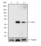 Blocking buffer: 5% NFDM/TBST
Primary Ab dilution: 1:2000
Primary Ab incubation condition: 2 hours at
room temperature
Secondary Ab: Goat Anti-Rabbit IgG H&L
(HRP)
Lysate: 1: THP-1 (Negative control), 2:
MOLT-4, 3: Jurkat
Protein loading quantity: 20 μg
Exposure time: 60 s
Predicted MW: 38 kDa
Observed MW: 43 kDa
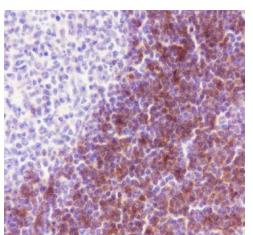 Tissue: Human tonsil
Section type: Formalin-fixed &
Paraffin-embedded section
Retrieval method: High temperature and high
pressure
Retrieval buffer: Tris/EDTA buffer, pH 9.0
Primary Ab dilution: 1:100
Primary Ab incubation condition: 1 hour at
room temperature
Secondary Ab: Anti-Rabbit and Mouse
Polymer HRP (Ready to use)
Counter stain: Hematoxylin (Blue)
Comment: Color brown is the positive signal for
SLM-60660R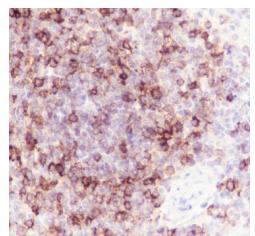 Tissue: Human spleen
Section type: Formalin-fixed &
Paraffin-embedded section
Retrieval method: High temperature and high
pressure
Retrieval buffer: Tris/EDTA buffer, pH 9.0
Primary Ab dilution: 1:100
Primary Ab incubation condition: 1 hour at
room temperature
Secondary Ab: Anti-Rabbit and Mouse
Polymer HRP (Ready to use)
Counter stain: Hematoxylin (Blue)
Comment: Color brown is the positive signal for
SLM-60660R
Cartpieces
Totalgoods,subtotals:¥Checkout
Bought notes(bought amounts latest0)
No one bought this product
User Comment(Total0User Comment Num)
- No comment


 +86 571 56623320
 +86 18668110335

