
Rabbit Anti-Periostin antibody
OSF 2; OSF2; osteoblast specific factor 2 (fasciclin I-like); Osteoblast specific factor 2; Osteoblast specific factor; PDLPOSTN; Periodontal ligament specific periostin; Periostin isoform thy2; Periostin isoform thy4; Periostin isoform thy6; Periostin is
View History [Clear]
Details
Product Name Periostin Chinese Name 成骨细胞特异性因子2Recombinant rabbit monoclonal anti Alias OSF 2; OSF2; osteoblast specific factor 2 (fasciclin I-like); Osteoblast specific factor 2; Osteoblast specific factor; PDLPOSTN; Periodontal ligament specific periostin; Periostin isoform thy2; Periostin isoform thy4; Periostin isoform thy6; Periostin isoform thy8; Periostin osteoblast specific factor; PN; POSTN; POSTN_HUMAN. Research Area Tumour Cardiovascular Cell biology immunology Signal transduction transcriptional regulatory factor Cell adhesion molecule vascular endothelial cell TumourCell biologyMaker Immunogen Species Rabbit Clonality Monoclonal React Species (predicted: Human, ) Applications WB=1:200-1000 IHC-P=1:100-500 Flow-Cyt=1:20-100 (Paraffin sections need antigen repair)
not yet tested in other applications.
optimal dilutions/concentrations should be determined by the end user.Theoretical molecular weight 100kDa Cellular localization Extracellular matrix Secretory protein Form Liquid Concentration 1mg/ml immunogen KLH conjugated synthetic peptide derived from human Periostin Lsotype IgG Purification affinity purified by Protein A Buffer Solution 0.01M TBS(pH7.4) with 1% BSA, 0.03% Proclin300 and 50% Glycerol. Storage 0.01M TBS(pH7.4) with 1% BSA, 0.03% Proclin300 and 50% Glycerol. Attention This product as supplied is intended for research use only, not for use in human, therapeutic or diagnostic applications. PubMed PubMed Product Detail Periostin is a disulfide linked 90 kDa, 811 amino acid protein originally isolated as a osteoblast-specific factor that functions as a cell adhesion molecule for preosteoblasts and is thought to be involved in osteoblast recruitment, attachment and spreading. Additionally, periostin expression has previously been shown to be significantly increased by both transforming growth factor beta 1(TGF beta 1) and bone morphogenetic protein (BMP2). Periostin has a typical signal sequence, followed by a cysteine-rich domain, a fourfold repeated domain and a C-terminal domain. The fourfold repeated domain of OSF2 shows homology with the insect protein fasciclin. Periostin mRNA is expressed in the developing mouse embryonic and fetal heart, and that it is localized to the endocardial cushions that ultimately divide the primitive heart tube into a four-chambered heart. Abnormal expression of periostin is also linked to angiogenesis and metastatsis in epithelial tumors.
SWISS:
Q15063
Gene ID:
10631
Database links:Entrez Gene: 10631 Human
Entrez Gene: 50706 Mouse
Omim: 608777 Human
SwissProt: Q15063 Human
SwissProt: Q62009 Mouse
Unigene: 136348 Human
Unigene: 721018 Human
Unigene: 236067 Mouse
Unigene: 30516 Rat
Product Picture 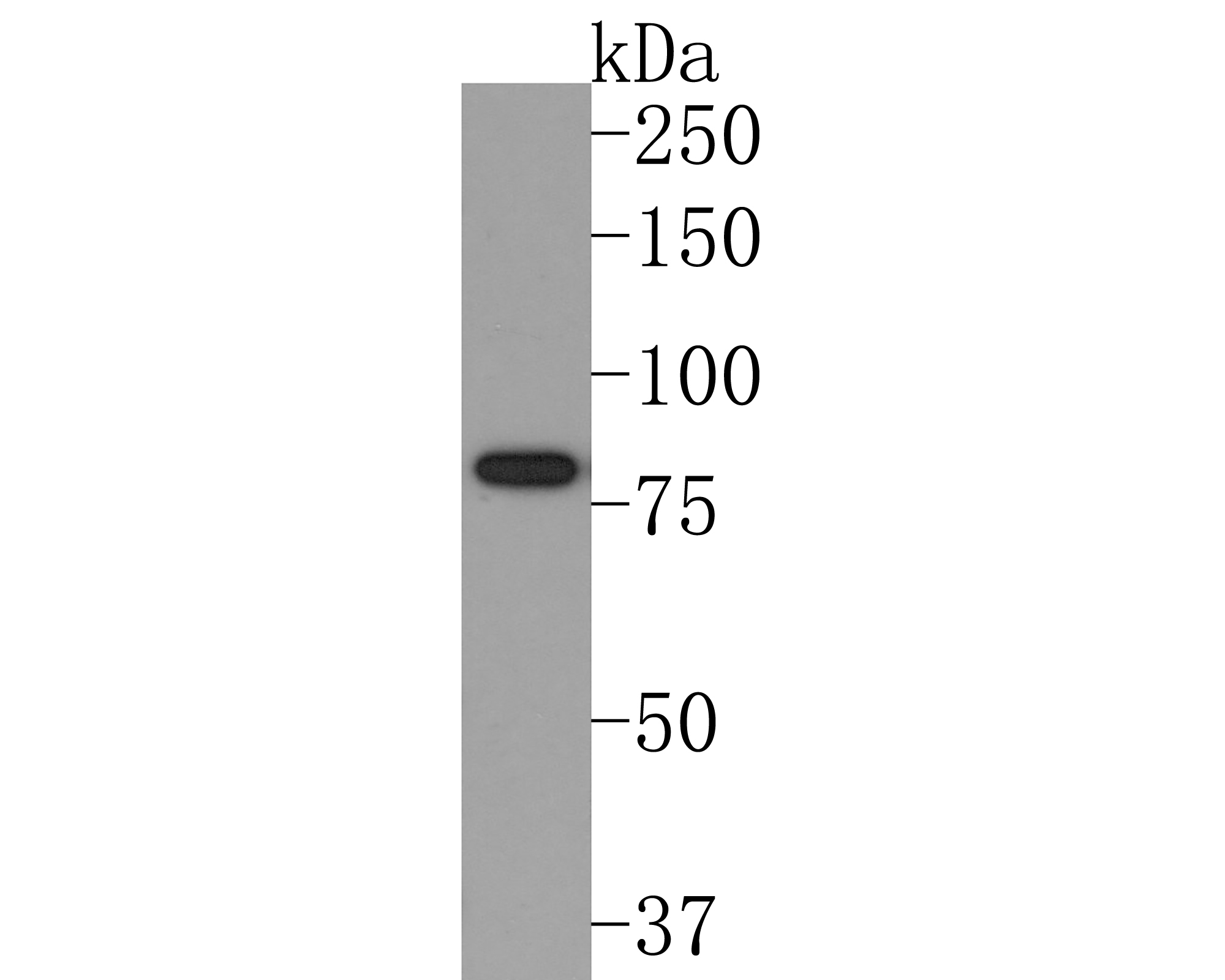 Western blot analysis of Periostin on human brain tissue lysates. Proteins were transferred to a PVDF membrane and blocked with 5% NFDM/TBST in PBS for 1 hour at room temperature. The primary antibody (SLM-54036R, 1/500) was used in 5% NFDM/TBST at room temperature for 2 hours. Goat Anti-Rabbit IgG - HRP Secondary Antibody at 1:200,000 dilution was used for 1 hour at room temperature.
Predicted band size: 93 kDa
Observed band size: 80 kDa
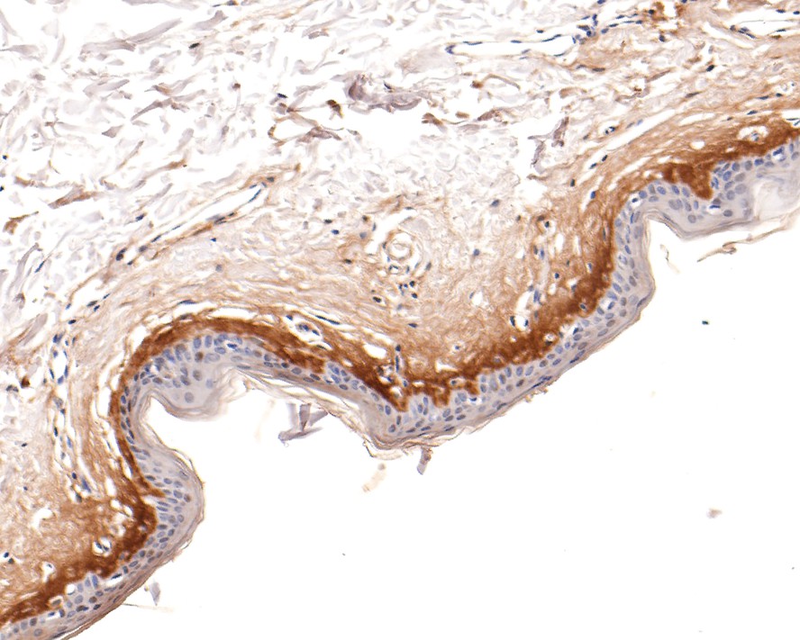 Immunohistochemical analysis of paraffin-embedded human skin tissue using anti-Periostin antibody. The section was pre-treated using heat mediated antigen retrieval with Tris-EDTA buffer (pH 9.0) for 20 minutes.The tissues were blocked in 1% BSA for 30 minutes at room temperature, washed with ddH2O and PBS, and then probed with the primary antibody (SLM-54036R, 1/400) for 30 minutes at room temperature. The detection was performed using an HRP conjugated compact polymer system. DAB was used as the chromogen. Tissues were counterstained with hematoxylin and mounted with DPX.
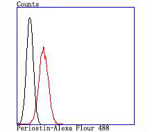 Flow cytometric analysis of Periostin was done on 293T cells. The cells were fixed, permeabilized and stained with the primary antibody (SLM-54036R, 1/50) (blue). After incubation of the primary antibody at room temperature for an hour, the cells were stained with a Alexa Fluor®488 conjugate-Goat anti-Rabbit IgG Secondary antibody at 1/1,000 dilution for 30 minutes.Unlabelled sample was used as a control (cells without incubation with primary antibody; red).
Cartpieces
Totalgoods,subtotals:¥Checkout
References (0)
No References
Bought notes(bought amounts latest0)
No one bought this product
User Comment(Total0User Comment Num)
- No comment


 +86 571 56623320
 +86 18668110335

