
Rabbit Anti-GATA1 antibody
GATA1; ERYF 1; ERYF1 antibody Erythroid transcription factor; Erythrold transcription factor 1; GATA 1; GATA binding factor 1; GATA binding protein 1; GF 1; GF1; Globin transcription factor 1; NF E1; NF E1 DNA binding protein; NFE 1; NFE1; GATA1_HUMAN; Er
View History [Clear]
Details
Product Name GATA1 Chinese Name 珠蛋白转录因子1抗体 Alias GATA1; ERYF 1; ERYF1 antibody Erythroid transcription factor; Erythrold transcription factor 1; GATA 1; GATA binding factor 1; GATA binding protein 1; GF 1; GF1; Globin transcription factor 1; NF E1; NF E1 DNA binding protein; NFE 1; NFE1; GATA1_HUMAN; Erythroid transcription factor; Eryf1; GATA-binding factor 1; GATA-1; GF-1; NF-E1 DNA-binding protein. Research Area Cell biology immunology transcriptional regulatory factor Cell Surface Molecule Immunogen Species Rabbit Clonality Monoclonal React Species (predicted: Human, ) Applications WB=1:500-1000 IHC-P=1:100-500 (Paraffin sections need antigen repair)
not yet tested in other applications.
optimal dilutions/concentrations should be determined by the end user.Theoretical molecular weight 45kDa Cellular localization The nucleus Form Liquid Concentration 1mg/ml immunogen Recombinant protein within human GATA1: 1-250/413 Lsotype IgG Purification affinity purified by Protein A Buffer Solution 0.01M TBS(pH7.4) with 1% BSA, 0.03% Proclin300 and 50% Glycerol. Storage Shipped at 4℃. Store at -20 °C for one year. Avoid repeated freeze/thaw cycles. Attention This product as supplied is intended for research use only, not for use in human, therapeutic or diagnostic applications. PubMed PubMed Product Detail GATA1 (Globin transcription factor 1) is a Cys2/Cys2 zinc finger DNA binding protein that is expressed primarily in erythroid, megakaryocytic, mast cells and eosinophilic cells. It belongs to the GATA family of transcription factors. GATA1 is a transcriptional activator which probably serves as a general switch factor for erythroid development. It binds to DNA sites with the consensus sequence [AT]GATA[AG] within regulatory regions of globin genes and of other genes expressed in erythroid cells. The protein also plays an important role in erythroid development by regulating the switch from fetal hemoglobin production to adult hemoglobin.
Function:
Transcriptional activator which probably serves as a general switch factor for erythroid development. It binds to DNA sites with the consensus sequence [AT]GATA[AG] within regulatory regions of globin genes and of other genes expressed in erythroid cells.
Subunit:
May form homodimers or heterodimers with other isoforms. Interacts (via the N-terminal zinc finger) with ZFPM1. Interacts with GFI1B. Interacts with PIAS4; the interaction enhances sumoylation and represses the transactivational activity in a sumoylation-independent manner. Interacts with LMCD1.
Subcellular Location:
Nucleus.
Tissue Specificity:
Erythrocytes.
Post-translational modifications:
Highly phosphorylated on serine residues. Phosphorylation on Ser-310 is enhanced on erythroid differentiation. Phosphorylation on Ser-142 promotes sumoylation on Lys-137.
DISEASE:
Defects in GATA1 are the cause of X-linked thrombocytopenia with beta-thalassemia (XLTT) [MIM:314050]; also knwon as thrombocytopenia, platelet dysfunction, hemolysis, and imbalanced globin synthesis. XLTT consists of an unusual form of thrombocytopenia with beta-thalassemia. Patients have splenomegaly and petechiae, moderate thrombocytopenia, prolonged bleeding time due to platelet dysfunction, reticulocytosis and unbalanced hemoglobin chain synthesis resembling that of beta-thalassemia minor.
Defects in GATA1 are the cause of anemia without thrombocytopenia X-linked (XLAWT) [MIM:300835]. XLAWT is a form of anemia characterized by abnormal morphology of erythrocytes and granulocytes in peripheral blood, bone marrow dysplasia with hypocellularity of erythroid and granulocytic lineages, and normal or increased number of megakaryocytes. Neutropenia of a variable degree is present in affected individuals.
Similarity:
Contains 2 GATA-type zinc fingers.
SWISS:
P15976
Gene ID:
2623
Database links:Entrez Gene: 2623 Human
Entrez Gene: 14460 Mouse
Omim: 305371 Human
SwissProt: P15976 Human
SwissProt: P17679 Mouse
Unigene: 765 Human
Unigene: 335973 Mouse
Unigene: 10024 Rat
Product Picture 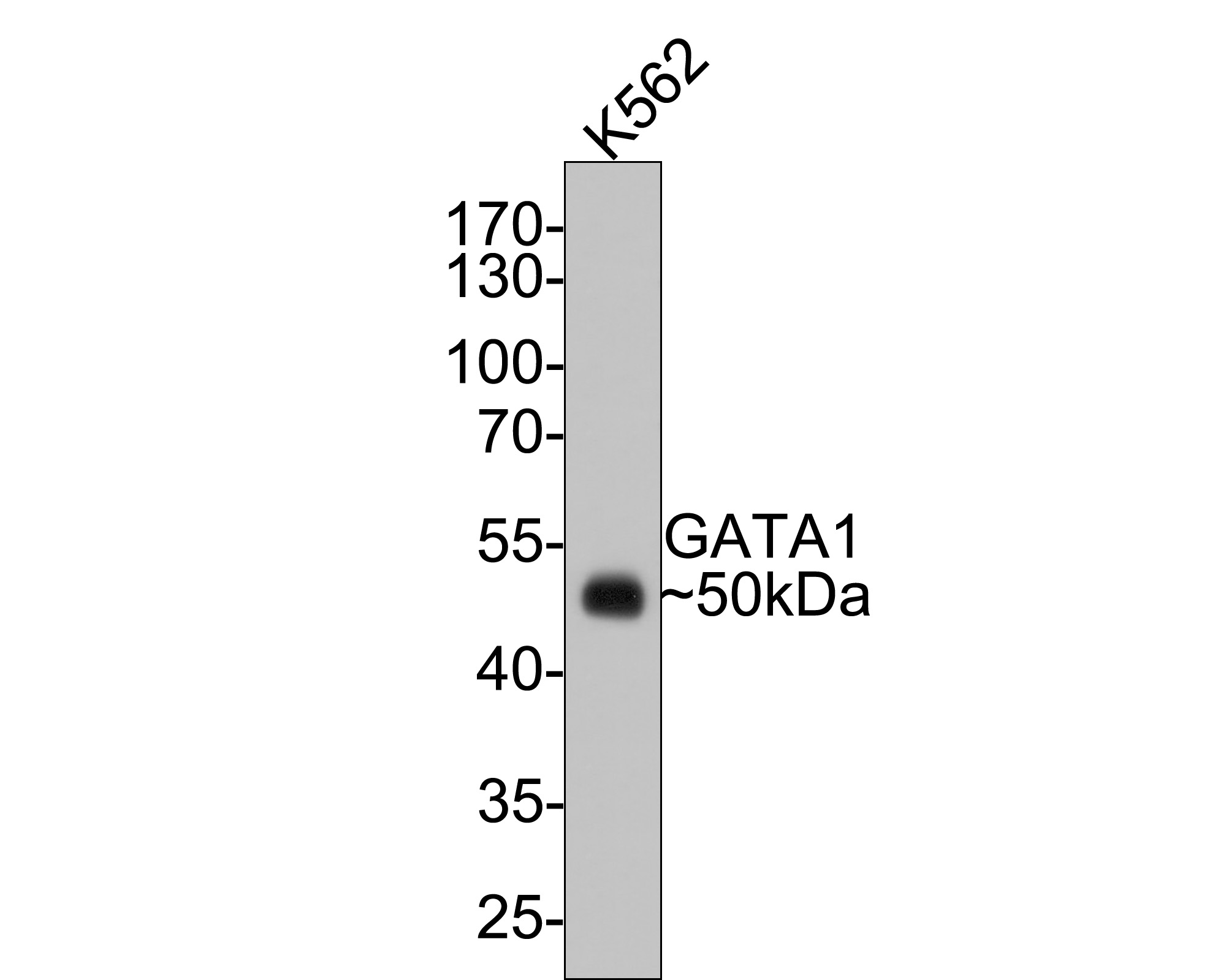 Western blot analysis of GATA1 on K562 cell lysates with Rabbit anti-GATA1 antibody (SLM-54013R) at 1/500 dilution.
Lysates/proteins at 10 µg/Lane.
Predicted band size: 43 kDa
Observed band size: 50 kDa
Exposure time: 30 seconds;
10% SDS-PAGE gel.
Proteins were transferred to a PVDF membrane and blocked with 5% NFDM/TBST for 1 hour at room temperature. The primary antibody (SLM-54013R) at 1/500 dilution was used in 5% NFDM/TBST at room temperature for 2 hours. Goat Anti-Rabbit IgG - HRP Secondary Antibody (HA1001) at 1:300,000 dilution was used for 1 hour at room temperature.
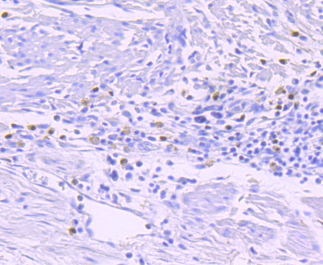 Immunohistochemical analysis of paraffin-embedded human liver tissue using anti-GATA1 antibody. The section was pre-treated using heat mediated antigen retrieval with Tris-EDTA buffer (pH 9.0) for 20 minutes.The tissues were blocked in 5% BSA for 30 minutes at room temperature, washed with ddH2O and PBS, and then probed with the primary antibody (SLM-54013R, 1/50) for 30 minutes at room temperature. The detection was performed using an HRP conjugated compact polymer system. DAB was used as the chromogen. Tissues were counterstained with hematoxylin and mounted with DPX
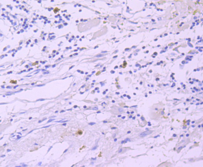 Immunohistochemical analysis of paraffin-embedded human breast carcinoma tissue using anti-GATA1 antibody. The section was pre-treated using heat mediated antigen retrieval with Tris-EDTA buffer (pH 9.0) for 20 minutes.The tissues were blocked in 5% BSA for 30 minutes at room temperature, washed with ddH2O and PBS, and then probed with the primary antibody (SLM-54013R, 1/50) for 30 minutes at room temperature. The detection was performed using an HRP conjugated compact polymer system. DAB was used as the chromogen. Tissues were counterstained with hematoxylin and mounted with DPX.
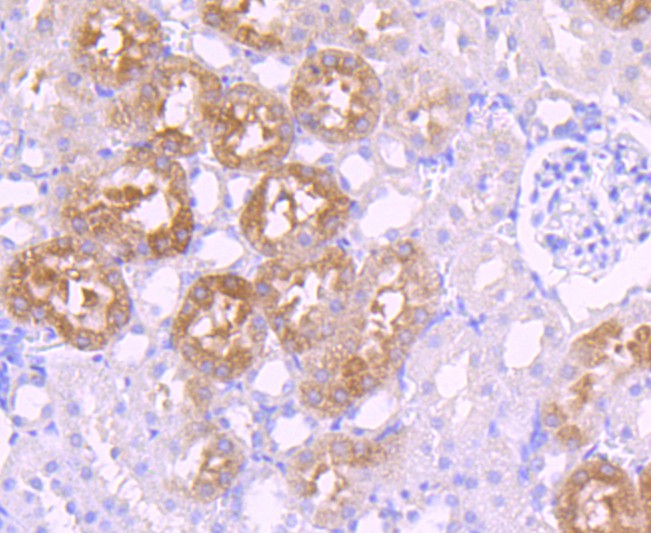 Immunohistochemical analysis of paraffin-embedded human kidney tissue using anti-GATA1 antibody. The section was pre-treated using heat mediated antigen retrieval with Tris-EDTA buffer (pH 9.0) for 20 minutes.The tissues were blocked in 5% BSA for 30 minutes at room temperature, washed with ddH2O and PBS, and then probed with the primary antibody (SLM-54013R, 1/50) for 30 minutes at room temperature. The detection was performed using an HRP conjugated compact polymer system. DAB was used as the chromogen. Tissues were counterstained with hematoxylin and mounted with DPX.
Cartpieces
Totalgoods,subtotals:¥Checkout
References (0)
No References
Bought notes(bought amounts latest0)
No one bought this product
User Comment(Total0User Comment Num)
- No comment


 +86 571 56623320
 +86 18668110335

