
Rabbit Anti-CFH antibody
CFAH_HUMAN; Complement factor H; HF; HF1; HF2; H factor 1; Factor H;
View History [Clear]
Details
Product Name CFH Chinese Name 补体因子HRecombinant rabbit monoclonal anti Alias CFAH_HUMAN; Complement factor H; HF; HF1; HF2; H factor 1; Factor H; Research Area Tumour immunology Growth factors and hormones Immunogen Species Rabbit Clonality Monoclonal React Species Human, Applications WB=1:500-2000 IHC-P=1:100-500 ICC=1:50-100 (Paraffin sections need antigen repair)
not yet tested in other applications.
optimal dilutions/concentrations should be determined by the end user.Theoretical molecular weight 51/137kDa Detection molecular weight 150 kDa Cellular localization Secretory protein Form Liquid Concentration 1mg/ml immunogen KLH conjugated synthetic peptide derived from human Factor H Lsotype IgG Purification affinity purified by Protein A Buffer Solution 0.01M TBS(pH7.4) with 1% BSA, 0.03% Proclin300 and 50% Glycerol. Storage Shipped at 4℃. Store at -20 °C for one year. Avoid repeated freeze/thaw cycles. Attention This product as supplied is intended for research use only, not for use in human, therapeutic or diagnostic applications. PubMed PubMed Product Detail This gene is a member of the Regulator of Complement Activation (RCA) gene cluster and encodes a protein with twenty short consensus repeat (SCR) domains. This protein is secreted into the bloodstream and has an essential role in the regulation of complement activation, restricting this innate defense mechanism to microbial infections. Mutations in this gene have been associated with hemolytic-uremic syndrome (HUS) and chronic hypocomplementemic nephropathy. Alternate transcriptional splice variants, encoding different isoforms, have been characterized. [provided by RefSeq, Oct 2011]
Function:
Factor H functions as a cofactor in the inactivation of C3b by factor I and also increases the rate of dissociation of the C3bBb complex (C3 convertase) and the (C3b)NBB complex (C5 convertase) in the alternative complement pathway.
Subcellular Location:
Secreted.
Tissue Specificity:
Expressed by the liver and secreted in plasma.
DISEASE:
Genetic variations in CFH are associated with basal laminar drusen (BLD) [MIM:126700]; also known as drusen of Bruch membrane or cuticular drusen or grouped early adult-onset drusen. Drusen are extracellular deposits that accumulate below the retinal pigment epithelium on Bruch membrane. Basal laminar drusen refers to an early adult-onset drusen phenotype that shows a pattern of uniform small, slightly raised yellow subretinal nodules randomly scattered in the macula. In later stages, these drusen often become more numerous, with clustered groups of drusen scattered throughout the retina. In time these small basal laminar drusen may expand and ultimately lead to a serous pigment epithelial detachment of the macula that may result in vision loss.
Defects in CFH are the cause of complement factor H deficiency (CFHD) [MIM:609814]. A disorder that can manifest as several different phenotypes, including asymptomatic, recurrent bacterial infections, and renal failure. Laboratory features usually include decreased serum levels of factor H, complement component C3, and a decrease in other terminal complement components, indicating activation of the alternative complement pathway. It is associated with a number of renal diseases with variable clinical presentation and progression, including membranoproliferative glomerulonephritis and atypical hemolytic uremic syndrome.
Defects in CFH are a cause of susceptibility to hemolytic uremic syndrome atypical type 1 (AHUS1) [MIM:235400]. An atypical form of hemolytic uremic syndrome. It is a complex genetic disease characterized by microangiopathic hemolytic anemia, thrombocytopenia, renal failure and absence of episodes of enterocolitis and diarrhea. In contrast to typical hemolytic uremic syndrome, atypical forms have a poorer prognosis, with higher death rates and frequent progression to end-stage renal disease. Note=Susceptibility to the development of atypical hemolytic uremic syndrome can be conferred by mutations in various components of or regulatory factors in the complement cascade system. Other genes may play a role in modifying the phenotype.
Genetic variation in CFH is associated with age-related macular degeneration type 4 (ARMD4) [MIM:610698]. ARMD is a multifactorial eye disease and the most common cause of irreversible vision loss in the developed world. In most patients, the disease is manifest as ophthalmoscopically visible yellowish accumulations of protein and lipid (known as drusen) that lie beneath the retinal pigment epithelium and within an elastin-containing structure known as Bruch membrane.
Similarity:
Contains 20 Sushi (CCP/SCR) domains.
SWISS:
P08603
Gene ID:
3075
Database links:Entrez Gene: 3075 Human
Entrez Gene: 12628 Mouse
SwissProt: P08603 Human
SwissProt: P06909 Mouse
Product Picture 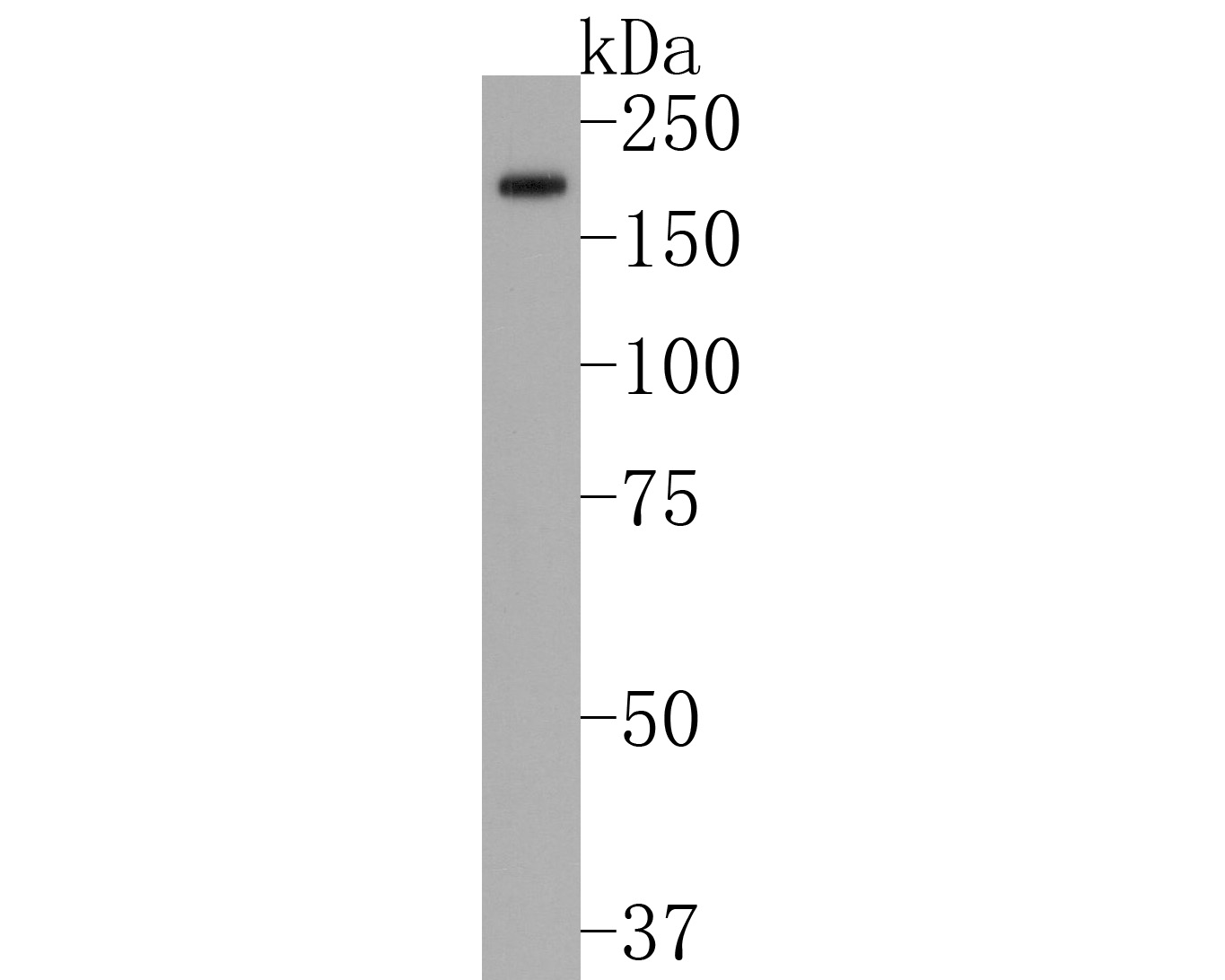 Western blot analysis of Factor H on human kidney tissue lysates. Proteins were transferred to a PVDF membrane and blocked with 5% BSA in PBS for 1 hour at room temperature. The primary antibody (SLM-54051R, 1/500) was used in 5% BSA at room temperature for 2 hours. Goat Anti-Rabbit IgG - HRP Secondary Antibody (HA1001) at 1:200,000 dilution was used for 1 hour at room temperature.
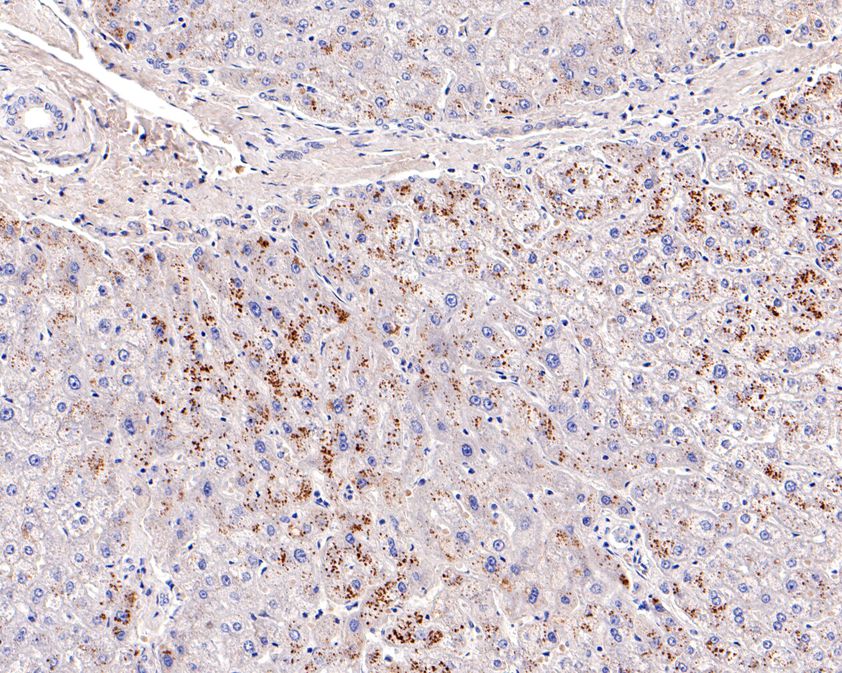 Immunohistochemical analysis of paraffin-embedded human liver tissue using anti-Factor H antibody. The section was pre-treated using heat mediated antigen retrieval with Tris-EDTA buffer (pH 8.0-8.4) for 20 minutes.The tissues were blocked in 5% BSA for 30 minutes at room temperature, washed with ddH2O and PBS, and then probed with the primary antibody (SLM-54051R, 1/100) for 30 minutes at room temperature. The detection was performed using an HRP conjugated compact polymer system. DAB was used as the chromogen. Tissues were counterstained with hematoxylin and mounted with DPX.
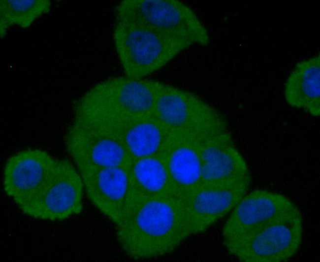 ICC staining of Factor H in HepG2 cells (green). Formalin fixed cells were permeabilized with 0.1% Triton X-100 in TBS for 10 minutes at room temperature and blocked with 1% Blocker BSA for 15 minutes at room temperature. Cells were probed with the primary antibody (SLM-54051R, 1/50) for 1 hour at room temperature, washed with PBS. Alexa Fluor®488 Goat anti-Rabbit IgG was used as the secondary antibody at 1/1,000 dilution. The nuclear counter stain is DAPI (blue).
 ICC staining of Factor H in Hela cells (green). Formalin fixed cells were permeabilized with 0.1% Triton X-100 in TBS for 10 minutes at room temperature and blocked with 1% Blocker BSA for 15 minutes at room temperature. Cells were probed with the primary antibody (SLM-54051R, 1/50) for 1 hour at room temperature, washed with PBS. Alexa Fluor®488 Goat anti-Rabbit IgG was used as the secondary antibody at 1/1,000 dilution. The nuclear counter stain is DAPI (blue).
Cartpieces
Totalgoods,subtotals:¥Checkout
References (0)
No References
Bought notes(bought amounts latest0)
No one bought this product
User Comment(Total0User Comment Num)
- No comment


 +86 571 56623320
 +86 18668110335

