
Rabbit Anti-TGFBI antibody
AI181842; AI747162; Beta ig; Beta ig h3; Beta ig-h3; BGH3_HUMAN; Big h3; BIGH3; CDB1; CDG2; CDGG1; CSD; CSD1; CSD2; CSD3; EBMD; Kerato epithelin; Kerato-epithelin; LCD1.
View History [Clear]
Details
Product Name TGFBI Chinese Name 角膜上皮蛋白TGFBIRecombinant rabbit monoclonal anti Alias AI181842; AI747162; Beta ig; Beta ig h3; Beta ig-h3; BGH3_HUMAN; Big h3; BIGH3; CDB1; CDG2; CDGG1; CSD; CSD1; CSD2; CSD3; EBMD; Kerato epithelin; Kerato-epithelin; LCD1. Research Area Cell biology Developmental biology Neurobiology Signal transduction Stem cells Growth factors and hormones Immunogen Species Rabbit Clonality Monoclonal React Species (predicted: Human, Mouse, Rat, ) Applications WB=1:500-1000 IHC-P=1:100-500 (Paraffin sections need antigen repair)
not yet tested in other applications.
optimal dilutions/concentrations should be determined by the end user.Theoretical molecular weight 72kDa Cellular localization Secretory protein Form Liquid Concentration 1mg/ml immunogen KLH conjugated synthetic peptide derived from human TGFBI Lsotype IgG Purification affinity purified by Protein A Buffer Solution 0.01M TBS(pH7.4) with 1% BSA, 0.03% Proclin300 and 50% Glycerol. Storage Shipped at 4℃. Store at -20 °C for one year. Avoid repeated freeze/thaw cycles. Attention This product as supplied is intended for research use only, not for use in human, therapeutic or diagnostic applications. PubMed PubMed Product Detail This gene encodes an RGD-containing protein that binds to type I, II and IV collagens. The RGD motif is found in many extracellular matrix proteins modulating cell adhesion and serves as a ligand recognition sequence for several integrins. This protein plays a role in cell-collagen interactions and may be involved in endochondrial bone formation in cartilage. The protein is induced by transforming growth factor-beta and acts to inhibit cell adhesion. Mutations in this gene are associated with multiple types of corneal dystrophy. [provided by RefSeq, Jul 2008]
Function:
Binds to type I, II, and IV collagens. This adhesion protein may play an important role in cell-collagen interactions. In cartilage, may be involved in endochondral bone formation.
Subcellular Location:
Secreted > extracellular space > extracellular matrix. May be associated both with microfibrils and with the cell surface. Tissue Specificity : Highly expressed in the corneal epithelium.
Post-translational modifications:
Gamma-carboxyglutamate residues are formed by vitamin K dependent carboxylation. These residues are essential for the binding of calcium.
DISEASE:
Defects in TGFBI are the cause of epithelial basement membrane corneal dystrophy (EBMD) [MIM:121820]; also known as Cogan corneal dystrophy or map-dot-fingerprint type corneal dystrophy. EBMD is a bilateral anterior corneal dystrophy characterized by grayish epithelial fingerprint lines, geographic map-like lines, and dots (or microcysts) on slit-lamp examination. Pathologic studies show abnormal, redundant basement membrane and intraepithelial lacunae filled with cellular debris. Although this disorder usually is not considered to be inherited, families with autosomal dominant inheritance have been identified.
Defects in TGFBI are the cause of corneal dystrophy Groenouw type 1 (CDGG1) [MIM:121900]; also known as corneal dystrophy granular type. Inheritance is autosomal dominant. Corneal dystrophies show progressive opacification of the cornea leading to severe visual handicap.
Defects in TGFBI are the cause of corneal dystrophy lattice type 1 (CDL1) [MIM:122200]. Inheritance is autosomal dominant.
Defects in TGFBI are a cause of corneal dystrophy Thiel-Behnke type (CDTB) [MIM:602082]; also known as corneal dystrophy of Bowman layer type 2 (CDB2). Defects in TGFBI are the cause of Reis-Buecklers corneal dystrophy (CDRB) [MIM:608470]; also known as corneal dystrophy of Bowman layer type 1 (CDB1). Defects in TGFBI are the cause of lattice corneal dystrophy type 3A (CDL3A) [MIM:608471]. CDL3A clinically resembles to lattice corneal dystrophy type 3, but differs in that its age of onset is 70 to 90 years. It has an autosomal dominant inheritance pattern.
Defects in TGFBI are the cause of Avellino corneal dystrophy (ACD) [MIM:607541]. ACD could be considered a variant of granular dystrophy with a significant amyloidogenic tendency. Inheritance is autosomal dominant.
Similarity:
Contains 1 EMI domain.
Contains 4 FAS1 domains.
SWISS:
Q15582
Gene ID:
7045
Product Picture 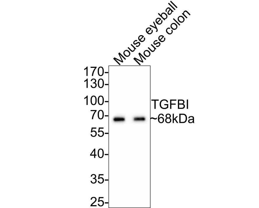 Western blot analysis of TGFBI on different lysates with Rabbit anti-TGFBI antibody (SLM-54085R) at 1/500 dilution.
Lane 1: Mouse eyeball tissue lysate
Lane 2: Mouse colon tissue lysate
Lysates/proteins at 20 µg/Lane.
Predicted band size: 75 kDa
Observed band size: 68 kDa
Exposure time: 2 minutes;
10% SDS-PAGE gel.
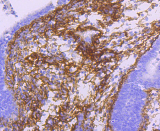 Immunohistochemical analysis of paraffin-embedded human uterus tissue using anti-TGFBI antibody. The section was pre-treated using heat mediated antigen retrieval with Tris-EDTA buffer (pH 8.0-8.4) for 20 minutes.The tissues were blocked in 5% BSA for 30 minutes at room temperature, washed with ddH2O and PBS, and then probed with the primary antibody (SLM-54085R, 1/50) for 30 minutes at room temperature. The detection was performed using an HRP conjugated compact polymer system. DAB was used as the chromogen. Tissues were counterstained with hematoxylin and mounted with DPX.
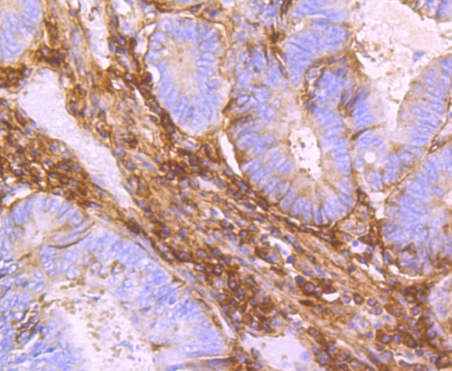 Immunohistochemical analysis of paraffin-embedded human colon carcinoma tissue using anti-TGFBI antibody. The section was pre-treated using heat mediated antigen retrieval with Tris-EDTA buffer (pH 8.0-8.4) for 20 minutes.The tissues were blocked in 5% BSA for 30 minutes at room temperature, washed with ddH2O and PBS, and then probed with the primary antibody (SLM-54085R, 1/50) for 30 minutes at room temperature. The detection was performed using an HRP conjugated compact polymer system. DAB was used as the chromogen. Tissues were counterstained with hematoxylin and mounted with DPX.
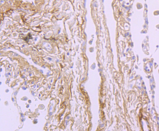 Immunohistochemical analysis of paraffin-embedded human lung carcinoma tissue using anti-TGFBI antibody. The section was pre-treated using heat mediated antigen retrieval with Tris-EDTA buffer (pH 8.0-8.4) for 20 minutes.The tissues were blocked in 5% BSA for 30 minutes at room temperature, washed with ddH2O and PBS, and then probed with the primary antibody (SLM-54085R, 1/50) for 30 minutes at room temperature. The detection was performed using an HRP conjugated compact polymer system. DAB was used as the chromogen. Tissues were counterstained with hematoxylin and mounted with DPX.
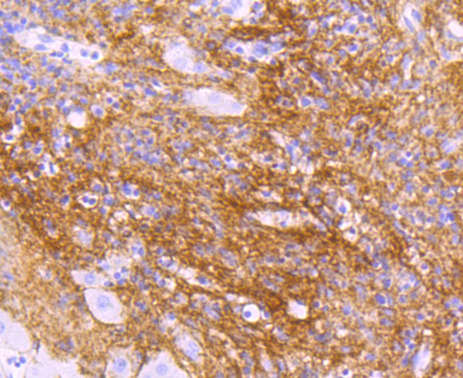 Immunohistochemical analysis of paraffin-embedded human liver carcinoma tissue using anti-TGFBI antibody. The section was pre-treated using heat mediated antigen retrieval with Tris-EDTA buffer (pH 8.0-8.4) for 20 minutes.The tissues were blocked in 5% BSA for 30 minutes at room temperature, washed with ddH2O and PBS, and then probed with the primary antibody (SLM-54085R, 1/50) for 30 minutes at room temperature. The detection was performed using an HRP conjugated compact polymer system. DAB was used as the chromogen. Tissues were counterstained with hematoxylin and mounted with DPX.
Cartpieces
Totalgoods,subtotals:¥Checkout
References (0)
No References
Bought notes(bought amounts latest0)
No one bought this product
User Comment(Total0User Comment Num)
- No comment


 +86 571 56623320
 +86 18668110335

