
Rabbit Anti-DNA PKcs antibody
PRKDC_HUMAN; DNA-dependent protein kinase catalytic subunit; EC:2.7.11.1; PRKDC; HYRC; HYRC1; DNA-PK catalytic subunit; DNA-PKcs; DNPK1; p460;
View History [Clear]
Details
Product Name DNA PKcs Chinese Name DNA依赖蛋白激酶催化亚基Recombinant rabbit monoclonal anti Alias PRKDC_HUMAN; DNA-dependent protein kinase catalytic subunit; EC:2.7.11.1; PRKDC; HYRC; HYRC1; DNA-PK catalytic subunit; DNA-PKcs; DNPK1; p460; Research Area Tumour Kinases and Phosphatases Immunogen Species Rabbit Clonality Monoclonal React Species (predicted: Human, Mouse, Rat, ) Applications WB=1:500-2000 IHC-P=1:100-500 ICC=1:50-200 IF=1:50-200 (Paraffin sections need antigen repair)
not yet tested in other applications.
optimal dilutions/concentrations should be determined by the end user.Theoretical molecular weight 454kDa Cellular localization The nucleus Form Liquid Concentration 1mg/ml immunogen KLH conjugated synthetic peptide derived from human DNA PKcs Lsotype IgG Purification affinity purified by Protein A Buffer Solution 0.01M TBS(pH7.4) with 1% BSA, 0.03% Proclin300 and 50% Glycerol. Storage Shipped at 4℃. Store at -20 °C for one year. Avoid repeated freeze/thaw cycles. Attention This product as supplied is intended for research use only, not for use in human, therapeutic or diagnostic applications. PubMed PubMed Product Detail This gene encodes the catalytic subunit of the DNA-dependent protein kinase (DNA-PK). It functions with the Ku70/Ku80 heterodimer protein in DNA double strand break repair and recombination. The protein encoded is a member of the PI3/PI4-kinase family.[provided by RefSeq, Jul 2010]
Function:
Serine/threonine-protein kinase that acts as a molecular sensor for DNA damage. Involved in DNA nonhomologous end joining (NHEJ) required for double-strand break (DSB) repair and V(D)J recombination. Must be bound to DNA to express its catalytic properties. Promotes processing of hairpin DNA structures in V(D)J recombination by activation of the hairpin endonuclease artemis DCLRE1C). The assembly of the DNA-PK complex at DNA ends is also required for the NHEJ ligation step. Required to protect and align broken ends of DNA. May also act as a scaffold protein to aid the localization of DNA repair proteins to the site of damage. Found at the ends of chromosomes, suggesting a further role in the maintenance of telomeric stability and the prevention of chromosomal end fusion. Also involved in modulation of transcription. Recognizes the substrate consensus sequence [ST]-Q. Phosphorylates 'Ser-139' of histone variant H2AX/H2AFX, thereby regulating DNA damage response mechanism. Phosphorylates DCLRE1C, c-Abl/ABL1, histone H1, HSPCA, c-jun/JUN, p53/TP53, PARP1, POU2F1, DHX9, SRF, XRCC1, XRCC1, XRCC4, XRCC5, XRCC6, WRN, MYC and RFA2. Can phosphorylate C1D not only in the presence of linear DNA but also in the presence of supercoiled DNA. Ability to phosphorylate p53/TP53 in the presence of supercoiled DNA is dependent on C1D.
Subunit:
DNA-PK is a heterotrimer of PRKDC and the Ku p70-p86 (XRCC6-XRCC5) dimer. Formation of this complex may be promoted by interaction with ILF3. Associates with the DNA-bound Ku heterodimer, but it can also bind to and be activated by free DNA. Interacts with DNA-PKcs-interacting protein (KIP) with the region upstream the kinase domain. PRKDC alone also interacts with and phosphorylates DCLRE1C, thereby activating the latent endonuclease activity of this protein. Interacts with C1D. Interacts with TTI1 and TELO2.
Subcellular Location:
Nucleus.
Post-translational modifications:
Phosphorylated upon DNA damage, probably by ATM or ATR. Autophosphorylated on Thr-2609, Thr-2638 and Thr-2647. Thr-2609 is a DNA damage-inducible phosphorylation site (inducible with ionizing radiation, IR). Autophosphorylation induces a conformational change that leads to remodeling of the DNA-PK complex, requisite for efficient end processing and DNA repair.
Similarity:
Belongs to the PI3/PI4-kinase family.
Contains 1 FAT domain.
Contains 1 FATC domain.
Contains 2 HEAT repeats.
Contains 1 PI3K/PI4K domain.
Contains 3 TPR repeats.
SWISS:
P78527
Gene ID:
5591
Database links:Entrez Gene: 5591 Human
Entrez Gene: 19090 Mouse
SwissProt: P78527 Human
SwissProt: P97313 Mouse
DNA依赖的蛋白激酶(DNA-PK)是一种DNA活化的核丝氨酸苏氨酸蛋白激酶. 参与体内DNA双链断裂缺口的修复, 在Tumour的预后、发生、发展、诊断和治疗等研究中引起科研人员越来越多的重视。Product Picture 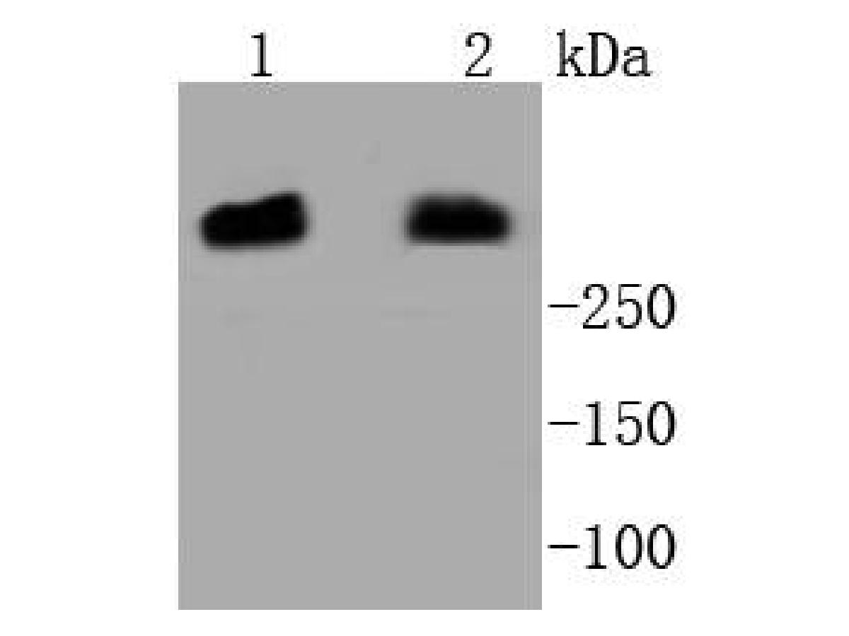 Western blot analysis of DNA PKcs on different lysates. Proteins were transferred to a PVDF membrane and blocked with 5% BSA in PBS for 1 hour at room temperature. The primary antibody (SLM-52493R, 1/500) was used in 5% BSA at room temperature for 2 hours. Goat Anti-Rabbit IgG - HRP Secondary Antibody (HA1001) at 1:5,000 dilution was used for 1 hour at room temperature.
Positive control:
Lane 1: Hela cell lysate
Lane 2: MCF-7 cell lysate
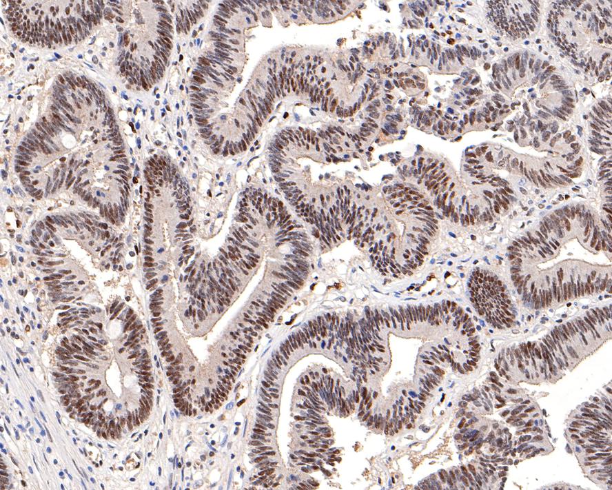 Immunohistochemical analysis of paraffin-embedded human colon carcinoma tissue with Rabbit anti-DNA PKcs antibody (SLM-52493R) at 1/200 dilution. The section was pre-treated using heat mediated antigen retrieval with sodium citrate buffer (pH 6.0) for 2 minutes. The tissues were blocked in 1% BSA for 20 minutes at room temperature, washed with ddH2O and PBS, and then probed with the primary antibody (SLM-52493R) at 1/200 dilution for 1 hour at room temperature. The detection was performed using an HRP conjugated compact polymer system. DAB was used as the chromogen. Tissues were counterstained with hematoxylin and mounted with DPX.
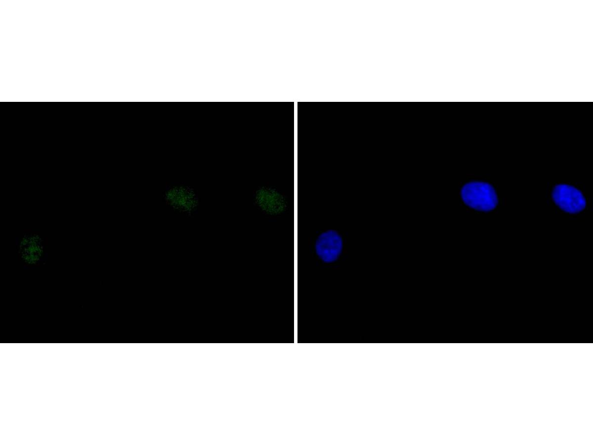 ICC staining of DNA PKcs in A549 cells (green). Formalin fixed cells were permeabilized with 0.1% Triton X-100 in TBS for 10 minutes at room temperature and blocked with 1% Blocker BSA for 15 minutes at room temperature. Cells were probed with the primary antibody (SLM-52493R, 1/50) for 1 hour at room temperature, washed with PBS. Alexa Fluor®488 Goat anti-Rabbit IgG was used as the secondary antibody at 1/1,000 dilution. The nuclear counter stain is DAPI (blue).
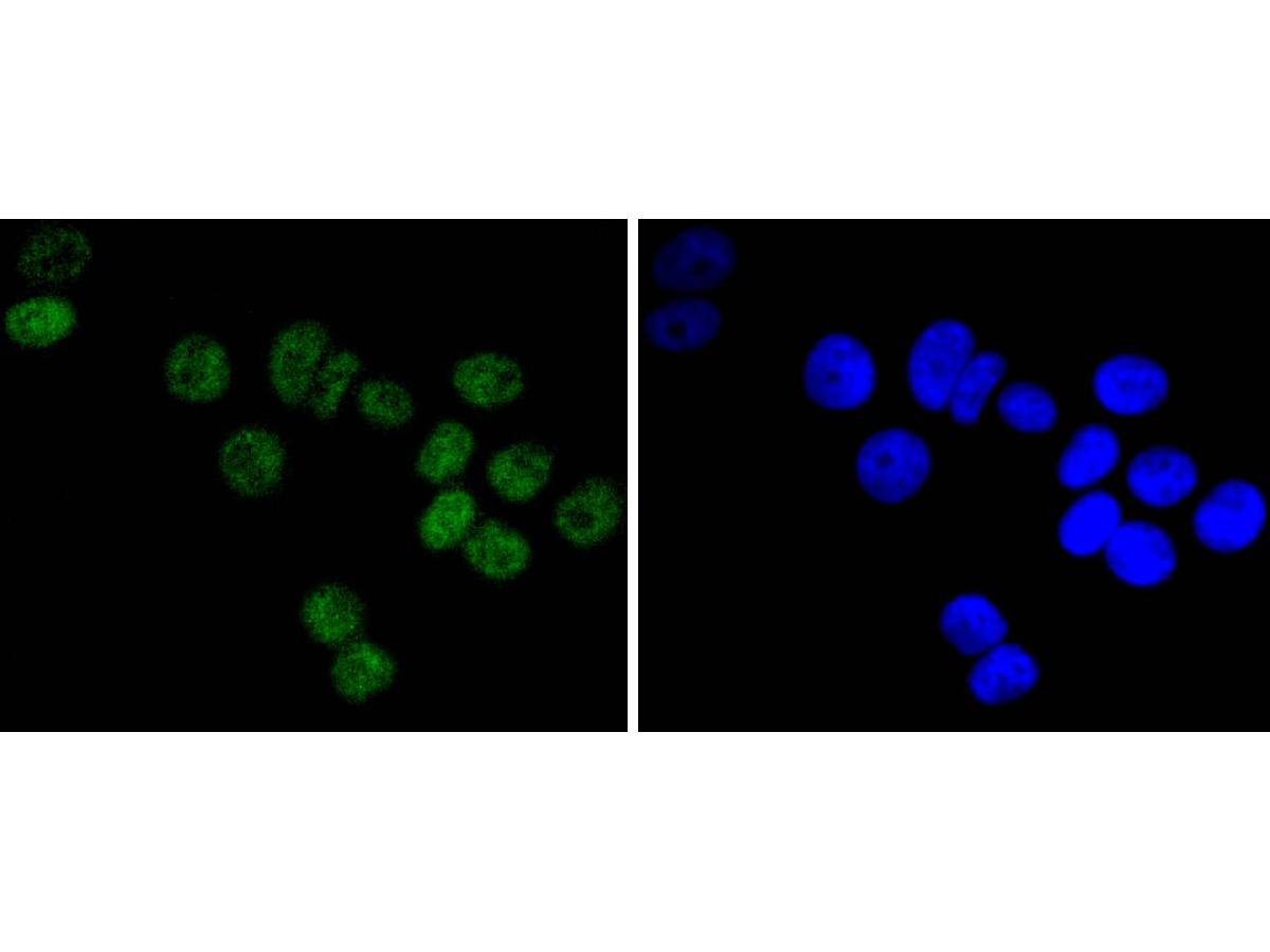 ICC staining of DNA PKcs in MCF-7 cells (green). Formalin fixed cells were permeabilized with 0.1% Triton X-100 in TBS for 10 minutes at room temperature and blocked with 1% Blocker BSA for 15 minutes at room temperature. Cells were probed with the primary antibody (SLM-52493R, 1/50) for 1 hour at room temperature, washed with PBS. Alexa Fluor®488 Goat anti-Rabbit IgG was used as the secondary antibody at 1/1,000 dilution. The nuclear counter stain is DAPI (blue).
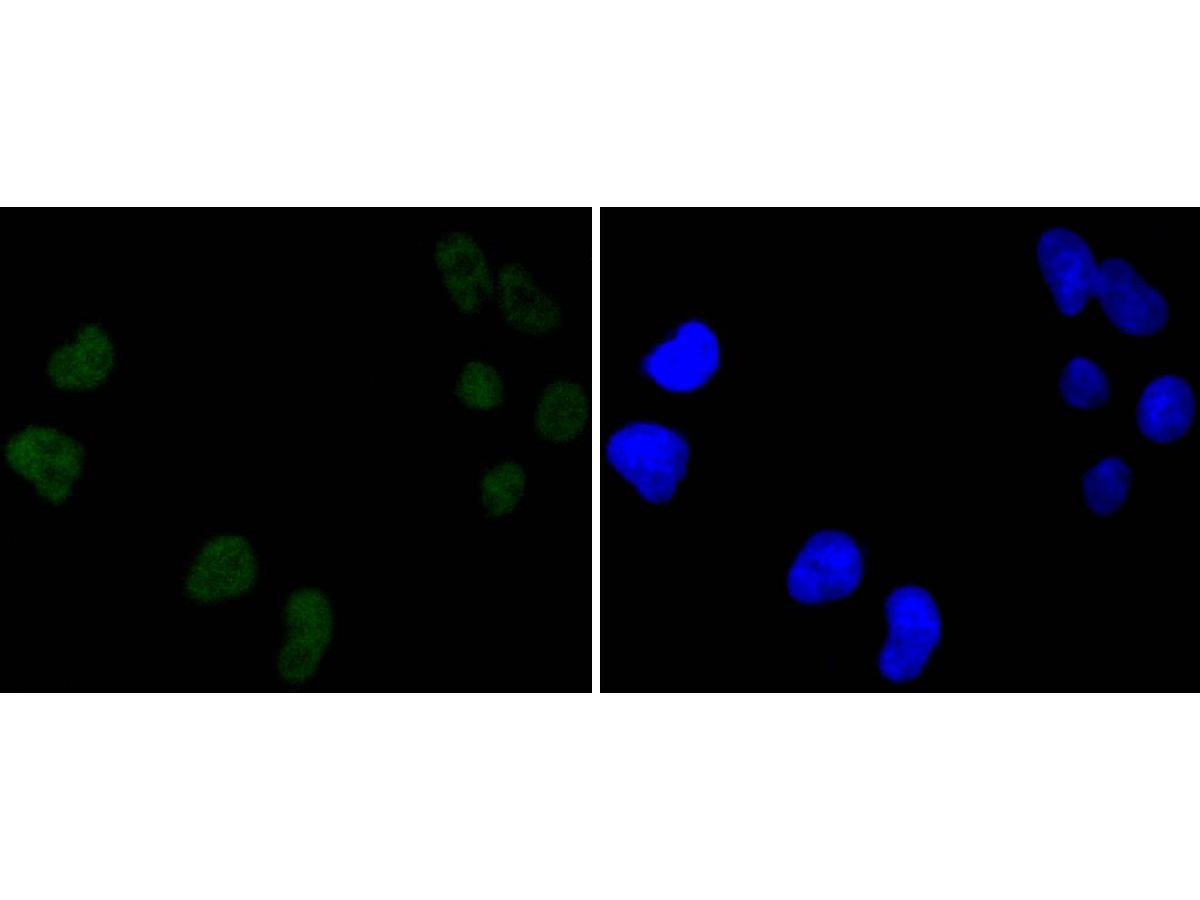 ICC staining of DNA PKcs in Hela cells (green). Formalin fixed cells were permeabilized with 0.1% Triton X-100 in TBS for 10 minutes at room temperature and blocked with 1% Blocker BSA for 15 minutes at room temperature. Cells were probed with the primary antibody (SLM-52493R, 1/50) for 1 hour at room temperature, washed with PBS. Alexa Fluor®488 Goat anti-Rabbit IgG was used as the secondary antibody at 1/1,000 dilution. The nuclear counter stain is DAPI (blue).
Cartpieces
Totalgoods,subtotals:¥Checkout
References (0)
No References
Bought notes(bought amounts latest0)
No one bought this product
User Comment(Total0User Comment Num)
- No comment


 +86 571 56623320
 +86 18668110335

