
Rabbit Anti-JAK1 antibody
JAK 1; JAK 1A; JAK 1B; JAK1; JAK1A; JAK1B; Janus kinase 1 (a protein tyrosine kinase); Janus kinase 1; JTK3; Tyrosine protein kinase JAK 1; Tyrosine protein kinase JAK1; JAK1_HUMAN.
View History [Clear]
Details
Product Name JAK1 Chinese Name 蛋白质酪氨酸激酶JAK-1Recombinant rabbit monoclonal anti Alias JAK 1; JAK 1A; JAK 1B; JAK1; JAK1A; JAK1B; Janus kinase 1 (a protein tyrosine kinase); Janus kinase 1; JTK3; Tyrosine protein kinase JAK 1; Tyrosine protein kinase JAK1; JAK1_HUMAN. Research Area Tumour Cell biology immunology Neurobiology Signal transduction Kinases and Phosphatases Immunogen Species Rabbit Clonality Monoclonal React Species (predicted: Human, Mouse, Rat, ) Applications WB=1:500-1000 IHC-P=1:100-500 (Paraffin sections need antigen repair)
not yet tested in other applications.
optimal dilutions/concentrations should be determined by the end user.Theoretical molecular weight 133kDa Cellular localization cytoplasmic Form Liquid Concentration 1mg/ml immunogen KLH conjugated synthetic peptide derived from human JAK1 Lsotype IgG Purification affinity purified by Protein A Buffer Solution 0.01M TBS(pH7.4) with 1% BSA, 0.03% Proclin300 and 50% Glycerol. Storage Shipped at 4℃. Store at -20 °C for one year. Avoid repeated freeze/thaw cycles. Attention This product as supplied is intended for research use only, not for use in human, therapeutic or diagnostic applications. PubMed PubMed Product Detail Janus kinase 1 (JAK1) is a member of a new class of non-receptor protein-tyrosine kinases (PTK) characterized by the presence of a second phosphotransferase-related domain immediately N-terminal to the PTK domain. The second phosphotransferase domain bears all the hallmarks of a protein kinase, although its structure differs significantly from that of the PTK and threonine/serine kinase family members. JAK1 is a large, widely expressed membrane-associated phosphoprotein. It is involved in the interferon-alpha/beta and -gamma signal transduction pathways. The reciprocal interdependence between JAK1 and TYK2 activities in the interferon-alpha pathway, and between JAK1 and JAK2 in the interferon-gamma pathway, may reflect a requirement for these kinases in the correct assembly of interferon recpeptor complexes. These kinases couple cytokine ligand binding to tyrosine phosphorylation of various known signaling proteins and a unique family of transcription factors termed the signal transducers and activators of transcription, or STATs.
Function:
Tyrosine kinase of the non-receptor type, involved in the IFN-alpha/beta/gamma signal pathway. Kinase partner for the interleukin (IL)-2 receptor.
Subunit:
Interacts with FER (By similarity). Interacts with IL31RA, IFNAR2, JAKMIP1 and SHB.
Subcellular Location:
Endomembrane system; Peripheral membrane protein. Note=Wholly intracellular, possibly membrane associated.
Tissue Specificity:
Expressed at higher levels in primary colon tumors than in normal colon tissue. The expression level in metastatic colon tumors is comparable to the expression level in normal colon tissue.
Similarity:
Belongs to the protein kinase superfamily. Tyr protein kinase family. JAK subfamily.
Contains 1 FERM domain.
Contains 2 protein kinase domains.
Contains 1 SH2 domain.
SWISS:
P23458
Gene ID:
3716
Database links:Entrez Gene: 395681 Chicken
Entrez Gene: 3716 Human
Entrez Gene: 16451 Mouse
Omim: 147795 Human
SwissProt: P23458 Human
SwissProt: P52332 Mouse
Unigene: 207538 Human
Unigene: 289657 Mouse
Unigene: 90191 Rat
Kinases and Phosphatases(Kinases and Phosphatases)Product Picture 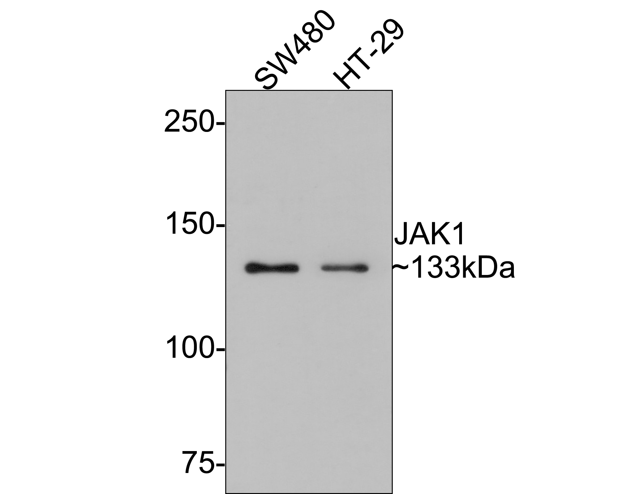 Western blot analysis of JAK1 on different lysates. Proteins were transferred to a PVDF membrane and blocked with 5% BSA in PBS for 1 hour at room temperature. The primary antibody (SLM-54138R, 1/500) was used in 5% BSA at room temperature for 2 hours. Goat Anti-Rabbit IgG - HRP Secondary Antibody (HA1001) at 1:5,000 dilution was used for 1 hour at room temperature. Positive control:
Lane 1: PC-12 cell lysate
Lane 2: Jurkat cell lysate
Lane 2: NIH/3T3 cell lysate
Western blot analysis of JAK1 on different lysates with Rabbit anti-JAK1 antibody (SLM-54138R) at 1/500 dilution.
Lane 1: SW480 cell lysate
Lane 2: HT-29 cell lysate
Lysates/proteins at 10 µg/Lane.
Predicted band size: 133 kDa
Observed band size: 133 kDa
Exposure time: 2 minutes;
6% SDS-PAGE gel.
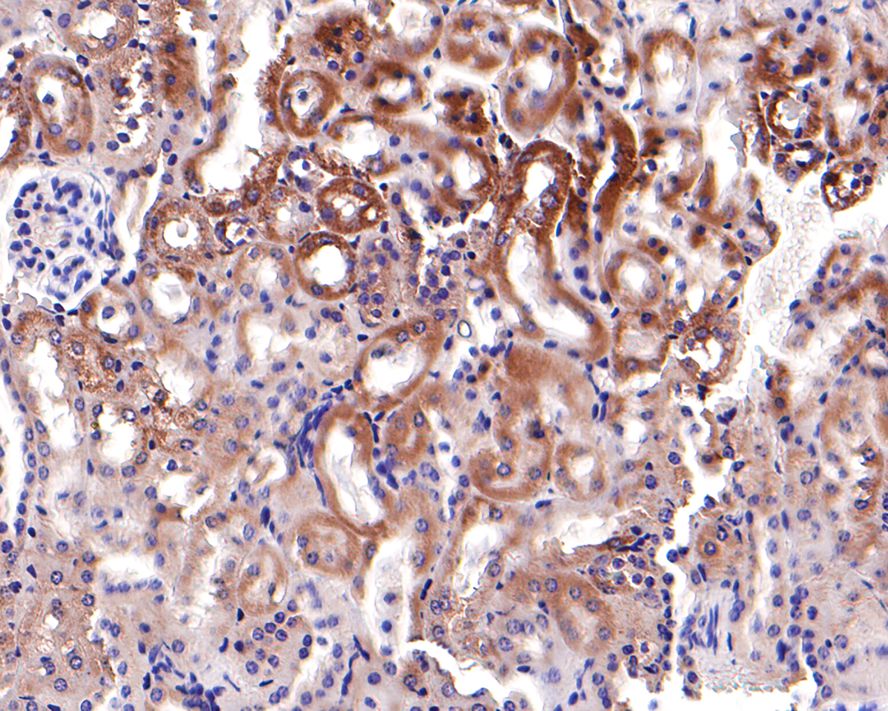 Immunohistochemical analysis of paraffin-embedded mouse kidney tissue with Rabbit anti-JAK1 antibody (SLM-54138R) at 1/500 dilution. The section was pre-treated using heat mediated antigen retrieval with Tris-EDTA buffer (pH 9.0) for 20 minutes. The tissues were blocked in 1% BSA for 20 minutes at room temperature, washed with ddH2O and PBS, and then probed with the primary antibody (SLM-54138R) at 1/500 dilution for 1 hour at room temperature. The detection was performed using an HRP conjugated compact polymer system. DAB was used as the chromogen. Tissues were counterstained with hematoxylin and mounted with DPX.
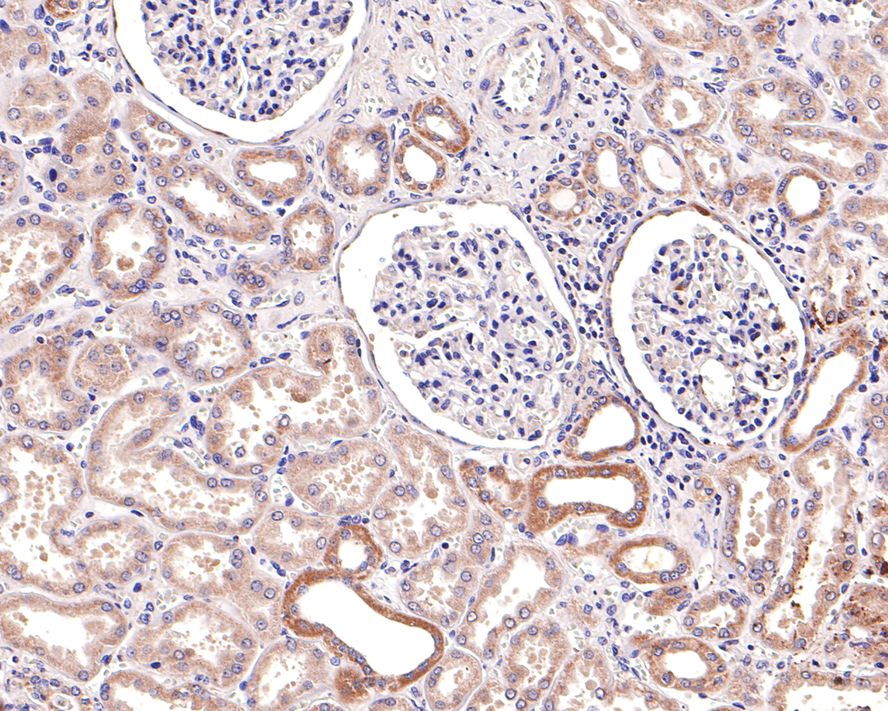 Immunohistochemical analysis of paraffin-embedded human kidney tissue with Rabbit anti-JAK1 antibody (SLM-54138R) at 1/200 dilution. The section was pre-treated using heat mediated antigen retrieval with Tris-EDTA buffer (pH 9.0) for 20 minutes. The tissues were blocked in 1% BSA for 20 minutes at room temperature, washed with ddH2O and PBS, and then probed with the primary antibody (SLM-54138R) at 1/200 dilution for 1 hour at room temperature. The detection was performed using an HRP conjugated compact polymer system. DAB was used as the chromogen. Tissues were counterstained with hematoxylin and mounted with DPX.
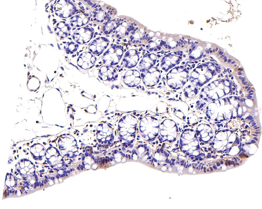 Immunohistochemical analysis of paraffin-embedded mouse large intestine tissue with Rabbit anti-JAK1 antibody (SLM-54138R) at 1/200 dilution. The section was pre-treated using heat mediated antigen retrieval with Tris-EDTA buffer (pH 9.0) for 20 minutes. The tissues were blocked in 1% BSA for 20 minutes at room temperature, washed with ddH2O and PBS, and then probed with the primary antibody (SLM-54138R) at 1/200 dilution for 1 hour at room temperature. The detection was performed using an HRP conjugated compact polymer system. DAB was used as the chromogen. Tissues were counterstained with hematoxylin and mounted with DPX.
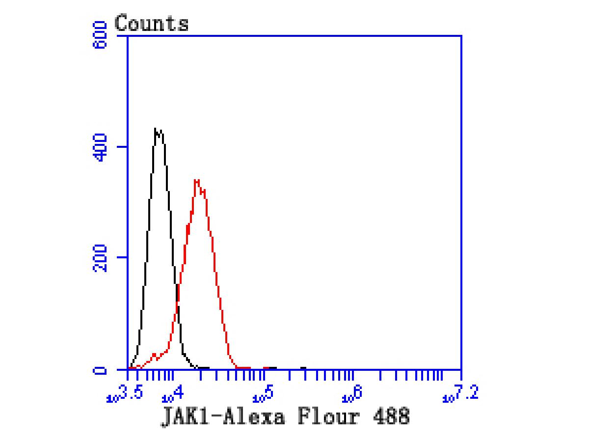 Flow cytometric analysis of JAK1 was done on SW480 cells. The cells were fixed, permeabilized and stained with the primary antibody (SLM-54138R, 1/50) (red). After incubation of the primary antibody at room temperature for an hour, the cells were stained with a Alexa Fluor 488-conjugated Goat anti-Rabbit IgG Secondary antibody at 1/1000 dilution for 30 minutes.Unlabelled sample was used as a control (cells without incubation with primary antibody; black).
Cartpieces
Totalgoods,subtotals:¥Checkout
References (0)
No References
Bought notes(bought amounts latest0)
No one bought this product
User Comment(Total0User Comment Num)
- No comment


 +86 571 56623320
 +86 18668110335

