
Rabbit Anti-HDAC9 antibody
HD 7; HD 7B; HD 9; HD7; HD7B; HD9; HDAC 7; HDAC 7B; HDAC 9; HDAC 9B; HDAC 9FL; HDAC; HDAC7; HDAC7B; HDAC9B; HDAC9FL; HDRP; Histone deacetylase 4/5 related protein; Histone deacetylase 7; Histone deacetylase 7B; Histone deacetylase 9; Histone deacetylase 9
View History [Clear]
Details
Product Name HDAC9 Chinese Name 组蛋白去乙酰化酶9Recombinant rabbit monoclonal anti Alias HD 7; HD 7B; HD 9; HD7; HD7B; HD9; HDAC 7; HDAC 7B; HDAC 9; HDAC 9B; HDAC 9FL; HDAC; HDAC7; HDAC7B; HDAC9B; HDAC9FL; HDRP; Histone deacetylase 4/5 related protein; Histone deacetylase 7; Histone deacetylase 7B; Histone deacetylase 9; Histone deacetylase 9A; Histone deacetylase related protein; KIAA0744; MEF2 interacting transcription repressor MITR; MEF2 interacting transcription repressor protein; MITR. DKFZp779K1053; HDAC9_HUMAN. Research Area Tumour immunology Signal transduction Apoptosis transcriptional regulatory factor Immunogen Species Rabbit Clonality Monoclonal React Species (predicted: Human, ) Applications WB=1:500-2000 IHC-P=1:100-500 ICC=1:50-200 (Paraffin sections need antigen repair)
not yet tested in other applications.
optimal dilutions/concentrations should be determined by the end user.Theoretical molecular weight 111kDa Cellular localization The nucleus Form Liquid Concentration 1mg/ml immunogen KLH conjugated synthetic peptide derived from human HDAC9 Lsotype IgG Purification affinity purified by Protein A Buffer Solution 0.01M TBS(pH7.4) with 1% BSA, 0.03% Proclin300 and 50% Glycerol. Storage Shipped at 4℃. Store at -20 °C for one year. Avoid repeated freeze/thaw cycles. Attention This product as supplied is intended for research use only, not for use in human, therapeutic or diagnostic applications. PubMed PubMed Product Detail HDAC9 (Histone Deacetylase 9) catalyses the deacetylation of lysine residues on the core histones (H2A, H2B, H3 and H4). It belongs to the Class IIa HDAC family, which are key regulators of several developmental and differentiation processes such as cardiac growth. HDAC9 represses MEF2 (myocyte enhancer factor 2)-dependent transcription, playing an important role in cardiac hypertrophy.
The HDAC9 isoform MITR/HDRP lacks active site residues and is therefore catalytically inactive. It represses MEF2-dependent transcription by interacting with HDAC1 and/or HDAC3. MITR/HDRP is thought to play a role in skeletal myogenesis and in heart development. It also protects neurons from apoptosis by inhibiting c-Jun phosphorylation by MAPK10 and by repressing c-Jun transcription through recruitment of HDAC1 to the c-Jun promoter.
Function:
Responsible for the deacetylation of lysine residues on the N-terminal part of the core histones (H2A, H2B, H3 and H4). Histone deacetylation gives a tag for epigenetic repression and plays an important role in transcriptional regulation, cell cycle progression and developmental events. Represses MEF2-dependent transcription.
Isoform 3 lacks active site residues and therefore is catalytically inactive. Represses MEF2-dependent transcription by recruiting HDAC1 and/or HDAC3. Seems to inhibit skeletal myogenesis and to be involved in heart development. Protects neurons from apoptosis, both by inhibiting JUN phosphorylation by MAPK10 and by repressing JUN transcription via HDAC1 recruitment to JUN promoter.
Subunit:
Homodimer. Interacts with CTBP1. The phosphorylated form interacts with 14-3-3. Interacts with HDAC1 and HDAC3, and probably with HDAC4 and HDAC5. Interacts with MEF2, MAPK10, ETV6, NCOR1 and BCL6.
Subcellular Location:
Nucleus.
Tissue Specificity:
Broadly expressed, with highest levels in brain, heart, muscle and testis. Isoform 3 is present in human bladder carcinoma cells (at protein level).
Post-translational modifications:
Phosphorylated on Ser-220 and Ser-450; which promotes 14-3-3-binding, impairs interaction with MEF2, and antagonizes antimyogenic activity. Phosphorylated on Ser-240; which impairs nuclear accumulation. Isoform 7 is phosphorylated on Tyr-1010. Phosphorylated by the PKC kinases PKN1 and PKN2, impairing nuclear import.
Sumoylated.
DISEASE:
Note=A chromosomal aberration involving HDAC9 is found in a family with Peters anomaly. Translocation t(1;7)(q41;p21) with TGFB2 resulting in lack of HDAC9 protein.
Similarity:
Belongs to the histone deacetylase family. HD type 2 subfamily.
SWISS:
Q9UKV0
Gene ID:
9734
Database links:Entrez Gene: 9734 Human
Entrez Gene: 79221 Mouse
Omim: 606543 Human
SwissProt: Q9UKV0 Human
SwissProt: Q99N13 Mouse
Unigene: 196054 Human
Unigene: 310551 Mouse
Unigene: 483009 Mouse
Product Picture 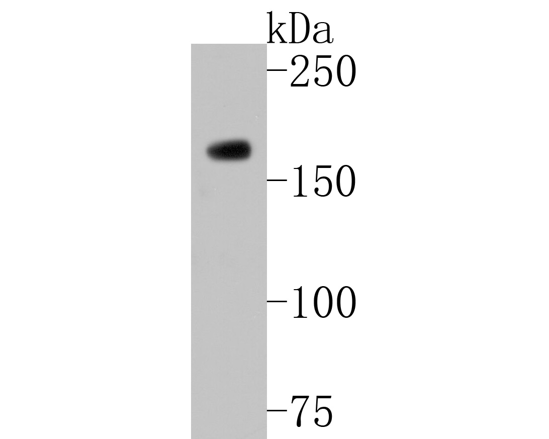 Western blot analysis of HDAC9 on K562 cell lysates. Proteins were transferred to a PVDF membrane and blocked with 5% BSA in PBS for 1 hour at room temperature. The primary antibody (SLM-54186R, 1/500) was used in 5% BSA at room temperature for 2 hours. Goat Anti-Rabbit IgG - HRP Secondary Antibody (HA1001) at 1:5,000 dilution was used for 1 hour at room temperature.
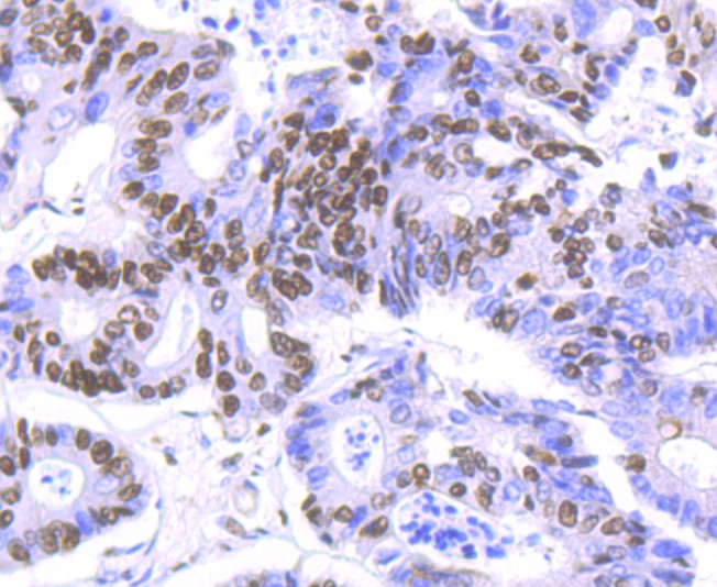 Immunohistochemical analysis of paraffin-embedded human colon carcinoma tissue using anti-HDAC9 antibody. The section was pre-treated using heat mediated antigen retrieval with Tris-EDTA buffer (pH 8.0-8.4) for 20 minutes.The tissues were blocked in 5% BSA for 30 minutes at room temperature, washed with ddH2O and PBS, and then probed with the primary antibody (SLM-54186R, 1/50) for 30 minutes at room temperature. The detection was performed using an HRP conjugated compact polymer system. DAB was used as the chromogen. Tissues were counterstained with hematoxylin and mounted with DPX.
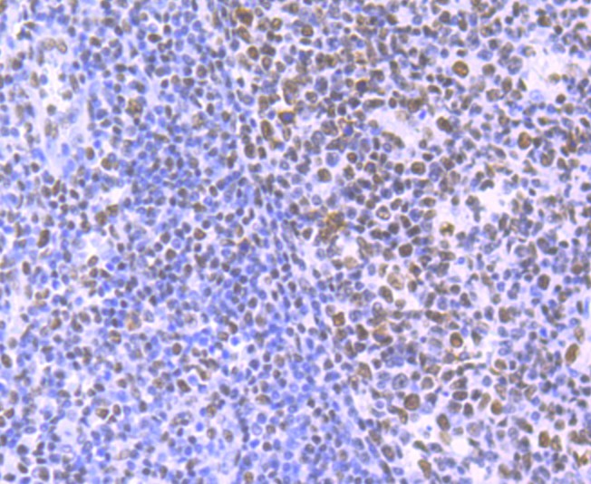 Immunohistochemical analysis of paraffin-embedded human tonsil tissue using anti-HDAC9 antibody. The section was pre-treated using heat mediated antigen retrieval with Tris-EDTA buffer (pH 8.0-8.4) for 20 minutes.The tissues were blocked in 5% BSA for 30 minutes at room temperature, washed with ddH2O and PBS, and then probed with the primary antibody (SLM-54186R, 1/50) for 30 minutes at room temperature. The detection was performed using an HRP conjugated compact polymer system. DAB was used as the chromogen. Tissues were counterstained with hematoxylin and mounted with DPX.
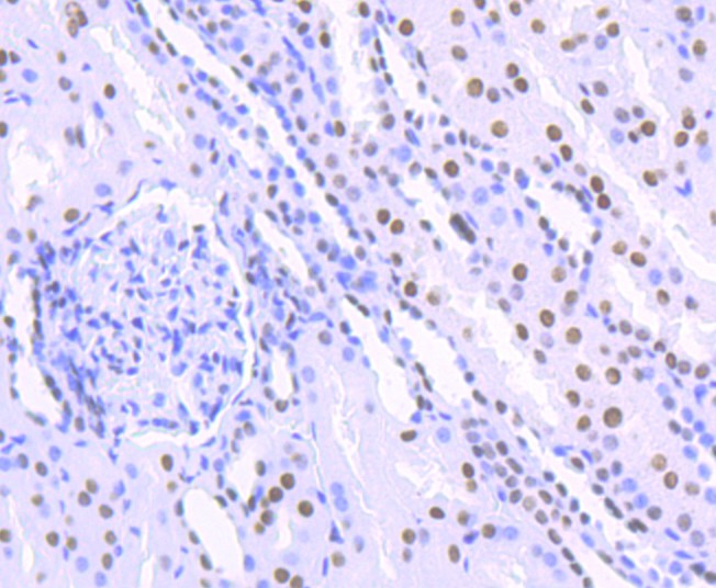 Immunohistochemical analysis of paraffin-embedded human kidney tissue using anti-HDAC9 antibody. The section was pre-treated using heat mediated antigen retrieval with Tris-EDTA buffer (pH 8.0-8.4) for 20 minutes.The tissues were blocked in 5% BSA for 30 minutes at room temperature, washed with ddH2O and PBS, and then probed with the primary antibody (SLM-54186R, 1/50) for 30 minutes at room temperature. The detection was performed using an HRP conjugated compact polymer system. DAB was used as the chromogen. Tissues were counterstained with hematoxylin and mounted with DPX.
 ICC staining of HDAC9 in PC-3M cells (green). Formalin fixed cells were permeabilized with 0.1% Triton X-100 in TBS for 10 minutes at room temperature and blocked with 1% Blocker BSA for 15 minutes at room temperature. Cells were probed with the primary antibody (SLM-54186R, 1/50) for 1 hour at room temperature, washed with PBS. Alexa Fluor®488 Goat anti-Rabbit IgG was used as the secondary antibody at 1/1,000 dilution. The nuclear counter stain is DAPI (blue).
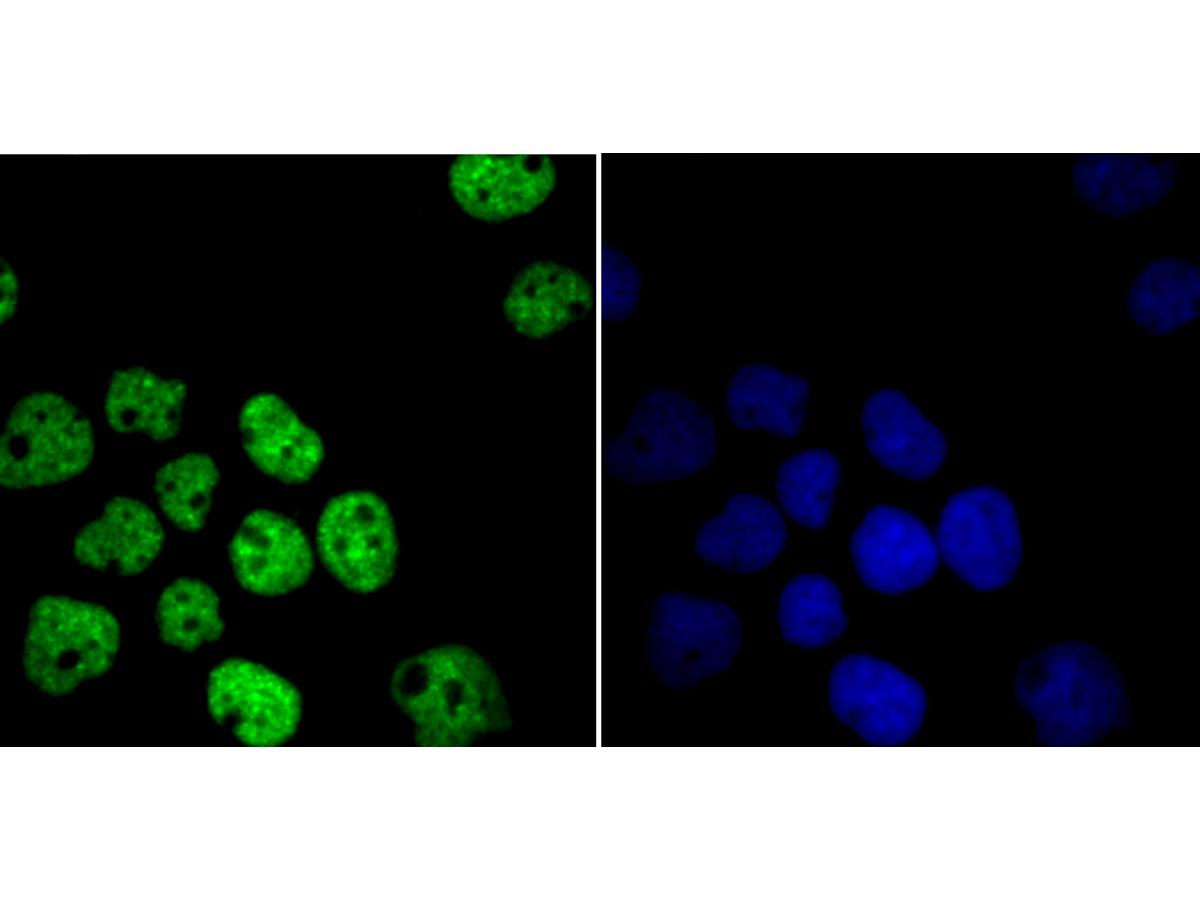 ICC staining of HDAC9 in A431 cells (green). Formalin fixed cells were permeabilized with 0.1% Triton X-100 in TBS for 10 minutes at room temperature and blocked with 1% Blocker BSA for 15 minutes at room temperature. Cells were probed with the primary antibody (SLM-54186R, 1/50) for 1 hour at room temperature, washed with PBS. Alexa Fluor®488 Goat anti-Rabbit IgG was used as the secondary antibody at 1/1,000 dilution. The nuclear counter stain is DAPI (blue).
Cartpieces
Totalgoods,subtotals:¥Checkout
References (0)
No References
Bought notes(bought amounts latest0)
No one bought this product
User Comment(Total0User Comment Num)
- No comment


 +86 571 56623320
 +86 18668110335

