
Rabbit Anti-phospho-Cyclin E1 (Thr77)antibody
Cyclin E1(T77); Cyclin E1 (phospho-Thr77); Cyclin E1 (phospho-T77); CCNE 1; CCNE; CCNE1; Cyclin Es; Cyclin Et; CyclinE; G1/S specific cyclin E; G1/S-specific cyclin-E1; CCNE1_HUMAN; pCCNE1.
View History [Clear]
Details
Product Name phospho-Cyclin E1 (Thr77) Chinese Name 磷酸化周期素E1Recombinant rabbit monoclonal anti Alias Cyclin E1(T77); Cyclin E1 (phospho-Thr77); Cyclin E1 (phospho-T77); CCNE 1; CCNE; CCNE1; Cyclin Es; Cyclin Et; CyclinE; G1/S specific cyclin E; G1/S-specific cyclin-E1; CCNE1_HUMAN; pCCNE1. Product Type Phosphorylated anti Recombinant rabbit monoclonal anti Research Area Tumour Cell biology Cyclin Immunogen Species Rabbit Clonality Monoclonal Clone NO. 4G10 React Species Human, Mouse, Rat, Applications WB=1:200-500 IHC-P=1:100-500 IHC-F=1:50-200 ICC=1:50-200 IF=1:50-200 (Paraffin sections need antigen repair)
not yet tested in other applications.
optimal dilutions/concentrations should be determined by the end user.Theoretical molecular weight 47kDa Cellular localization The nucleus Form Liquid Concentration 1mg/ml immunogen KLH conjugated Synthesised phosphopeptide derived from human Cyclin E around the phosphorylation site of Thr77: IP(p-T)PD Lsotype IgG Purification affinity purified by Protein A Buffer Solution 0.01M TBS(pH7.4) with 1% BSA, 0.03% Proclin300 and 50% Glycerol. Storage Shipped at 4℃. Store at -20 °C for one year. Avoid repeated freeze/thaw cycles. Attention This product as supplied is intended for research use only, not for use in human, therapeutic or diagnostic applications. PubMed PubMed Product Detail Cyclin E is a regulatory subunit of Cdk2 and controls G1 / S transition during the mammalian cell cycle. Multiple isoforms of Cyclin E are only expressed in tumors but not in normal tissue, suggesting a post transcriptional regulation of Cyclin E. In vitro analyses indicated that these truncated variant isoforms of Cyclin E are able to phosphorylate histone H1. Alterations in the Cyclin E protein have been implicated as indicators of worse prognosis in various cancers.
Function:
Essential for the control of the cell cycle at the G1/S (start) transition.
Subunit:
Interacts with a member of the CDK2/CDK protein kinases to form a serine/threonine kinase holoenzyme complex. The cyclin subunit imparts substrate specificity to the complex. Found in a complex with CDK2, CABLES1 and CCNA1. Part of a complex consisting of UHRF2, CDK2 and CCNE1. Interacts directly with UHRF2; the interaction ubiquitinates CCNE1 and appears to occur independently of CCNE1 phosphorylation.
Subcellular Location:
Nucleus.
Tissue Specificity:
Highly expressed in testis and placenta. Low levels in bronchial epithelial cells.
Post-translational modifications:
Phosphorylation of Thr-395 by GSK3 and of Ser-399 by CDK2 accelerates degradation via the ubiquitin proteasome pathway. Phosphorylated upon DNA damage, probably by ATM or ATR.
Ubiquitinated by UHRF2; appears to occur independently of phosphorylation.
Similarity:
Belongs to the cyclin family. Cyclin E subfamily.
SWISS:
P24864
Gene ID:
898
Database links:Entrez Gene: 898 Human
Entrez Gene: 12447 Mouse
Omim: 123837 Human
SwissProt: P24864 Human
SwissProt: Q61457 Mouse
Unigene: 244723 Human
Unigene: 16110 Mouse
Unigene: 15455 Rat
Product Picture 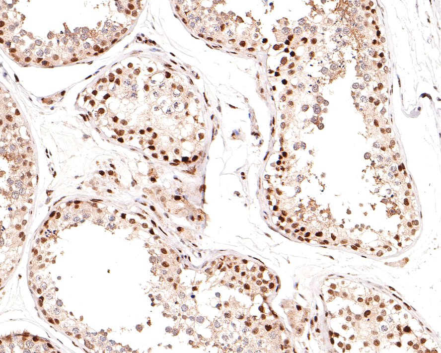 Immunohistochemical analysis of paraffin-embedded human testis tissue using anti-Phospho-Cyclin E1(T77) antibody. The section was pre-treated using heat mediated antigen retrieval with sodium citrate buffer (pH 6.0) for 20 minutes. The tissues were blocked in 1% BSA for 30 minutes at room temperature, washed with ddH2O and PBS, and then probed with the primary antibody (SLM-52146R, 1/400) for 30 minutes at room temperature. The detection was performed using an HRP conjugated compact polymer system. DAB was used as the chromogen. Tissues were counterstained with hematoxylin and mounted with DPX.
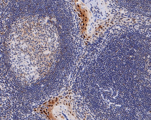 Immunohistochemical analysis of paraffin-embedded human tonsil tissue using anti-Phospho-Cyclin E1(T77) antibody. The section was pre-treated using heat mediated antigen retrieval with sodium citrate buffer (pH 6.0) for 20 minutes. The tissues were blocked in 5% BSA for 30 minutes at room temperature, washed with ddH2O and PBS, and then probed with the primary antibody (SLM-52146R, 1/50) for 30 minutes at room temperature. The detection was performed using an HRP conjugated compact polymer system. DAB was used as the chromogen. Tissues were counterstained with hematoxylin and mounted with DPX.
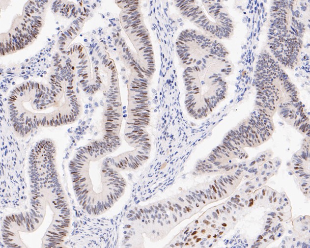 Immunohistochemical analysis of paraffin-embedded human colon carcinoma tissue using anti-Phospho-Cyclin E1(T77) antibody. The section was pre-treated using heat mediated antigen retrieval with sodium citrate buffer (pH 6.0) for 20 minutes. The tissues were blocked in 5% BSA for 30 minutes at room temperature, washed with ddH2O and PBS, and then probed with the primary antibody (SLM-52146R, 1/50) for 30 minutes at room temperature. The detection was performed using an HRP conjugated compact polymer system. DAB was used as the chromogen. Tissues were counterstained with hematoxylin and mounted with DPX.
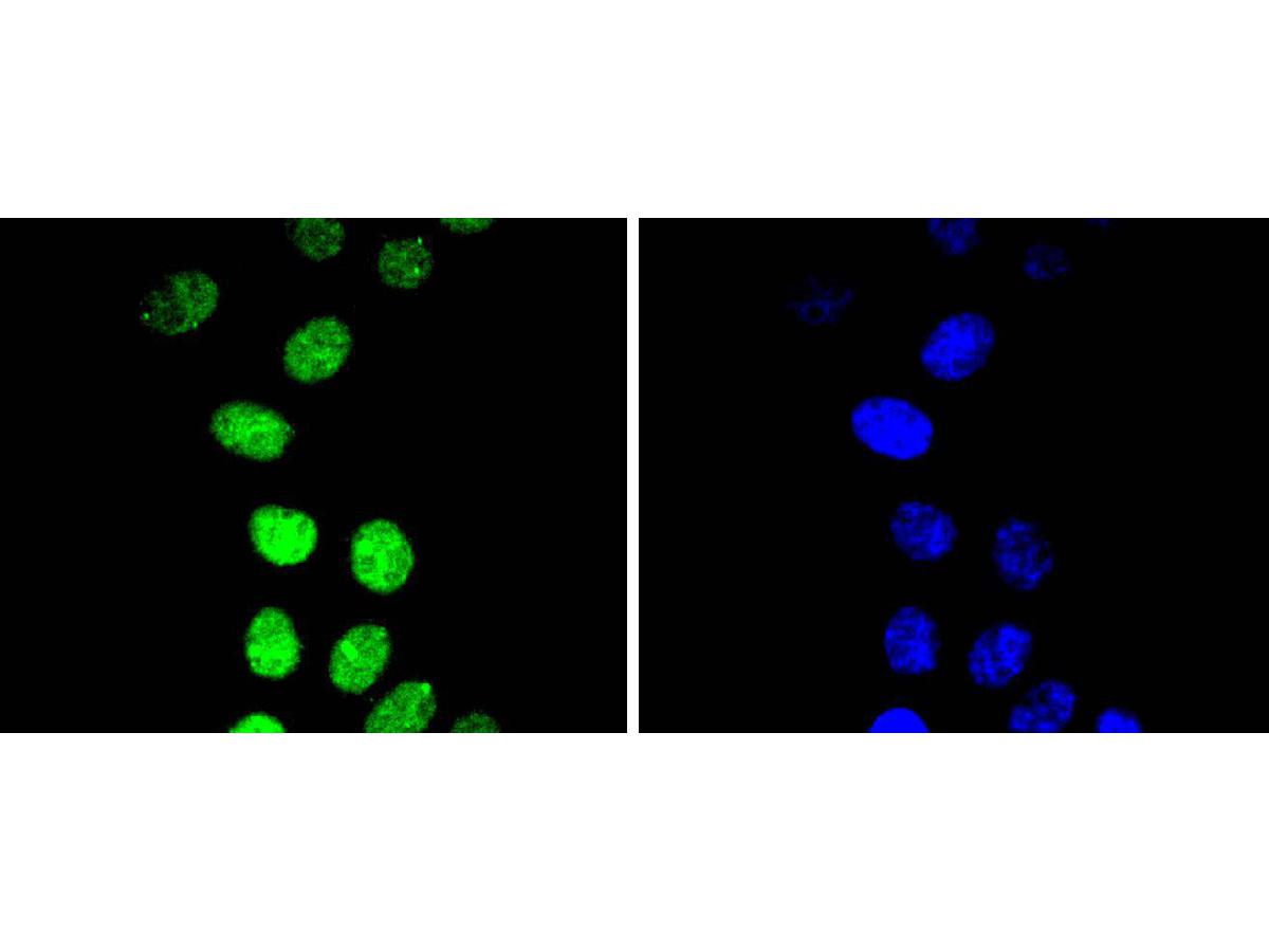 ICC staining of Phospho-Cyclin E1(T77) in HepG2 cells (green). Formalin fixed cells were permeabilized with 0.1% Triton X-100 in TBS for 10 minutes at room temperature and blocked with 1% Blocker BSA for 15 minutes at room temperature. Cells were probed with the primary antibody (SLM-52146R, 1/50) for 1 hour at room temperature, washed with PBS. Alexa Fluor®488 Goat anti-Rabbit IgG was used as the secondary antibody at 1/1,000 dilution. The nuclear counter stain is DAPI (blue).
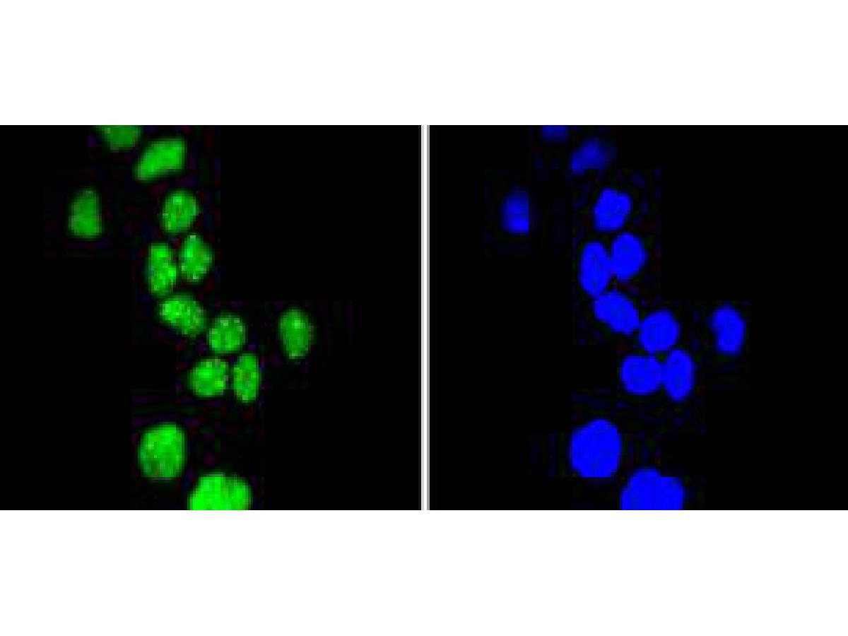 ICC staining of Phospho-Cyclin E1(T77) in Hela cells (green). Formalin fixed cells were permeabilized with 0.1% Triton X-100 in TBS for 10 minutes at room temperature and blocked with 1% Blocker BSA for 15 minutes at room temperature. Cells were probed with the primary antibody (SLM-52146R, 1/50) for 1 hour at room temperature, washed with PBS. Alexa Fluor®488 Goat anti-Rabbit IgG was used as the secondary antibody at 1/1,000 dilution. The nuclear counter stain is DAPI (blue).
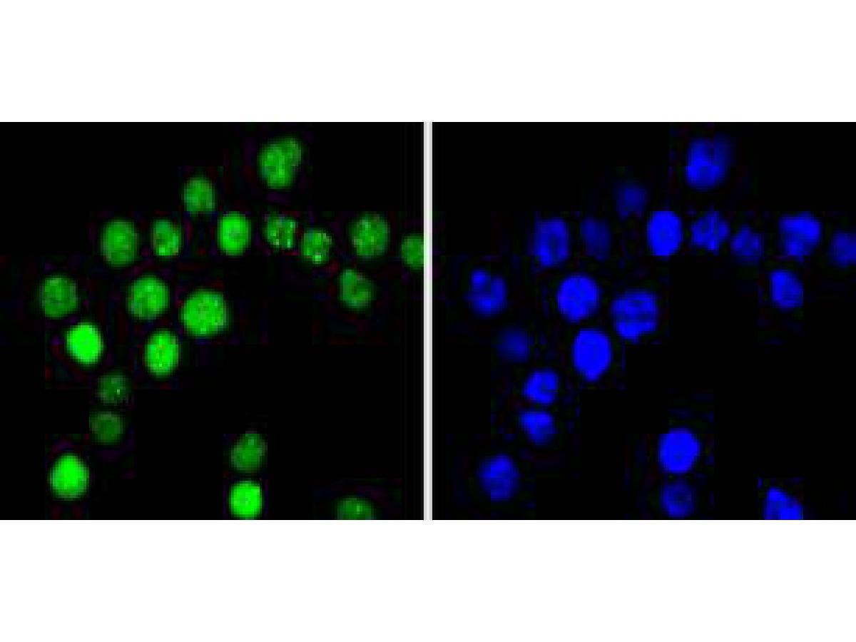 ICC staining of Phospho-Cyclin E1(T77) in SW480 cells (green). Formalin fixed cells were permeabilized with 0.1% Triton X-100 in TBS for 10 minutes at room temperature and blocked with 1% Blocker BSA for 15 minutes at room temperature. Cells were probed with the primary antibody (SLM-52146R, 1/50) for 1 hour at room temperature, washed with PBS. Alexa Fluor®488 Goat anti-Rabbit IgG was used as the secondary antibody at 1/1,000 dilution. The nuclear counter stain is DAPI (blue).
Cartpieces
Totalgoods,subtotals:¥Checkout
Bought notes(bought amounts latest0)
No one bought this product
User Comment(Total0User Comment Num)
- No comment


 +86 571 56623320
 +86 18668110335

