
Rabbit Anti-Integrin beta 1 antibody
Integrin beta-1; beta 1 subunit; CD 29; CD-29; CD29; CD29 antigen; Fibrinogen Receptor beta subunit; Fibronectin receptor subunit beta; Fibronectin receptor, beta subunit; FNRB; GPIIA; GP IIa; Integrin VLA 4 subunit beta; ITGB; ITGB1; ITGB 1; MDF2; MSK12;
View History [Clear]
Details
Product Name Integrin beta 1 Chinese Name 整合素β1(CD29)Recombinant rabbit monoclonal anti Alias Integrin beta-1; beta 1 subunit; CD 29; CD-29; CD29; CD29 antigen; Fibrinogen Receptor beta subunit; Fibronectin receptor subunit beta; Fibronectin receptor, beta subunit; FNRB; GPIIA; GP IIa; Integrin VLA 4 subunit beta; ITGB; ITGB1; ITGB 1; MDF2; MSK12; Very Late Activation Protein beta Polypeptide; Glycoprotein IIa; Integrin VLA 4 subunit beta; integrin VLA-4 beta subunit; ITB1_HUMAN; very late activation protein, beta polypeptide; VLA BETA; VLA-4 subunit beta; VLA-BETA; VLAB; VLAbeta; Integrin β1. Research Area Cell biology immunology Cell adhesion molecule Immunogen Species Rabbit Clonality Monoclonal React Species (predicted: Human, Mouse, Rat, ) Applications WB=1:1000-2000 IHC-P=1:100-500 (Paraffin sections need antigen repair)
not yet tested in other applications.
optimal dilutions/concentrations should be determined by the end user.Theoretical molecular weight 88kDa Cellular localization cytoplasmic The cell membrane Form Liquid Concentration 1mg/ml immunogen KLH conjugated synthetic peptide derived from human Integrin beta 1 Lsotype IgG Purification affinity purified by Protein A Buffer Solution 0.01M TBS(pH7.4) with 1% BSA, 0.03% Proclin300 and 50% Glycerol. Storage Shipped at 4℃. Store at -20 °C for one year. Avoid repeated freeze/thaw cycles. Attention This product as supplied is intended for research use only, not for use in human, therapeutic or diagnostic applications. PubMed PubMed Product Detail Integrins alpha-1/beta-1, alpha-2/beta-1, alpha-10/beta-1 and alpha-11/beta-1 are receptors for collagen. Integrins alpha-1/beta-1 and alpha-2/beta-2 recognize the proline-hydroxylated sequence G-F-P-G-E-R in collagen. Integrins alpha-2/beta-1, alpha-3/beta-1, alpha-4/beta-1, alpha-5/beta-1, alpha-8/beta-1, alpha-10/beta-1, alpha-11/beta-1 and alpha-V/beta-1 are receptors for fibronectin. Alpha-4/beta-1 recognizes one or more domains within the alternatively spliced CS-1 and CS-5 regions of fibronectin. Integrin alpha-5/beta-1 is a receptor for fibrinogen. Integrin alpha-1/beta-1, alpha-2/beta-1, alpha-6/beta-1 and alpha-7/beta-1 are receptors for lamimin. Integrin alpha-4/beta-1 is a receptor for VCAM1. It recognizes the sequence Q-I-D-S in VCAM1. Integrin alpha-9/beta-1 is a receptor for VCAM1, cytotactin and osteopontin. It recognizes the sequence A-E-I-D-G-I-E-L in cytotactin. Integrin alpha-3/beta-1 is a receptor for epiligrin, thrombospondin and CSPG4. Alpha-3/beta-1 may mediate with LGALS3 the stimulation by CSPG4 of endothelial cells migration. Integrin alpha-V/beta-1 is a receptor for vitronectin. Beta-1 integrins recognize the sequence R-G-D in a wide array of ligands. Isoform beta-1B interferes with isoform beta-1A resulting in a dominant negative effect on cell adhesion and migration (in vitro). In case of HIV-1 infection, the interaction with extracellular viral Tat protein seems to enhance angiogenesis in Kaposi's sarcoma lesions.
Function:
Integrins alpha-1/beta-1, alpha-2/beta-1, alpha-10/beta-1 and alpha-11/beta-1 are receptors for collagen. Integrins alpha-1/beta-1 and alpha-2/beta-2 recognize the proline-hydroxylated sequence G-F-P-G-E-R in collagen. Integrins alpha-2/beta-1, alpha-3/beta-1, alpha-4/beta-1, alpha-5/beta-1, alpha-8/beta-1, alpha-10/beta-1, alpha-11/beta-1 and alpha-V/beta-1 are receptors for fibronectin. Alpha-4/beta-1 recognizes one or more domains within the alternatively spliced CS-1 and CS-5 regions of fibronectin. Integrin alpha-5/beta-1 is a receptor for fibrinogen. Integrin alpha-1/beta-1, alpha-2/beta-1, alpha-6/beta-1 and alpha-7/beta-1 are receptors for lamimin. Integrin alpha-4/beta-1 is a receptor for VCAM1. It recognizes the sequence Q-I-D-S in VCAM1. Integrin alpha-9/beta-1 is a receptor for VCAM1, cytotactin and osteopontin. It recognizes the sequence A-E-I-D-G-I-E-L in cytotactin. Integrin alpha-3/beta-1 is a receptor for epiligrin, thrombospondin and CSPG4. Alpha-3/beta-1 may mediate with LGALS3 the stimulation by CSPG4 of endothelial cells migration. Integrin alpha-V/beta-1 is a receptor for vitronectin. Beta-1 integrins recognize the sequence R-G-D in a wide array of ligands. Isoform beta-1B interferes with isoform beta-1A resulting in a dominant negative effect on cell adhesion and migration (in vitro). In case of HIV-1 infection, the interaction with extracellular viral Tat protein seems to enhance angiogenesis in Kaposi's sarcoma lesions.
Subunit:
Heterodimer of an alpha and a beta subunit. Beta-1 associates with either alpha-1, alpha-2, alpha-3, alpha-4, alpha-5, alpha-6, alpha-7, alpha-8, alpha-9, alpha-10, alpha-11 or alpha-V. Binds LGALS3BP and ITGB1BP3, when associated with alpha-7, but not with alpha-5. Interacts with FLNA, FLNB and RANBP9. Isoform Beta-1D interacts with ACE2. Isoform Beta-1A interacts with the C-terminal region of FLNC. Interacts with KRT1 in the presence of GNB2L1 and SRC. Interacts with HIV-1 Tat. Binds to human echoviruses 1 and 8 capsid proteins and acts as a receptor for these viruses. Interacts with RAB21. Interacts (via the cytoplasmic region) with RAB25 (via the hypervariable C-terminal region). Interacts with FGR and HCK (By similarity). Interacts with MYO10.
Subcellular Location:
Cell membrane; Single-pass type I membrane protein. Melanosome. Cleavage furrow. Note=Isoform beta-1B does not localize to focal adhesions. Highly enriched in stage I melanosomes. Located on plasma membrane of neuroblastoma NMB7 cells. In a lung cancer cell line, in prometaphase and metaphase, localizes diffusely at the membrane and in afew intracellular vesicles. In early telophase, detected mainly on the matrix-facing side of the cells. By mid-telophase, concentrated to the ingressing cleavage furrow, mainly to the basal side of the furrow. In late telophase, concentrated to the extending protrusions formed at the opposite ends of the spreading daughter cells, in vesicles at the base of the lamellipodia formed by the separating daughter cells.
Tissue Specificity:
Isoform beta-1A is widely expressed, other isoforms are generally coexpressed with a more restricted distribution. Isoform beta-1B is expressed in skin, liver, skeletal muscle, cardiac muscle, placenta, umbilical vein endothelial cells, neuroblastoma cells, lymphoma cells, hepatoma cells and astrocytoma cells. Isoform beta-1C and isoform beta-1C-2 are expressed in muscle, kidney, liver, placenta, cervical epithelium, umbilical vein endothelial cells, fibroblast cells, embryonal kidney cells, platelets and several blood cell lines. Isoform beta-C-2, rather than isoform beta-1C, is selectively expressed in peripheral T-cells. Isoform beta-1C is expressed in non-proliferating and differentiated prostate gland epithelial cells and in platelets, on the surface of erythroleukemia cells and in various hematopoietic cell lines. Isoform beta-1D is expressed specifically in striated muscle (skeletal and cardiac muscle).
Post-translational modifications:
The cysteine residues are involved in intrachain disulfide bonds.
Similarity:
Belongs to the integrin beta chain family.
Contains 1 VWFA domain.
SWISS:
P05556
Gene ID:
3688
Database links:Entrez Gene: 3688 Human
Entrez Gene: 16412 Mouse
Omim: 135630 Human
SwissProt: P05556 Human
SwissProt: P09055 Mouse
Unigene: 643813 Human
Unigene: 263396 Mouse
Unigene: 25733 Rat
Product Picture 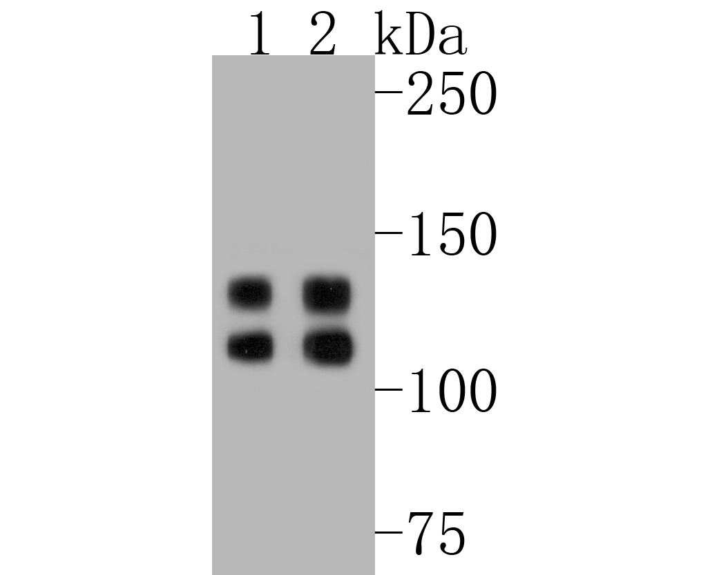 Western blot analysis of Integrin beta 1 on different lysates. Proteins were transferred to a PVDF membrane and blocked with 5% BSA in PBS for 1 hour at room temperature. The primary antibody (SLM-52266R, 1/1,000) was used in 5% BSA at room temperature for 2 hours. Goat Anti-Rabbit IgG - HRP Secondary Antibody (HA1001) at 1:5,000 dilution was used for 1 hour at room temperature.
Positive control:
Lane 1: CRC cell lysate
Lane 2: HepG2 cell lysate
 Blocking buffer: 5% NFDM/TBST
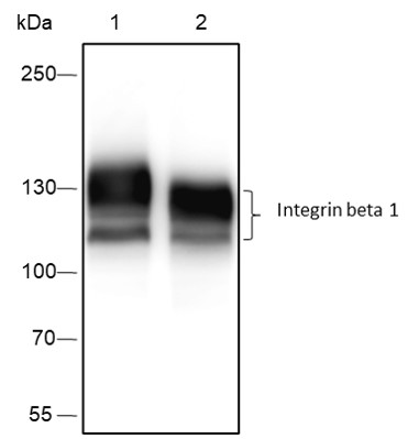
Primary Ab dilution: 1:2000
Primary Ab incubation condition: 2 hours at room temperature
Secondary Ab: Goat Anti-Rabbit IgG H&L (HRP)
Lysate: 1: A431, 2: U87-MG
Protein loading quantity: 20 μg
Exposure time: 10 s
Predicted MW: 88 kDa
Observed MW: 120, 130 kDa
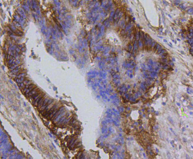 Immunohistochemical analysis of paraffin-embedded human colon carcinoma tissue using anti-Integrin beta 1 antibody. The section was pre-treated using heat mediated antigen retrieval with Tris-EDTA buffer (pH 8.0-8.4) for 20 minutes.The tissues were blocked in 5% BSA for 30 minutes at room temperature, washed with ddH2O and PBS, and then probed with the primary antibody (SLM-52266R, 1/50) for 30 minutes at room temperature. The detection was performed using an HRP conjugated compact polymer system. DAB was used as the chromogen. Tissues were counterstained with hematoxylin and mounted with DPX.
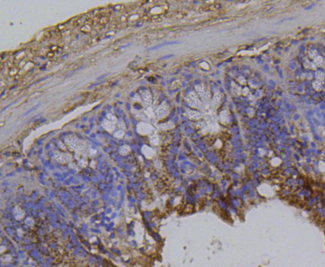 Immunohistochemical analysis of paraffin-embedded mouse colon tissue using anti-Integrin beta 1 antibody. The section was pre-treated using heat mediated antigen retrieval with Tris-EDTA buffer (pH 8.0-8.4) for 20 minutes.The tissues were blocked in 5% BSA for 30 minutes at room temperature, washed with ddH2O and PBS, and then probed with the primary antibody (SLM-52266R, 1/50) for 30 minutes at room temperature. The detection was performed using an HRP conjugated compact polymer system. DAB was used as the chromogen. Tissues were counterstained with hematoxylin and mounted with DPX.
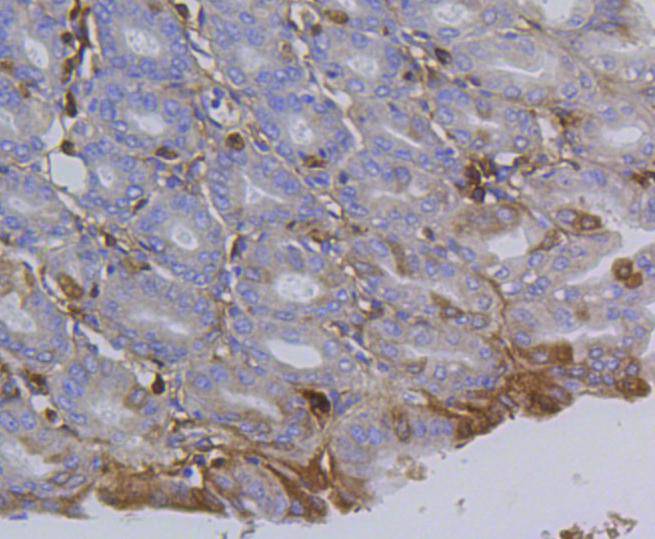 Immunohistochemical analysis of paraffin-embedded mouse stomach tissue using anti-Integrin beta 1 antibody. The section was pre-treated using heat mediated antigen retrieval with Tris-EDTA buffer (pH 8.0-8.4) for 20 minutes.The tissues were blocked in 5% BSA for 30 minutes at room temperature, washed with ddH2O and PBS, and then probed with the primary antibody (SLM-52266R, 1/50) for 30 minutes at room temperature. The detection was performed using an HRP conjugated compact polymer system. DAB was used as the chromogen. Tissues were counterstained with hematoxylin and mounted with DPX.
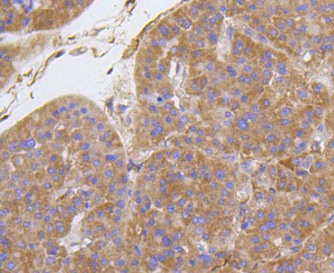 Immunohistochemical analysis of paraffin-embedded human liver carcinoma tissue using anti-Integrin beta 1 antibody. The section was pre-treated using heat mediated antigen retrieval with Tris-EDTA buffer (pH 8.0-8.4) for 20 minutes.The tissues were blocked in 5% BSA for 30 minutes at room temperature, washed with ddH2O and PBS, and then probed with the primary antibody (SLM-52266R, 1/50) for 30 minutes at room temperature. The detection was performed using an HRP conjugated compact polymer system. DAB was used as the chromogen. Tissues were counterstained with hematoxylin and mounted with DPX.
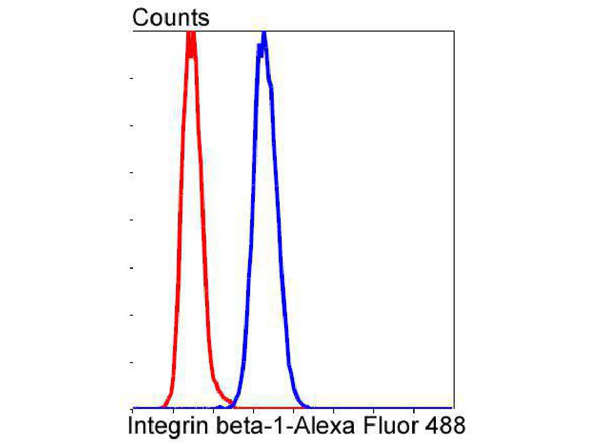 Flow cytometric analysis of Integrin beta 1 was done on Hela cells. The cells were fixed, permeabilized and stained with the primary antibody (SLM-52266R, 1/50) (blue). After incubation of the primary antibody at room temperature for an hour, the cells were stained with a Alexa Fluor 488-conjugated Goat anti-Rabbit IgG Secondary antibody at 1/1,000 dilution for 30 minutes.Unlabelled sample was used as a control (cells without incubation with primary antibody; red).
Cartpieces
Totalgoods,subtotals:¥Checkout
References (0)
No References
Bought notes(bought amounts latest0)
No one bought this product
User Comment(Total0User Comment Num)
- No comment


 +86 571 56623320
 +86 18668110335

