
Rabbit Anti-CHMP2B antibody
Charged multivesicular body protein 2b; CHM2B_HUMAN; CHMP2.5; CHMP2b; Chromatin modifying protein 2b; Chromatin-modifying protein 2b; DMT1; hVps2-2; Vacuolar protein sorting 2-2; VPS2 homolog B; Vacuolar protein sorting-associated protein 2-2; Vps2-2; VPS
View History [Clear]
Details
Product Name CHMP2B Chinese Name 染色质修饰蛋白2BRecombinant rabbit monoclonal anti Alias Charged multivesicular body protein 2b; CHM2B_HUMAN; CHMP2.5; CHMP2b; Chromatin modifying protein 2b; Chromatin-modifying protein 2b; DMT1; hVps2-2; Vacuolar protein sorting 2-2; VPS2 homolog B; Vacuolar protein sorting-associated protein 2-2; Vps2-2; VPS2B. Research Area Tumour Cell biology Neurobiology Signal transduction Immunogen Species Rabbit Clonality Monoclonal React Species (predicted: Human, Mouse, Rat, ) Applications WB=1:500-1000 IHC-P=1:100-500 ICC=1:50-100 (Paraffin sections need antigen repair)
not yet tested in other applications.
optimal dilutions/concentrations should be determined by the end user.Theoretical molecular weight 24kDa Cellular localization cytoplasmic Form Liquid Concentration 1mg/ml immunogen Recombinant protein within C-terminal Human CHMP2B. Lsotype IgG Purification affinity purified by Protein A Buffer Solution 0.01M TBS(pH7.4) with 1% BSA, 0.03% Proclin300 and 50% Glycerol. Storage Shipped at 4℃. Store at -20 °C for one year. Avoid repeated freeze/thaw cycles. Attention This product as supplied is intended for research use only, not for use in human, therapeutic or diagnostic applications. PubMed PubMed Product Detail The charged multivesicular body proteins, commonly designated CHMPs, belong to the vacuolar sorting protein family and function as chromatin-modifying proteins. CHMP1-6 are all components of ESCRT (endosomal sorting complex required for transport) I, II or III complexes. These complexes are crucial for sorting endosomal articles into multivesicular bodies (MVBs), and are also required for the formation of these bodies. CHMP2B, also known as CHMP2.5 or vacuolar protein-sorting-associated protein 2-2, is a 213 amino acid cytosolic protein. Widely expressed in brain, heart, skeletal muscle, small intestine, pancreas, lung, placenta and leukocytes, CHMP2B associates directly with CHMP2A and vps4 for the disassembly of the ESCRT-III complex. Defects in the gene encoding CHMP2B have been shown to cause chromosome 3-linked frontotemporal dementia (FTD3).
Function:
Probable core component of the endosomal sorting required for transport complex III (ESCRT-III) which is involved in multivesicular bodies (MVBs) formation and sorting of endosomal cargo proteins into MVBs. MVBs contain intraluminal vesicles (ILVs) that are generated by invagination and scission from the limiting membrane of the endosome and mostly are delivered to lysosomes enabling degradation of membrane proteins, such as stimulated growth factor receptors, lysosomal enzymes and lipids. The MVB pathway appears to require the sequential function of ESCRT-O, -I,-II and -III complexes. ESCRT-III proteins mostly dissociate from the invaginating membrane before the ILV is released. The ESCRT machinery also functions in topologically equivalent membrane fission events, such as the terminal stages of cytokinesis and the budding of enveloped viruses (HIV-1 and other lentiviruses). ESCRT-III proteins are believed to mediate the necessary vesicle extrusion and/or membrane fission activities, possibly in conjunction with the AAA ATPase VPS4.
Subunit:
Probable core component of the endosomal sorting required for transport complex III (ESCRT-III). ESCRT-III components are thought to multimerize to form a flat lattice on the perimeter membrane of the endosome. Several assembly forms of ESCRT-III may exist that interact and act sequentally. Interacts with CHMP2A. Interacts with VPS4A. Interacts with VPS4B; the interaction is direct.
Subcellular Location:
Cytoplasm
Tissue Specificity:
Widely expressed. Expressed in brain, heart, skeletal muscle, spleen, kidney, liver, small intestine, pancreas, lung, placenta and leukocytes. In brain, it is expressed in cerebellum, cerebral cortex, medulla, spinal chord, occipital lobe, frontal lobe, temporal lobe and putamen.
DISEASE:
Defects in CHMP2B are the cause of frontotemporal dementia, chromosome 3-linked (FTD3) [MIM:600795]. FTD3 is characterized by an onset of dementia in the late 50's initially characterized by behavioral and personality changes including apathy, restlessness, disinhibition and hyperorality, progressing to stereotyped behaviors, non-fluent aphasia, mutism and dystonia, with a marked lack of insight. The brains of individuals with FTD3 have no distinctive neuropathological features. They show global cortical and central atrophy, but no beta-amyloid deposits.
Similarity:
Belongs to the SNF7 family.
SWISS:
Q9UQN3
Gene ID:
25978
Database links:Entrez Gene: 25978 Human
Entrez Gene: 68942 Mouse
Omim: 609512 Human
SwissProt: Q9UQN3 Human
SwissProt: Q8BJF9 Mouse
Unigene: 476930 Human
Unigene: 432944 Mouse
Product Picture 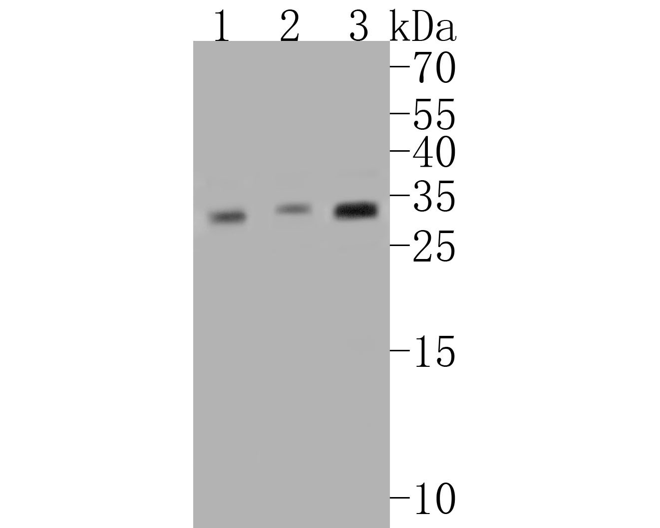 .1 Western blot analysis of CHMP2B on different lysates. Proteins were transferred to a PVDF membrane and blocked with 5% BSA in PBS for 1 hour at room temperature. The primary antibody (SLM-54727R, 1/500) was used in 5% BSA at room temperature for 2 hours. Goat Anti-Rabbit IgG - HRP Secondary Antibody at 1:5,000 dilution was used for 1 hour at room temperature.
Positive control:
Lane 1: Mouse bone marrow tissue lysate
Lane 2: Rat bone marrow tissue lysate
Lane 3: Human skeletal muscle tissue lysate
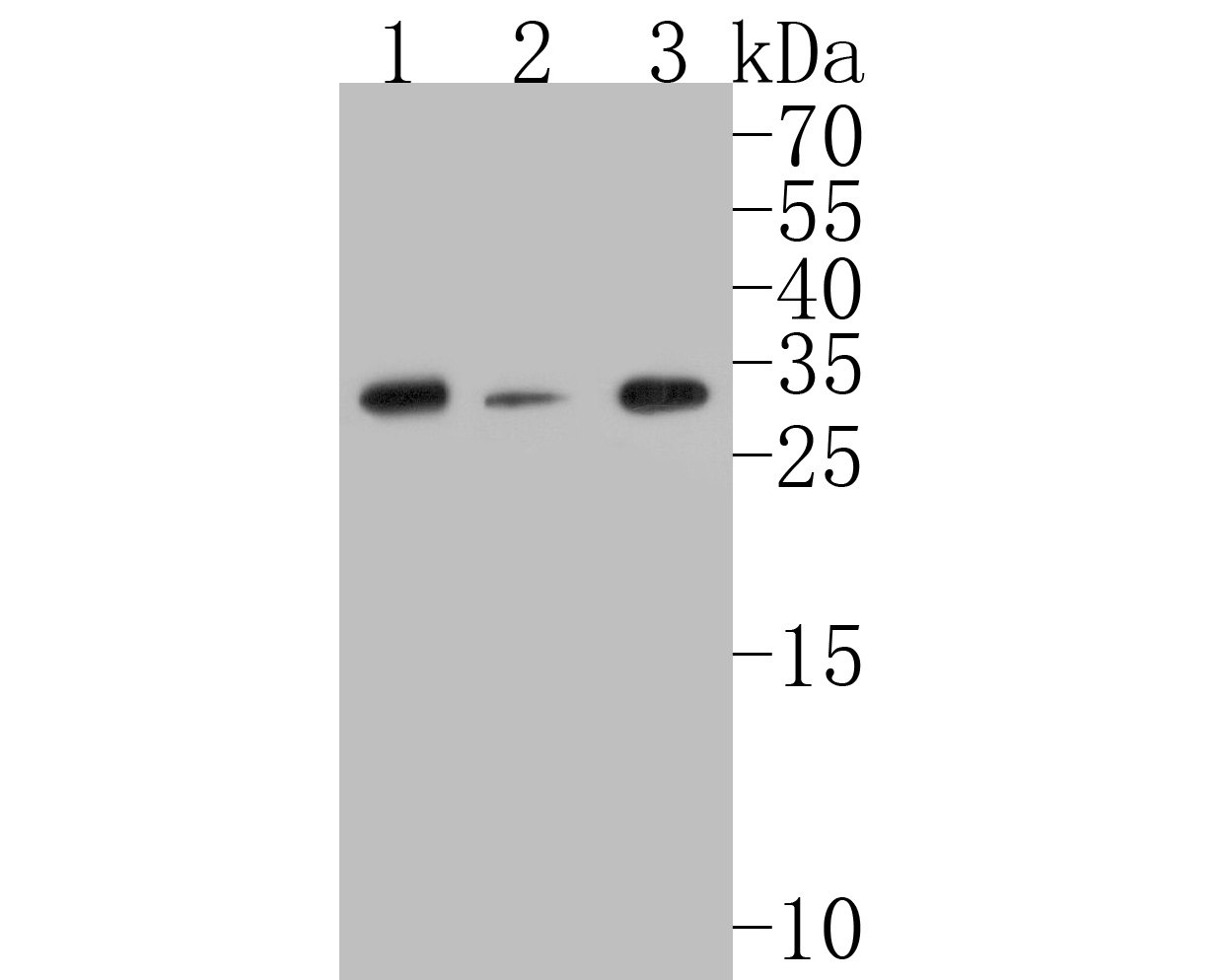 Western blot analysis of CHMP2B on different lysates. Proteins were transferred to a PVDF membrane and blocked with 5% BSA in PBS for 1 hour at room temperature. The primary antibody (SLM-54727R, 1/500) was used in 5% BSA at room temperature for 2 hours. Goat Anti-Rabbit IgG - HRP Secondary Antibody at 1:5,000 dilution was used for 1 hour at room temperature.
Positive control:
Lane 1: A549 cell lysate
Lane 2: A431 cell lysate
Lane 3: Hela cell lysate
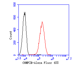 Flow cytometric analysis of CHMP2B was done on A549 cells. The cells were fixed, permeabilized and stained with the primary antibody (SLM-54727R, 1/50) (red). After incubation of the primary antibody at room temperature for an hour, the cells were stained with a Alexa Fluor 488-conjugated Goat anti-Rabbit IgG Secondary antibody at 1/1000 dilution for 30 minutes.Unlabelled sample was used as a control (cells without incubation with primary antibody; black).
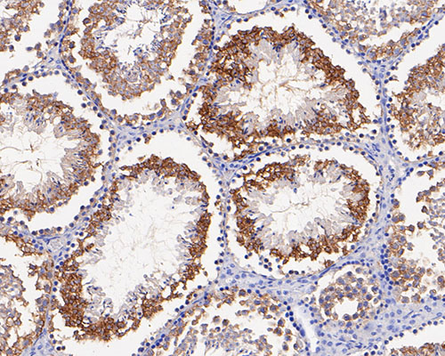 Immunohistochemical analysis of paraffin-embedded mouse testis tissue using anti-CHMP2B antibody. The section was pre-treated using heat mediated antigen retrieval with Tris-EDTA buffer (pH 8.0-8.4) for 20 minutes.The tissues were blocked in 5% BSA for 30 minutes at room temperature, washed with ddH2O and PBS, and then probed with the primary antibody (SLM-54727R, 1/100) for 30 minutes at room temperature. The detection was performed using an HRP conjugated compact polymer system. DAB was used as the chromogen. Tissues were counterstained with hematoxylin and mounted with DPX.
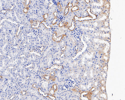 Immunohistochemical analysis of paraffin-embedded mouse kidney tissue using anti-CHMP2B antibody. The section was pre-treated using heat mediated antigen retrieval with Tris-EDTA buffer (pH 8.0-8.4) for 20 minutes.The tissues were blocked in 5% BSA for 30 minutes at room temperature, washed with ddH2O and PBS, and then probed with the primary antibody (SLM-54727R, 1/100) for 30 minutes at room temperature. The detection was performed using an HRP conjugated compact polymer system. DAB was used as the chromogen. Tissues were counterstained with hematoxylin and mounted with DPX.
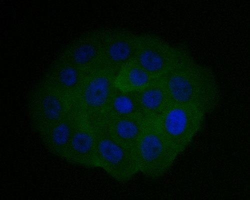 ICC staining of CHMP2B in SW1990 cells (green). Formalin fixed cells were permeabilized with 0.1% Triton X-100 in TBS for 10 minutes at room temperature and blocked with 1% Blocker BSA for 15 minutes at room temperature. Cells were probed with the primary antibody (SLM-54727R, 1/50) for 1 hour at room temperature, washed with PBS. Alexa Fluor®488 Goat anti-Rabbit IgG was used as the secondary antibody at 1/1,000 dilution. The nuclear counter stain is DAPI (blue).
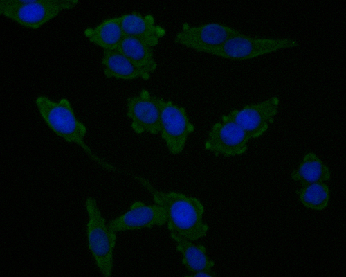 ICC staining of CHMP2B in SW620 cells (green). Formalin fixed cells were permeabilized with 0.1% Triton X-100 in TBS for 10 minutes at room temperature and blocked with 1% Blocker BSA for 15 minutes at room temperature. Cells were probed with the primary antibody (SLM-54727R, 1/50) for 1 hour at room temperature, washed with PBS. Alexa Fluor®488 Goat anti-Rabbit IgG was used as the secondary antibody at 1/1,000 dilution. The nuclear counter stain is DAPI (blue).
Cartpieces
Totalgoods,subtotals:¥Checkout
References (0)
No References
Bought notes(bought amounts latest0)
No one bought this product
User Comment(Total0User Comment Num)
- No comment


 +86 571 56623320
 +86 18668110335

