
Rabbit Anti-TAK1/MAP3K7 antibody
Mitogen-activated protein kinase kinase kinase 7; Transforming growth factor-beta-activated kinase 1; TGF-beta-activated kinase 1; MAP3K 7; MAPKKK7; Mitogen activated protein kinase kinase kinase 7; TAK1; TGF beta activated kinase 1; TGF1a; Transforming g
View History [Clear]
Details
Product Name TAK1/MAP3K7 Chinese Name 转化生长因子β活化激酶1抗体 Alias Mitogen-activated protein kinase kinase kinase 7; Transforming growth factor-beta-activated kinase 1; TGF-beta-activated kinase 1; MAP3K 7; MAPKKK7; Mitogen activated protein kinase kinase kinase 7; TAK1; TGF beta activated kinase 1; TGF1a; Transforming growth factor beta activated kinase 1; M3K7_HUMAN; Map3k7; MEKK7; TGF-beta-activated kinase 1; TGF1a; Transforming growth factor-beta-activated kinase 1. Research Area Tumour immunology Signal transduction Apoptosis transcriptional regulatory factor Kinases and Phosphatases Immunogen Species Rabbit Clonality Monoclonal React Species (predicted: Human, Mouse, ) Applications WB=1:500-1000 ICC=1:50-100 IF=1:50-100
not yet tested in other applications.
optimal dilutions/concentrations should be determined by the end user.Theoretical molecular weight 67kDa Cellular localization cytoplasmic The cell membrane Form Liquid Concentration 1mg/ml immunogen KLH conjugated synthetic peptide derived from human TAK1/MAP3K7 Lsotype IgG Purification affinity purified by Protein A Buffer Solution 0.01M TBS(pH7.4) with 1% BSA, 0.03% Proclin300 and 50% Glycerol. Storage Shipped at 4℃. Store at -20 °C for one year. Avoid repeated freeze/thaw cycles. Attention This product as supplied is intended for research use only, not for use in human, therapeutic or diagnostic applications. PubMed PubMed Product Detail The protein encoded by this gene is a member of the serine/threonine protein kinase family. This kinase mediates the signaling transduction induced by TGF beta and morphogenetic protein (BMP), and controls a variety of cell functions including transcription regulation and apoptosis. In response to IL-1, this protein forms a kinase complex including TRAF6, MAP3K7P1/TAB1 and MAP3K7P2/TAB2; this complex is required for the activation of nuclear factor kappa B. This kinase can also activate MAPK8/JNK, MAP2K4/MKK4, and thus plays a role in the cell response to environmental stresses. Four alternatively spliced transcript variants encoding distinct isoforms have been reported. [provided by RefSeq, Jul 2008]
Function:
Component of a protein kinase signal transduction cascade. Mediator of TRAF6 and TGF-beta signal transduction. Activates IKBKB and MAPK8 in response to TRAF6 signaling. Stimulates NF-kappa-B activation and the p38 MAPK pathway. In osmotic stress signaling, plays a major role in the activation of MAPK8/JNK, but not that of NF-kappa-B.
Subunit:
Binds both upstream activators and downstream substrates in multimolecular complexes. Interacts with TAB1/MAP3K7IP1 and TAB2/MAP3K7IP2. Identified in the TRIKA2 complex composed of MAP3K7, TAB1/MAP3K7IP1 and TAB2/MAP3K7IP2. Interacts with PPM1L. Interaction with PP2A and PPP6C leads to its repressed activity. Interacts with TRAF6 and TAB1/MAP3K7IP1; during IL-1 signaling. Interacts with TAOK1 and TAOK2; interaction with TAOK2 interferEs with MAP3K7 interaction with IKKA, thus preventing NF-kappa-B activation. Interacts with WDR34 (via WD domains). Interacts with RBCK1. Interacts with CYLD.
Subcellular Location:
Cytoplasm. Cell membrane; Peripheral membrane protein; Cytoplasmic side. Note=Although the majority of MAP3K7/TAK1 is found in the cytosol, when complexed with TAB1/MAP3K7IP1 and TAB2/MAP3K7IP2, it is also localized at the cell membrane.
Tissue Specificity:
Isoform 1A is the most abundant in ovary, skeletal muscle, spleen and blood mononuclear cells. Isoform 1B is highly expressed in brain, kidney and small intestine. Isoform 1C is the major form in prostate. Isoform 1D is the less abundant form.
Post-translational modifications:
Association with TAB1/MAP3K7IP1 promotes autophosphorylation and subsequent activation. Association with TAB2/MAP3K7IP2, itself associated with free unanchored Lys-63 polyubiquitin chain, promotes autophosphorylation and subsequent activation of MAP3K7. Dephosphorylation at Thr-187 by PP2A and PPP6C leads to inactivation.
Ubiquitinated, leading to proteasomal degradation. Requires 'Lys-63'-linked polyubiquitination for autophosphorylation and subsequent activation. 'Lys-63'-linked ubiquitination does not lead to proteasomal degradation. Deubiquitinated by CYLD, a protease that selectively cleaves 'Lys-63'-linked ubiquitin chains.
Similarity:
Belongs to the protein kinase superfamily. STE Ser/Thr protein kinase family. MAP kinase kinase kinase subfamily.
Contains 1 protein kinase domain.
SWISS:
O43318
Gene ID:
6885
Product Picture 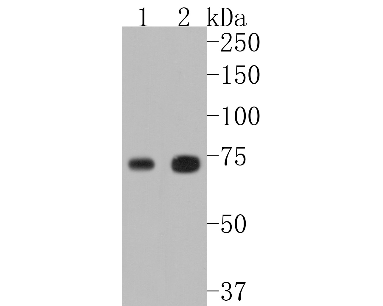 Western blot analysis of TAK1 on different lysates. Proteins were transferred to a PVDF membrane and blocked with 5% BSA in PBS for 1 hour at room temperature. The primary antibody (SLM-54071R, 1/500) was used in 5% BSA at room temperature for 2 hours. Goat Anti-Rabbit IgG - HRP Secondary Antibody (HA1001) at 1:5,000 dilution was used for 1 hour at room temperature. Positive control:
Lane 1: MCF-7 cell lysate
Lane 2: A431 cell lysate
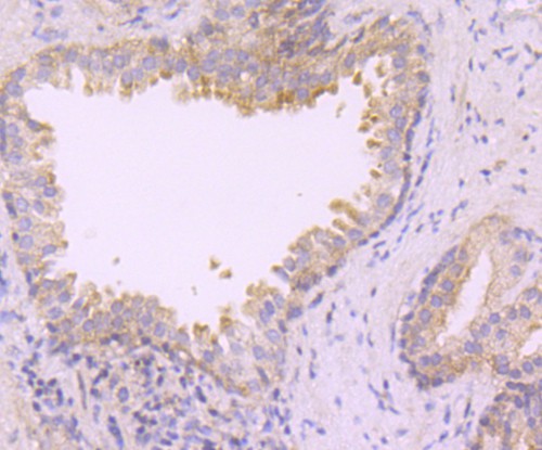 Immunohistochemical analysis of paraffin-embedded human prostate tissue using anti-TAK1 antibody. The section was pre-treated using heat mediated antigen retrieval with Tris-EDTA buffer (pH 8.0-8.4) for 20 minutes.The tissues were blocked in 5% BSA for 30 minutes at room temperature, washed with ddH2O and PBS, and then probed with the primary antibody (SLM-54071R, 1/50) for 30 minutes at room temperature. The detection was performed using an HRP conjugated compact polymer system. DAB was used as the chromogen. Tissues were counterstained with hematoxylin and mounted with DPX.
 Immunohistochemical analysis of paraffin-embedded human placenta tissue using anti-TAK1 antibody. The section was pre-treated using heat mediated antigen retrieval with Tris-EDTA buffer (pH 8.0-8.4) for 20 minutes.The tissues were blocked in 5% BSA for 30 minutes at room temperature, washed with ddH2O and PBS, and then probed with the primary antibody (SLM-54071R, 1/50) for 30 minutes at room temperature. The detection was performed using an HRP conjugated compact polymer system. DAB was used as the chromogen. Tissues were counterstained with hematoxylin and mounted with DPX.
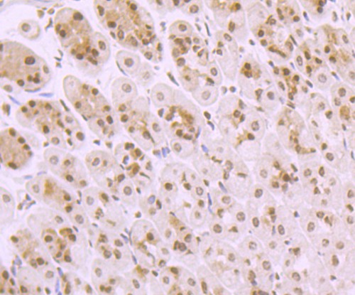 Immunohistochemical analysis of paraffin-embedded mouse stomach tissue using anti-TAK1 antibody. The section was pre-treated using heat mediated antigen retrieval with Tris-EDTA buffer (pH 8.0-8.4) for 20 minutes.The tissues were blocked in 5% BSA for 30 minutes at room temperature, washed with ddH2O and PBS, and then probed with the primary antibody (SLM-54071R, 1/50) for 30 minutes at room temperature. The detection was performed using an HRP conjugated compact polymer system. DAB was used as the chromogen. Tissues were counterstained with hematoxylin and mounted with DPX.ICC staining of TAK1 in N2A cells (green). Formalin fixed cells were permeabilized with 0.1% Triton X-100 in TBS for 10 minutes at room temperature and blocked with 1% Blocker BSA for 15 minutes at room temperature. Cells were probed with the primary antibody (SLM-54071R, 1/50) for 1 hour at room temperature, washed with PBS. Alexa Fluor®488 Goat anti-Rabbit IgG was used as the secondary antibody at 1/1,000 dilution. The nuclear counter stain is DAPI (blue).
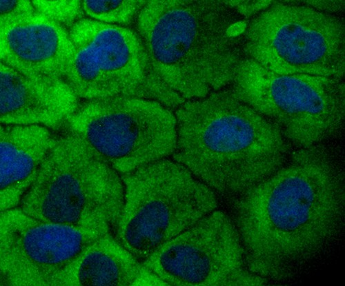 ICC staining of TAK1 in A431 cells (green). Formalin fixed cells were permeabilized with 0.1% Triton X-100 in TBS for 10 minutes at room temperature and blocked with 1% Blocker BSA for 15 minutes at room temperature. Cells were probed with the primary antibody (SLM-54071R, 1/50) for 1 hour at room temperature, washed with PBS. Alexa Fluor®488 Goat anti-Rabbit IgG was used as the secondary antibody at 1/1,000 dilution. The nuclear counter stain is DAPI (blue).
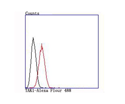 Flow cytometric analysis of TAK1 was done on A431 cells. The cells were fixed, permeabilized and stained with the primary antibody (SLM-54071R, 1/50) (red). After incubation of the primary antibody at room temperature for an hour, the cells were stained with a Alexa Fluor 488-conjugated Goat anti-Rabbit IgG Secondary antibody at 1/1,000 dilution for 30 minutes.Unlabelled sample was used as a control (cells without incubation with primary antibody; black).
Cartpieces
Totalgoods,subtotals:¥Checkout
References (0)
No References
Bought notes(bought amounts latest0)
No one bought this product
User Comment(Total0User Comment Num)
- No comment


 +86 571 56623320
 +86 18668110335

