
Rabbit Anti-Tyrosine Hydroxylase antibody
DYT14; DYT5b; ple; Protein Pale; c; The; TYH; Tyrosine 3 hydroxylase; Tyrosine 3 monooxygenase; Tyk2; TY3H_HUMAN; Tyrosine 3-monooxygenase; Tyrosine 3-hydroxylase; TH.
View History [Clear]
Details
Product Name Tyrosine Hydroxylase Chinese Name 酪氨酸羟化酶兔单克隆抗体 Alias DYT14; DYT5b; ple; Protein Pale; c; The; TYH; Tyrosine 3 hydroxylase; Tyrosine 3 monooxygenase; Tyk2; TY3H_HUMAN; Tyrosine 3-monooxygenase; Tyrosine 3-hydroxylase; TH. Research Area Tumour immunology Neurobiology Signal transduction The new supersedes the old Immunogen Species Rabbit Clonality Monoclonal Clone NO. 8B6 React Species Mouse, Rat, (predicted: Human, ) Applications WB=1:500-2000 IHC-P=1:100-500 ICC=1:50-200 (Paraffin sections need antigen repair)
not yet tested in other applications.
optimal dilutions/concentrations should be determined by the end user.Theoretical molecular weight 60kDa Cellular localization cytoplasmic The cell membrane Form Liquid Concentration 1mg/ml immunogen KLH conjugated synthetic peptide derived from human TH Lsotype IgG Purification affinity purified by Protein A Buffer Solution 0.01M TBS(pH7.4) with 1% BSA, 0.03% Proclin300 and 50% Glycerol. Storage Shipped at 4℃. Store at -20 °C for one year. Avoid repeated freeze/thaw cycles. Attention This product as supplied is intended for research use only, not for use in human, therapeutic or diagnostic applications. PubMed PubMed Product Detail The protein encoded by this gene is involved in the conversion of tyrosine to dopamine. It is the rate-limiting enzyme in the synthesis of catecholamines, hence plays a key role in the physiology of adrenergic neurons. Mutations in this gene have been associated with autosomal recessive Segawa syndrome. Alternatively spliced transcript variants encoding different isoforms have been noted for this gene. [provided by RefSeq, Jul 2008]
Function:
Plays an important role in the physiology of adrenergic neurons.
Tissue Specificity:
Mainly expressed in the brain and adrenal glands.
DISEASE:
Defects in TH are the cause of Segawa syndrome autosomal recessive (ARSEGS) [MIM:605407]. A form of DOPA-responsive dystonia presenting in infancy or early childhood. Dystonia is defined by the presence of sustained involuntary muscle contractions, often leading to abnormal postures. Some cases present with parkinsonian symptoms in infancy. Unlike all other forms of dystonia, it is an eminently treatable condition, due to a favorable response to L-DOPA.
Note=May play a role in the pathogenesis of Parkinson disease (PD). A genome-wide copy number variation analysis has identified a 34 kilobase deletion over the TH gene in a PD patient but not in any controls.
Similarity:
Belongs to the biopterin-dependent aromatic amino acid hydroxylase family.
SWISS:
P07101
Gene ID:
7054
Database links:Entrez Gene: 7054 Human
Entrez Gene: 21823 Mouse
Omim: 191290 Human
SwissProt: P07101 Human
SwissProt: P24529 Mouse
Unigene: 435609 Human
Unigene: 1292 Mouse
Unigene: 11082 Rat
神经细胞Maker
酪氨酸羟化酶(TH)是儿茶酚胺类神经递质即多巴胺、去甲肾上腺素、肾上腺素生物合成过程所需的限速酶,它以四氢生物喋呤啶(BH4)为辅酶,催化酪氨酸的羟化而生成多巴(DOPA)。
已知在患帕金森病(Parkinson disease,PD)时,脑内多巴胺(dopamine,DA)的减少与此酶活性低下有关。因此对PD模型动物来说,若将TH基因植入脑内,便可以提高脑内DA水平而达到基因治疗目的。Product Picture 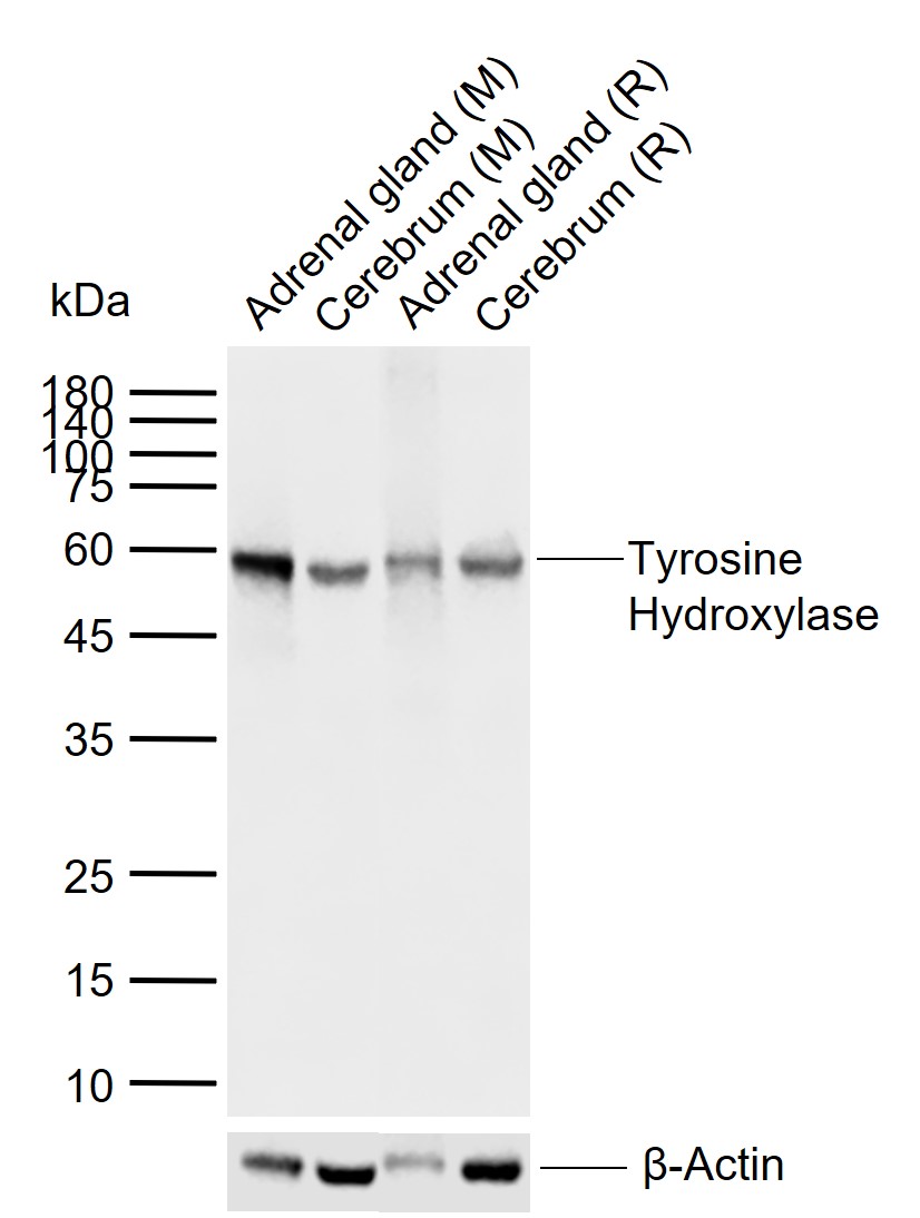 Sample:
Lane 1: Mouse Adrenal gland tissue lysates
Lane 2: Mouse Cerebrum tissue lysates
Lane 3: Rat Adrenal gland tissue lysates
Lane 4: Rat Cerebrum tissue lysates
Primary: Anti-Tyrosine Hydroxylase (SLM-52574R) at 1/1000 dilution
Secondary: IRDye800CW Goat Anti-Rabbit IgG at 1/20000 dilution
Predicted band size: 60 kDa
Observed band size: 60 kDa
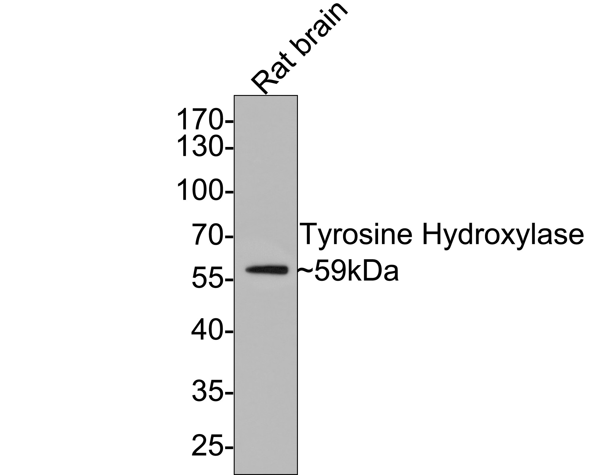 Western blot analysis of Tyrosine Hydroxylase on rat brain tissue lysates with Rabbit anti-Tyrosine Hydroxylase antibody (SLM-52574R) at 1/1,000 dilution.
Lysates/proteins at 20 µg/Lane.
Predicted band size: 59 kDa
Observed band size: 59 kDa
Exposure time: 2 minutes;
10% SDS-PAGE gel.
Proteins were transferred to a PVDF membrane and blocked with 5% NFDM/TBST for 1 hour at room temperature. The primary antibody (SLM-52574R) at 1/1,000 dilution was used in 5% NFDM/TBST at room temperature for 2 hours. Goat Anti-Rabbit IgG - HRP Secondary Antibody at 1:300,000 dilution was used for 1 hour at room temperature.
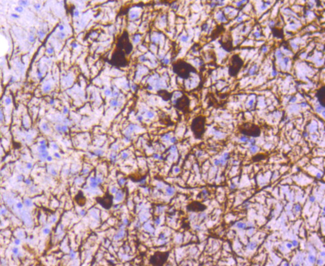 Immunohistochemical analysis of paraffin-embedded mouse brain tissue using anti-Tyrosine Hydroxylase antibody. The section was pre-treated using heat mediated antigen retrieval with Tris-EDTA buffer (pH 8.0-8.4) for 20 minutes.The tissues were blocked in 5% BSA for 30 minutes at room temperature, washed with ddH2O and PBS, and then probed with the primary antibody (SLM-52574R, 1/50) for 30 minutes at room temperature. The detection was performed using an HRP conjugated compact polymer system. DAB was used as the chromogen. Tissues were counterstained with hematoxylin and mounted with DPX
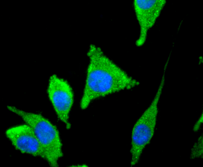 ICC staining of Tyrosine Hydroxylase in SH-SY5Y cells (green). Formalin fixed cells were permeabilized with 0.1% Triton X-100 in TBS for 10 minutes at room temperature and blocked with 1% Blocker BSA for 15 minutes at room temperature. Cells were probed with the primary antibody (SLM-52574R, 1/50) for 1 hour at room temperature, washed with PBS. Alexa Fluor®488 Goat anti-Rabbit IgG was used as the secondary antibody at 1/1,000 dilution. The nuclear counter stain is DAPI (blue).
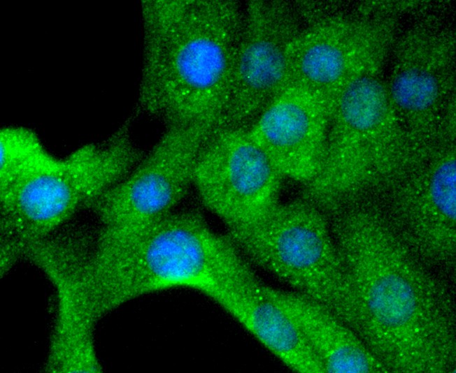 ICC staining of Tyrosine Hydroxylase in NIH/3T3 cells (green). Formalin fixed cells were permeabilized with 0.1% Triton X-100 in TBS for 10 minutes at room temperature and blocked with 1% Blocker BSA for 15 minutes at room temperature. Cells were probed with the primary antibody (SLM-52574R, 1/50) for 1 hour at room temperature, washed with PBS. Alexa Fluor®488 Goat anti-Rabbit IgG was used as the secondary antibody at 1/1,000 dilution. The nuclear counter stain is DAPI (blue).
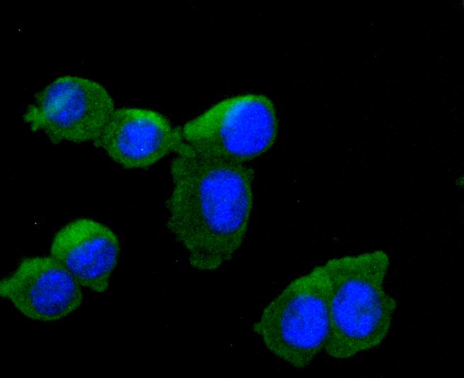 ICC staining of Tyrosine Hydroxylase in N2A cells (green). Formalin fixed cells were permeabilized with 0.1% Triton X-100 in TBS for 10 minutes at room temperature and blocked with 1% Blocker BSA for 15 minutes at room temperature. Cells were probed with the primary antibody (SLM-52574R, 1/50) for 1 hour at room temperature, washed with PBS. Alexa Fluor®488 Goat anti-Rabbit IgG was used as the secondary antibody at 1/1,000 dilution. The nuclear counter stain is DAPI (blue).
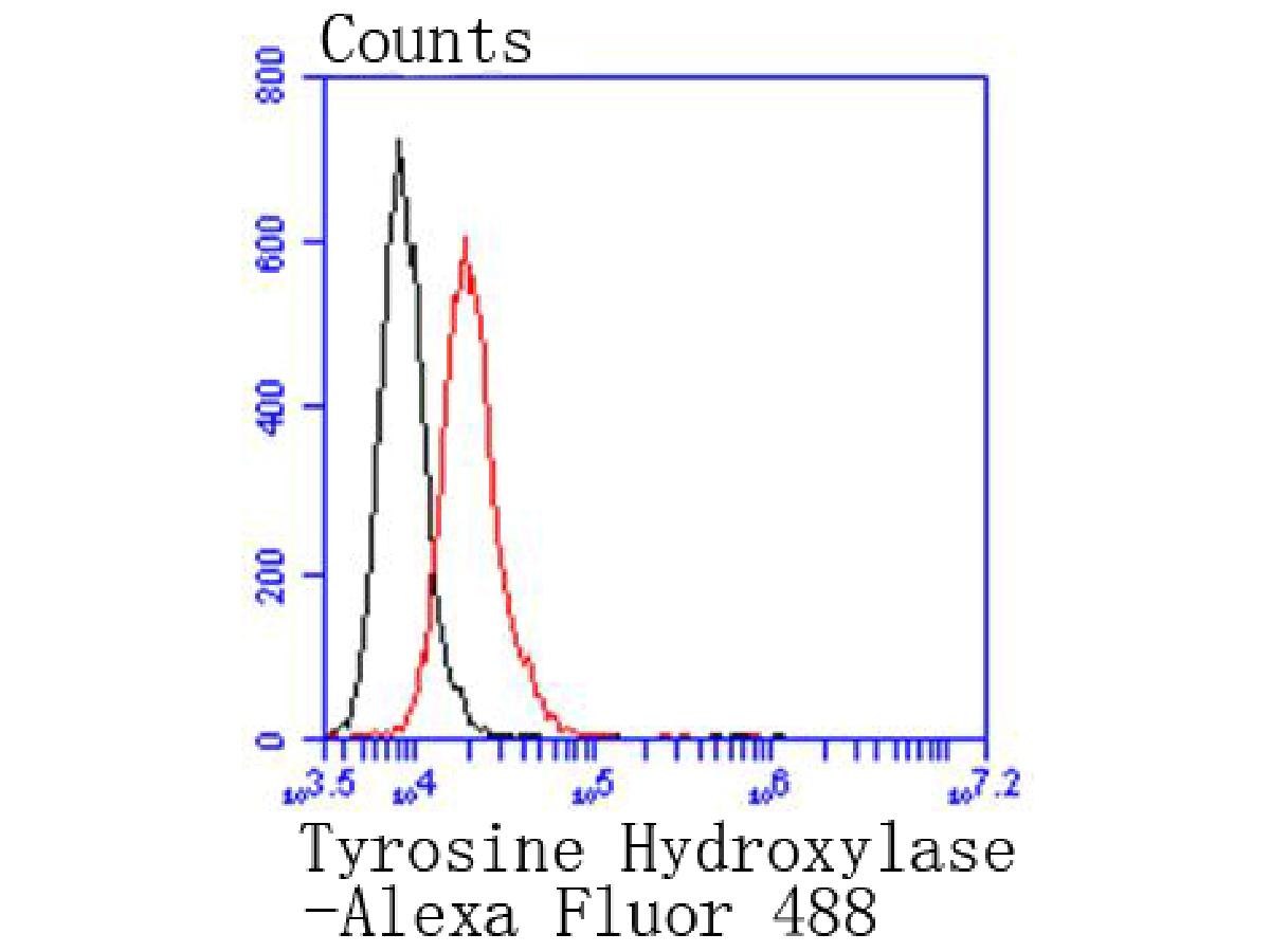 Flow cytometric analysis of Tyrosine Hydroxylase was done on N2A cells. The cells were fixed, permeabilized and stained with the primary antibody (SLM-52574R, 1/50) (red). After incubation of the primary antibody at room temperature for an hour, the cells were stained with a Alexa Fluor 488-conjugated Goat anti-Rabbit IgG Secondary antibody at 1/1,000 dilution for 30 minutes.Unlabelled sample was used as a control (cells without incubation with primary antibody; black).
Cartpieces
Totalgoods,subtotals:¥Checkout
References (0)
No References
Bought notes(bought amounts latest0)
No one bought this product
User Comment(Total0User Comment Num)
- No comment


 +86 571 56623320
 +86 18668110335

