
Rabbit Anti-TCF7L2 antibody
E2 2; HMG box transcription factor 4; hTCF 4; Immunoglobulin transcription factor 2; ITF2; SEF2 1; SEF2 1A; SEF2 1B; SEF2; T cell factor 4; T cell specific HMG box; T cell specific transcription factor 4; TCF 4; TCF4; TCF7L2; TCF7L2 protein; hTCF-4; T-cel
View History [Clear]
Details
Product Name TCF7L2 Chinese Name 转录因子7类似物2Recombinant rabbit monoclonal anti Alias E2 2; HMG box transcription factor 4; hTCF 4; Immunoglobulin transcription factor 2; ITF2; SEF2 1; SEF2 1A; SEF2 1B; SEF2; T cell factor 4; T cell specific HMG box; T cell specific transcription factor 4; TCF 4; TCF4; TCF7L2; TCF7L2 protein; hTCF-4; T-cell factor 4; T-cell-specific transcription factor 4; TCF-4; TF7L2_HUMAN; Transcription factor 7-like 2; Transcription factor 4; Transcription factor 7 like 2; Transcription factor 7 like 2 T cell specific HMG box. literatures Research Area Cardiovascular Chromatin and nuclear signals transcriptional regulatory factor Epigenetics Immunogen Species Rabbit Clonality Monoclonal Clone NO. 2C2 React Species Human, (predicted: Mouse, Rat, ) Applications WB=1:500-1000 IP=1:20-100 IHC-P=1:100-500 Flow-Cyt=1:50 ICC=1:100 IF=1:50-200 (Paraffin sections need antigen repair)
not yet tested in other applications.
optimal dilutions/concentrations should be determined by the end user.Theoretical molecular weight 68kDa Cellular localization The nucleus Form Liquid Concentration 1mg/ml immunogen KLH conjugated synthetic peptide derived from human TCF7L2 Lsotype IgG Purification affinity purified by Protein A Buffer Solution 0.01M TBS(pH7.4) with 1% BSA, 0.03% Proclin300 and 50% Glycerol. Storage Shipped at 4℃. Store at -20 °C for one year. Avoid repeated freeze/thaw cycles. Attention This product as supplied is intended for research use only, not for use in human, therapeutic or diagnostic applications. PubMed PubMed Product Detail TCF-4, transcription factor 4, is a basic helix-turn-helix transcription factor. This protein recognizes an Ephrussi-box ('E-box') binding site ('CANNTG') - a motif first identified in immunoglobulin enhancers. The gene for TCF-4 is expressed predominantly in pre-B-cells, although it is found in other tissues as well. Multiple alternatively spliced transcript variants that encode different proteins have been described. TCF4, also known as TCF7L2, is expressed widely during development. Gene targeting study indicates that it is required to maintain the crypt stem cells of the small intestine. TCF4 has many different splicing isoforms and they are expressed differentially in tissues and in cancers of different stages. Studies also indicate that variant of the TCF4 gene confers an increased risk of type 2 diabetes.
Function:
Participates in the Wnt signaling pathway and modulates MYC expression by binding to its promoter in a sequence-specific manner. Acts as repressor in the absence of CTNNB1, and as activator in its presence. Activates transcription from promoters with several copies of the Tcf motif 5'-CCTTTGATC-3' in the presence of CTNNB1. TLE1, TLE2, TLE3 and TLE4 repress transactivation mediated by TCF7L2/TCF4 and CTNNB1. Expression of dominant-negative mutants results in cell-cycle arrest in G1. Necessary for the maintenance of the epithelial stem-cell compartment of the small intestine.
Subunit:
Interacts with TGFB1I1 (By similarity). Interacts with CTNNB1 (via the armadillo repeat); forms stable transcription complex. Interacts with EP300. Interacts with NLK. Interacts with CCDC85B (probably through the HMG box); prevents interaction with CTNNB1. Interacts with TNIK. Interacts with MAD2L2; prevents TCF7L2/TCF4 binding to promZIPK/DAPK3oters, negatively modulating its transcriptional activity. Interacts with ZIPK/DAPK3. Interacts with XIAP/BIRC4 and TLE3. Interacts with DDIT3/CHOP.
Subcellular Location:
Nucleus, PML body. Note=Diffuse pattern. Colocalizes with SUMO1 and PIAS4 in a subset of PML (promyelocytic leukemia) nuclear bodies.
Tissue Specificity:
Detected in epithelium from small intestine, with the highest expression at the top of the crypts and a gradient of expression from crypt to villus. Detected in colon epithelium and colon cancer, and in epithelium from mammary gland and carcinomas derived therefrom.
Post-translational modifications:
In vitro, phosphorylated by TNIK.
Phosphorylated at Thr-201 and/or Thr-212 by NLK. Phosphorylation by NLK at these sites inhibits DNA-binding by TCF7L2/TCF4, thereby preventing transcriptional activation of target genes of the canonical Wnt/beta-catenin signaling pathway.
Polysumoylated. Sumoylation is enhanced by PIAS family members and desumoylation is enhanced by SENP2. Sumoylation/desumoylation regulates TCF7L2/TCF4 transcription activity in the Wnt/beta-catenin signaling pathway without altering interaction with CTNNB1 nor binding to DNA.
DISEASE:
Note=Constitutive activation and subsequent transactivation of target genes may lead to the maintenance of stem-cell characteristics (cycling and longevity) in cells that should normally undergo terminal differentiation and constitute the primary transforming event in colorectal cancer (CRC).
Diabetes mellitus, non-insulin-dependent (NIDDM) [MIM:125853]: A multifactorial disorder of glucose homeostasis caused by a lack of sensitivity to the body's own insulin. Affected individuals usually have an obese body habitus and manifestations of a metabolic syndrome characterized by diabetes, insulin resistance, hypertension and hypertriglyceridemia. The disease results in long-term complications that affect the eyes, kidneys, nerves, and blood vessels. Note=Disease susceptibility is associated with variations affecting the gene represented in this entry.
Similarity:
Belongs to the TCF/LEF family.
Contains 1 HMG box DNA-binding domain.
SWISS:
Q9NQB0
Gene ID:
6934
Database links:Entrez Gene: 6934 Human
Entrez Gene: 21416 Mouse
Omim: 602228 Human
SwissProt: Q9NQB0 Human
SwissProt: Q924A0 Mouse
Unigene: 593995 Human
Unigene: 139815 Mouse
Product Picture 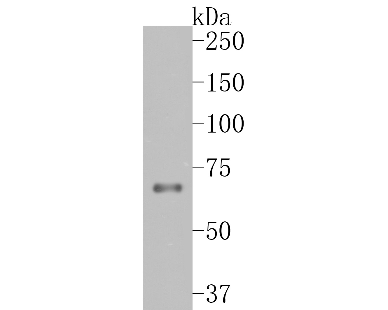 Western blot analysis of TCF7L2 on JAR cell lysates. Proteins were transferred to a PVDF membrane and blocked with 5% BSA in PBS for 1 hour at room temperature. The primary antibody (SLM-52543R, 1/500) was used in 5% BSA at room temperature for 2 hours. Goat Anti-Rabbit IgG - HRP Secondary Antibody at 1:5,000 dilution was used for 1 hour at room temperature.
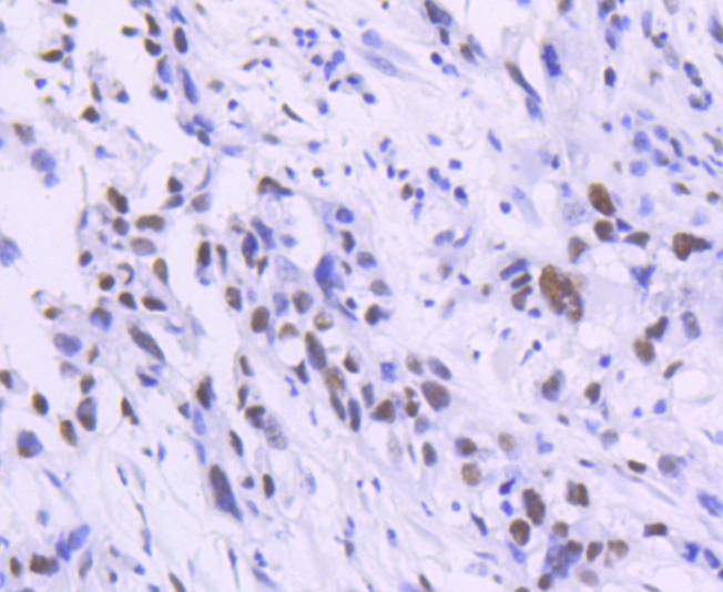 Immunohistochemical analysis of paraffin-embedded human breast carcinoma tissue using anti-TCF7L2 antibody. The section was pre-treated using heat mediated antigen retrieval with Tris-EDTA buffer (pH 8.0-8.4) for 20 minutes.The tissues were blocked in 5% BSA for 30 minutes at room temperature, washed with ddH2O and PBS, and then probed with the primary antibody (SLM-52543R, 1/50) for 30 minutes at room temperature. The detection was performed using an HRP conjugated compact polymer system. DAB was used as the chromogen. Tissues were counterstained with hematoxylin and mounted with DPX.
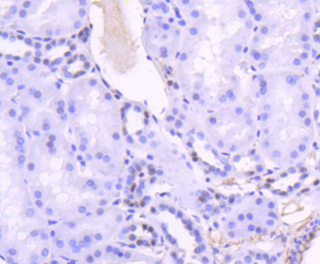 Immunohistochemical analysis of paraffin-embedded human kidney tissue using anti-TCF7L2 antibody. The section was pre-treated using heat mediated antigen retrieval with Tris-EDTA buffer (pH 8.0-8.4) for 20 minutes.The tissues were blocked in 5% BSA for 30 minutes at room temperature, washed with ddH2O and PBS, and then probed with the primary antibody (SLM-52543R, 1/50) for 30 minutes at room temperature. The detection was performed using an HRP conjugated compact polymer system. DAB was used as the chromogen. Tissues were counterstained with hematoxylin and mounted with DPX.
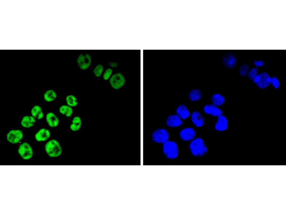 ICC staining of TCF7L2 in SW480 cells (green). Formalin fixed cells were permeabilized with 0.1% Triton X-100 in TBS for 10 minutes at room temperature and blocked with 1% Blocker BSA for 15 minutes at room temperature. Cells were probed with the primary antibody (SLM-52543R, 1/50) for 1 hour at room temperature, washed with PBS. Alexa Fluor®488 Goat anti-Rabbit IgG was used as the secondary antibody at 1/1,000 dilution. The nuclear counter stain is DAPI (blue).
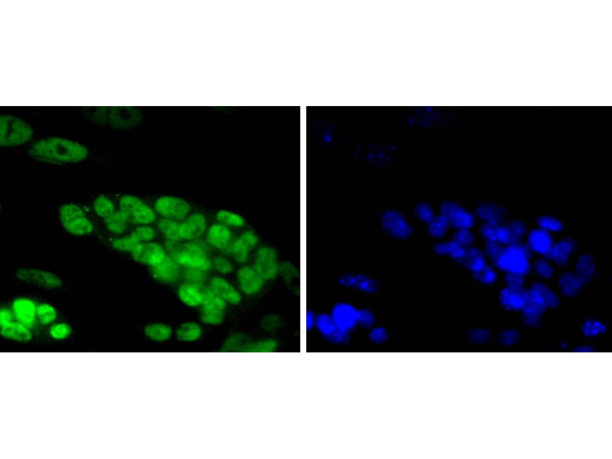 ICC staining of TCF7L2 in D3 cells (green). Formalin fixed cells were permeabilized with 0.1% Triton X-100 in TBS for 10 minutes at room temperature and blocked with 1% Blocker BSA for 15 minutes at room temperature. Cells were probed with the primary antibody (SLM-52543R, 1/50) for 1 hour at room temperature, washed with PBS. Alexa Fluor®488 Goat anti-Rabbit IgG was used as the secondary antibody at 1/1,000 dilution. The nuclear counter stain is DAPI (blue).
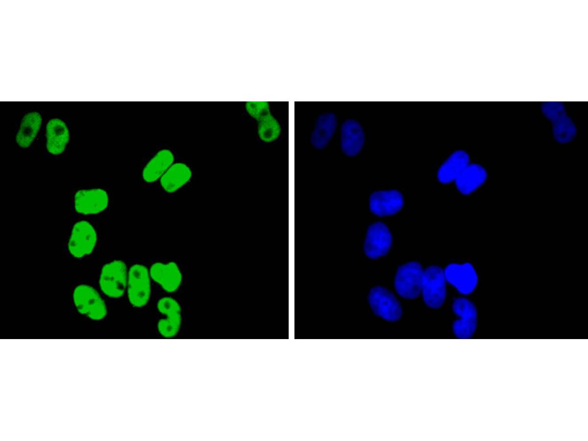 ICC staining of TCF7L2 in Hela cells (green). Formalin fixed cells were permeabilized with 0.1% Triton X-100 in TBS for 10 minutes at room temperature and blocked with 1% Blocker BSA for 15 minutes at room temperature. Cells were probed with the primary antibody (SLM-52543R, 1/50) for 1 hour at room temperature, washed with PBS. Alexa Fluor®488 Goat anti-Rabbit IgG was used as the secondary antibody at 1/1,000 dilution. The nuclear counter stain is DAPI (blue).
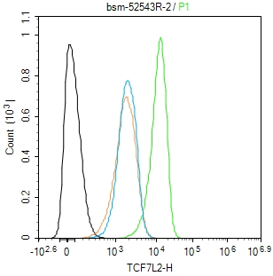 Blank control(black line):HepG2.
Primary Antibody (green line): Mouse Anti-TCF7L2 antibody (SLM-52543R)
Dilution:1:50;
Secondary Antibody(white blue line): Goat anti-Mouse IgG-AF488
Dilution: 0.5ug/Test.
Isotype control(orange line): Normal Rabbit IgG
Protocol
The cells were fixed with 4% PFA (10min at room temperature)and then permeabilized with 90% ice-cold methanol for 20 min at -20℃, The cells were then incubated in 5%BSA to block non-specific protein-protein interactions for 30 min at room temperature .Cells stained with Primary Antibody for 30 min at room temperature. The secondary antibody used for 40 min at room temperature. Acquisition of 20,000 events was performed.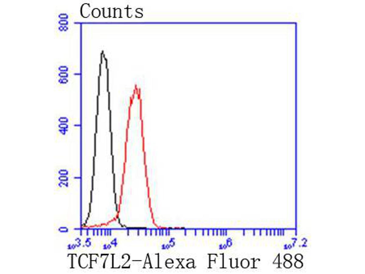 Flow cytometric analysis of TCF7L2 was done on Jurkat cells. The cells were fixed, permeabilized and stained with the primary antibody (SLM-52543R, 1/50) (red). After incubation of the primary antibody at room temperature for an hour, the cells were stained with a Alexa Fluor 488-conjugated Goat anti-Rabbit IgG Secondary antibody at 1/1000 dilution for 30 minutes.Unlabelled sample was used as a control (cells without incubation with primary antibody; black).
Cartpieces
Totalgoods,subtotals:¥Checkout
References (0)
No References
Bought notes(bought amounts latest0)
No one bought this product
User Comment(Total0User Comment Num)
- No comment


 +86 571 56623320
 +86 18668110335

