
Rabbit Anti-PSMB8 antibody
Proteasome 20S LMP7; D6S216; D6S216E; Large multifunctional peptidase 7; Large multifunctional protease 7; LMP 7; LMP-7; LMP7; Low molecular mass protein 7; Low molecular weight protein 7; Macropain subunit C13; MGC1491; Multicatalytic endopeptidase compl
View History [Clear]
Details
Product Name PSMB8 Chinese Name 蛋白酶体20S亚基LMP7Recombinant rabbit monoclonal anti Alias Proteasome 20S LMP7; D6S216; D6S216E; Large multifunctional peptidase 7; Large multifunctional protease 7; LMP 7; LMP-7; LMP7; Low molecular mass protein 7; Low molecular weight protein 7; Macropain subunit C13; MGC1491; Multicatalytic endopeptidase complex subunit C13; Protease component C13; Proteasome (prosome macropain) subunit beta type 8; Proteasome (prosome, macropain) subunit, beta type, 8 (large multifunctional peptidase 7); Proteasome beta 8 subunit; Proteasome catalytic subunit 3i; Proteasome component C13; Proteasome related gene 7; Proteasome subunit beta 5i; Proteasome subunit beta type 8; Proteasome subunit beta type; Proteasome subunit beta type-8; Proteasome subunit beta-5i; Proteasome subunit Y2; PSB8_HUMAN; PSMB 8; PSMB5i; Really interesting new gene 10 protein; RING 10; RING10; Y2; ALDD; D6S216E; JMP; NKJO; PSMB5i; RING10. Research Area Cell biology immunology Immunogen Species Rabbit Clonality Monoclonal React Species (predicted: Human, Mouse, Rat, ) Applications WB=1:500-2000 IHC-P=1:100-500 Flow-Cyt=1:50-100 ICC=1:50-100 IF=1:50-100 (Paraffin sections need antigen repair)
not yet tested in other applications.
optimal dilutions/concentrations should be determined by the end user.Theoretical molecular weight 23kDa Cellular localization The nucleus cytoplasmic Form Liquid Concentration 1mg/ml immunogen Recombinant human PSMB8 protein Lsotype IgG Purification affinity purified by Protein A Buffer Solution 0.01M TBS(pH7.4) with 1% BSA, 0.03% Proclin300 and 50% Glycerol. Storage Shipped at 4℃. Store at -20 °C for one year. Avoid repeated freeze/thaw cycles. Attention This product as supplied is intended for research use only, not for use in human, therapeutic or diagnostic applications. PubMed PubMed Product Detail The proteasome is a multicatalytic proteinase complex with a highly ordered ring-shaped 20S core structure. The core structure is composed of 4 rings of 28 non-identical subunits; 2 rings are composed of 7 alpha subunits and 2 rings are composed of 7 beta subunits. Proteasomes are distributed throughout eukaryotic cells at a high concentration and cleave peptides in an ATP/ubiquitin-dependent process in a non-lysosomal pathway. An essential function of a modified proteasome, the immunoproteasome, is the processing of class I MHC peptides. This gene encodes a member of the proteasome B-type family, also known as the T1B family, that is a 20S core beta subunit. This gene is located in the class II region of the MHC (major histocompatibility complex). Expression of this gene is induced by gamma interferon and this gene product replaces catalytic subunit 3 (proteasome beta 5 subunit) in the immunoproteasome. Proteolytic processing is required to generate a mature subunit. Two alternative transcripts encoding two isoforms have been identified; both isoforms are processed to yield the same mature subunit. [provided by RefSeq, Jul 2008].
Function:
The proteasome is a multicatalytic proteinase complex which is characterized by its ability to cleave peptides with Arg, Phe, Tyr, Leu, and Glu adjacent to the leaving group at neutral or slightly basic pH. The proteasome has an ATP-dependent proteolytic activity. This subunit is involved in antigen processing to generate class I binding peptides. Replacement of PSMB5 by PSMB8 increases the capacity of the immunoproteasome to cleave model peptides after hydrophobic and basic residues. Acts as a major component of interferon gamma-induced sensitivity. Plays a key role in apoptosis via the degradation of the apoptotic inhibitor MCL1. May be involved in the inflammatory response pathway. In cancer cells, substitution of isoform 1 (E2) by isoform 2 (E1) results in immunoproteasome deficiency. Required for the differentiation of preadipocytes into adipocytes.
Subunit:
The 26S proteasome consists of a 20S proteasome core and two 19S regulatory subunits. The 20S proteasome core is composed of 28 subunits that are arranged in four stacked rings, resulting in a barrel-shaped structure. The two end rings are each formed by seven alpha subunits, and the two central rings are each formed by seven beta subunits. The catalytic chamber with the active sites is on the inside of the barrel. This subunit is part of the immunoproteasome where it displaces the equivalent housekeeping subunit PSMB5. Directly interacts with POMP. Interacts with HIV-1 TAT protein. Interacts with TAP1.
Subcellular Location:
Cytoplasm. Nucleus.
Tissue Specificity:
Highly expressed in immature dendritic cells (at protein level).
Post-translational modifications:
Autocleaved. The resulting N-terminal Thr residue of the mature subunit is responsible for the nucleophile proteolytic activity.
DISEASE:
Defects in PSMB8 are the cause of Nakajo syndrome (NKJO) [MIM:256040]; also called joint contractures muscular atrophy microcytic anemia and panniculitis-induced lipodystrophy. An autosomal recessive autoinflammatory disorder characterized by childhood onset of recurrent fever, joint stiffness and severe contractures of the hands and feet, erythematous skin lesions with subsequent development of lipodystrophy, and laboratory evidence of immune dysregulation. Accompanying features include muscle weakness and atrophy, hepatosplenomegaly, and microcytic anemia.
Note=Mutation Met-75 has been found in chronic atypical neutrophilic dermatosis with lipodystrophy and elevated temperature syndrome (CANDLE syndrome). CANDLE patients have some overlapping features with NKJO patients, including a cutaneous eruption and lipodystrophy. They show a characteristic neutrophilic dermatosis with a mononuclear interstitial infiltrate in the dermis that seems pathognomonic for CANDLE syndrome (PubMed:21953331).
Similarity:
Belongs to the peptidase T1B family.
SWISS:
P28062
Gene ID:
5696
Product Picture 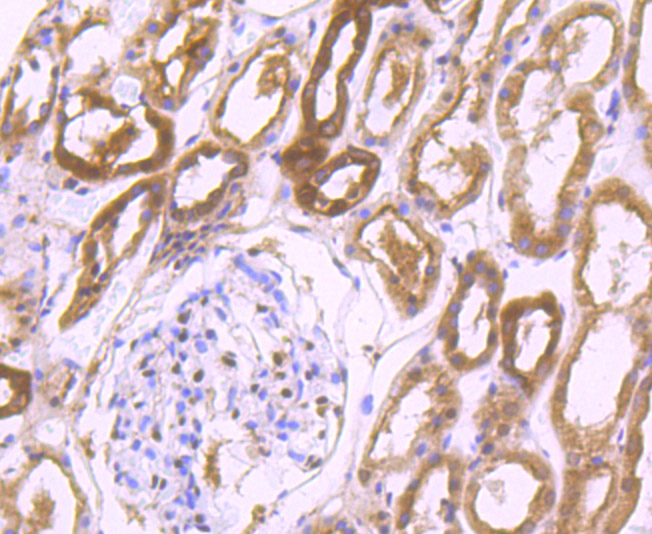 Immunohistochemical analysis of paraffin-embedded human kidney tissue using anti-Proteasome 20S LMP7 antibody. The section was pre-treated using heat mediated antigen retrieval with Tris-EDTA buffer (pH 9.0) for 20 minutes.The tissues were blocked in 1% BSA for 30 minutes at room temperature, washed with ddH2O and PBS, and then probed with the primary antibody (SLM-54282R , 1/50) for 30 minutes at room temperature. The detection was performed using an HRP conjugated compact polymer system. DAB was used as the chromogen. Tissues were counterstained with hematoxylin and mounted with DPX.
 Immunohistochemical analysis of paraffin-embedded mouse small intestine tissue using anti-Proteasome 20S LMP7 antibody. The section was pre-treated using heat mediated antigen retrieval with Tris-EDTA buffer (pH 9.0) for 20 minutes.The tissues were blocked in 1% BSA for 30 minutes at room temperature, washed with ddH2O and PBS, and then probed with the primary antibody (SLM-54282R , 1/50) for 30 minutes at room temperature. The detection was performed using an HRP conjugated compact polymer system. DAB was used as the chromogen. Tissues were counterstained with hematoxylin and mounted with DPX.
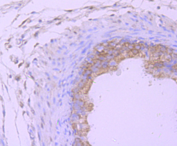 Immunohistochemical analysis of paraffin-embedded rat epididymis tissue using anti-Proteasome 20S LMP7 antibody. The section was pre-treated using heat mediated antigen retrieval with Tris-EDTA buffer (pH 9.0) for 20 minutes.The tissues were blocked in 1% BSA for 30 minutes at room temperature, washed with ddH2O and PBS, and then probed with the primary antibody (ET7107-36, 1/50) for 30 minutes at room temperature. The detection was performed using an HRP conjugated compact polymer system. DAB was used as the chromogen. Tissues were counterstained with hematoxylin and mounted with DPX.
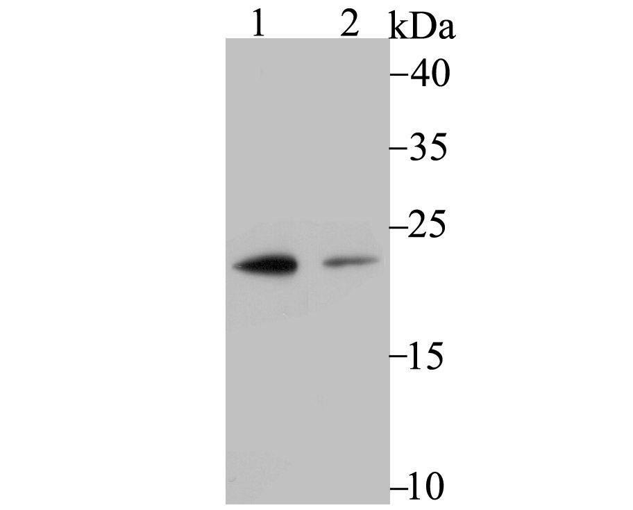 Western blot analysis of Proteasome 20S LMP7 on different lysates. Proteins were transferred to a PVDF membrane and blocked with 5% BSA in PBS for 1 hour at room temperature. The primary antibody (SLM-54282R, 1/500) was used in 5% BSA at room temperature for 2 hours. Goat Anti-Rabbit IgG - HRP Secondary Antibody at 1:200,000 dilution was used for 1 hour at room temperature. Positive control: Lane 1: U937 cell lysate Lane 2: A431 cell lysate
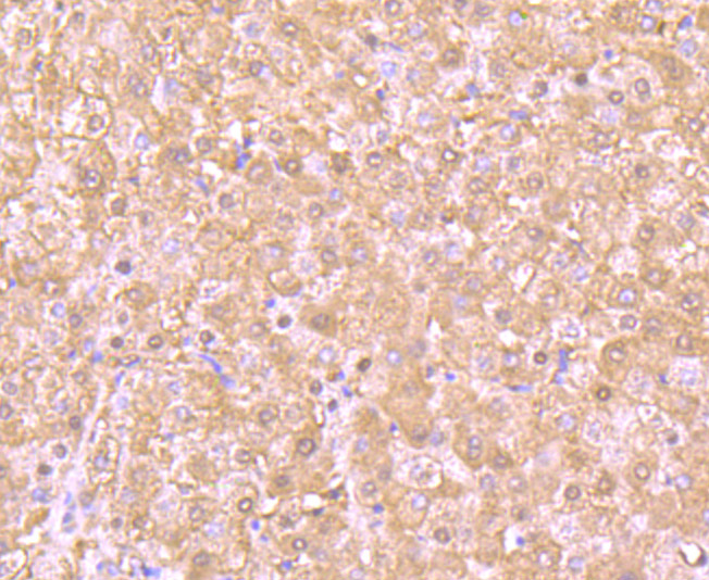 Immunohistochemical analysis of paraffin-embedded human liver tissue using anti-Proteasome 20S LMP7 antibody. The section was pre-treated using heat mediated antigen retrieval with Tris-EDTA buffer (pH 9.0) for 20 minutes.The tissues were blocked in 1% BSA for 30 minutes at room temperature, washed with ddH2O and PBS, and then probed with the primary antibody (SLM-54282R , 1/50) for 30 minutes at room temperature. The detection was performed using an HRP conjugated compact polymer system. DAB was used as the chromogen. Tissues were counterstained with hematoxylin and mounted with DPX.
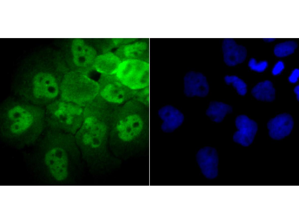 ICC staining of Proteasome 20S LMP7 in A431 cells (green). Formalin fixed cells were permeabilized with 0.1% Triton X-100 in TBS for 10 minutes at room temperature and blocked with 10% negative goat serum for 15 minutes at room temperature. Cells were probed with the primary antibody (ET7107-36 , 1/50) for 1 hour at room temperature, washed with PBS. Alexa Fluor®488 conjugate-Goat anti-Rabbit IgG was used as the secondary antibody at 1/1,000 dilution. The nuclear counter stain is DAPI (blue).
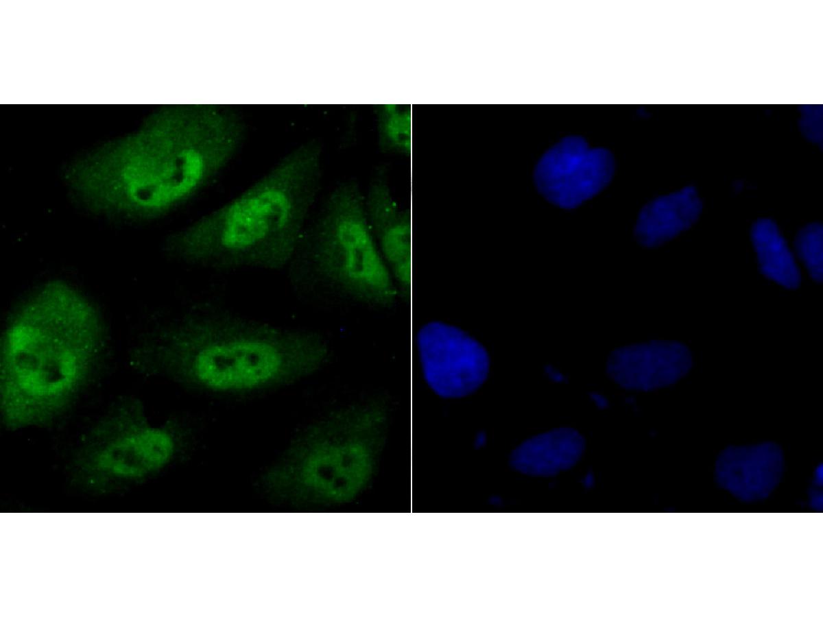 ICC staining of Proteasome 20S LMP7 in HUVEC cells (green). Formalin fixed cells were permeabilized with 0.1% Triton X-100 in TBS for 10 minutes at room temperature and blocked with 10% negative goat serum for 15 minutes at room temperature. Cells were probed with the primary antibody (SLM-54282R, 1/50) for 1 hour at room temperature, washed with PBS. Alexa Fluor®488 conjugate-Goat anti-Rabbit IgG was used as the secondary antibody at 1/1,000 dilution. The nuclear counter stain is DAPI (blue).
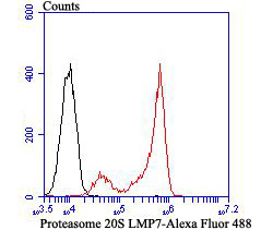 Flow cytometric analysis of Proteasome 20S LMP7 was done on Daudi cells. The cells were fixed, permeabilized and stained with the primary antibody (SLM-54282R , 1/50) (red). After incubation of the primary antibody at room temperature for an hour, the cells were stained with a Alexa Fluor®488 conjugate-Goat anti-Rabbit IgG Secondary antibody at 1/1,000 dilution for 30 minutes.Unlabelled sample was used as a control (cells without incubation with primary antibody; black).
Cartpieces
Totalgoods,subtotals:¥Checkout
References (0)
No References
Bought notes(bought amounts latest0)
No one bought this product
User Comment(Total0User Comment Num)
- No comment


 +86 571 56623320
 +86 18668110335

