
Rabbit Anti-IGF2 antibody
IGF2_HUMAN; Insulin-like growth factor II; IGF-II; IGF II; Somatomedin-A; T3M-11-derived growth factor; Insulin-like growth factor II; Insulin-like growth factor II Ala-25 Del; Preptin; PP1446; Insulin like Growth Factor 2;
View History [Clear]
Details
Product Name IGF2 Chinese Name 胰岛素样生长因子-IIRecombinant rabbit monoclonal anti Alias IGF2_HUMAN; Insulin-like growth factor II; IGF-II; IGF II; Somatomedin-A; T3M-11-derived growth factor; Insulin-like growth factor II; Insulin-like growth factor II Ala-25 Del; Preptin; PP1446; Insulin like Growth Factor 2; Research Area immunology Developmental biology Signal transduction Growth factors and hormones Diabetes Immunogen Species Rabbit Clonality Monoclonal React Species Human, Applications WB=1:1000 IHC-P=1:100-500 IF=1:100-500 (Paraffin sections need antigen repair)
not yet tested in other applications.
optimal dilutions/concentrations should be determined by the end user.Theoretical molecular weight 11kDa Cellular localization Secretory protein Form Liquid Concentration 1mg/ml immunogen Recombinant human IGF2 protein: 25-121 /180 Lsotype IgG Purification affinity purified by Protein A Buffer Solution 0.01M TBS(pH7.4) with 1% BSA, 0.03% Proclin300 and 50% Glycerol. Storage Shipped at 4℃. Store at -20 °C for one year. Avoid repeated freeze/thaw cycles. Attention This product as supplied is intended for research use only, not for use in human, therapeutic or diagnostic applications. PubMed PubMed Product Detail The IGF2 gene encodes a member of the insulin family of polypeptide growth factors that is involved in development and growth. It is expressed only from the paternally inherited allele and is an imprinted gene which is a candidate gene for eating disorders. There is a read-through, INS-IGF2, which aligns to this gene at the 3' region and to the upstream INS gene at the 5' region. Two alternatively spliced transcript variants encoding the same protein have been found for this gene. Insulin Like Growth Factor-2 (IGF-2) is a polypeptide growth factor, which stimulates the proliferation of a wide range of cell types.
Function:
The insulin-like growth factors possess growth-promoting activity. In vitro, they are potent mitogens for cultured cells. IGF-II is influenced by placental lactogen and may play a role in fetal development.
Preptin undergoes glucose-mediated co-secretion with insulin, and acts as physiological amplifier of glucose-mediated insulin secretion. Exhibits osteogenic properties by increasing osteoblast mitogenic activity through phosphoactivation of MAPK1 and MAPK3.
Subcellular Location:
Secreted.
Post-translational modifications:
O-glycosylated with core 1 or possibly core 8 glycans. Thr-96 is a minor glycosylation site compared to Thr-99.
DISEASE:
Epigenetic changes of DNA hypomethylation in IGF2 are a cause of Silver-Russell syndrome (SRS) [MIM:180860]. A clinically heterogeneous condition characterized by severe intrauterine growth retardation, poor postnatal growth, craniofacial features such as a triangular shaped face and a broad forehead, body asymmetry, and a variety of minor malformations. The phenotypic expression changes during childhood and adolescence, with the facial features and asymmetry usually becoming more subtle with age.
Similarity:
Belongs to the insulin family.
SWISS:
P01344
Gene ID:
3481
Database links:Entrez Gene: 3481 Human
Entrez Gene: 16002 Mouse
SwissProt: P01344 Human
SwissProt: P09535 Mouse
胰岛素样生长因子2(IGF-2又称Somatomedin A),是一种生长调节激素,由肝分泌并入血液循环的中性多肽,具有调节生长和代谢、胰岛素样及促细胞分裂的活性,为主要的胚胎生长因子。Product Picture 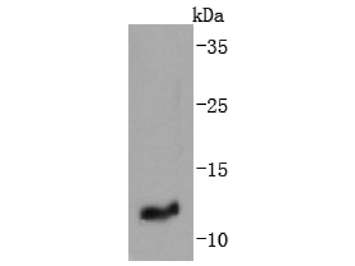 Western blot analysis of IGF2 on human placenta tissue lysates. Proteins were transferred to a PVDF membrane and blocked with 5% BSA in PBS for 1 hour at room temperature. The primary antibody (SLM-52776R, 1/500) was used in 5% BSA at room temperature for 2 hours. Goat Anti-Rabbit IgG - HRP Secondary Antibody (HA1001) at 1:200,000 dilution was used for 1 hour at room temperature.
Predicted band size: 20 kDa
Observed band size: 11 kDa
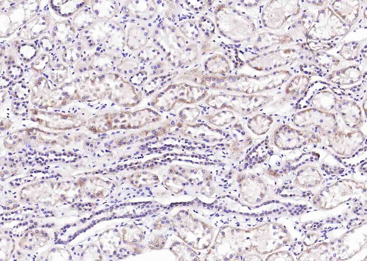 Paraformaldehyde-fixed, paraffin embedded (Human kidney); Antigen retrieval by boiling in sodium citrate buffer (pH6.0) for 15min; Block endogenous peroxidase by 3% hydrogen peroxide for 20 minutes; Blocking buffer (normal goat serum) at 37°C for 30min; Antibody incubation with (IGF2) Monoclonal Antibody, Unconjugated (SLM-52776R) at 1:200 overnight at 4°C, followed by operating according to SP Kit(Rabbit) (sp-0023) instructionsand DAB staining.
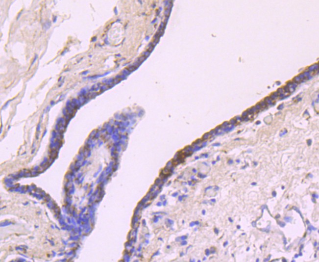 Immunohistochemical analysis of paraffin-embedded human breast carcinoma tissue using anti-IGF2 antibody. The section was pre-treated using heat mediated antigen retrieval with Tris-EDTA buffer (pH 9.0) for 20 minutes.The tissues were blocked in 1% BSA for 30 minutes at room temperature, washed with ddH2O and PBS, and then probed with the primary antibody (SLM-52776R, 1/50) for 30 minutes at room temperature. The detection was performed using an HRP conjugated compact polymer system. DAB was used as the chromogen. Tissues were counterstained with hematoxylin and mounted with DPX.
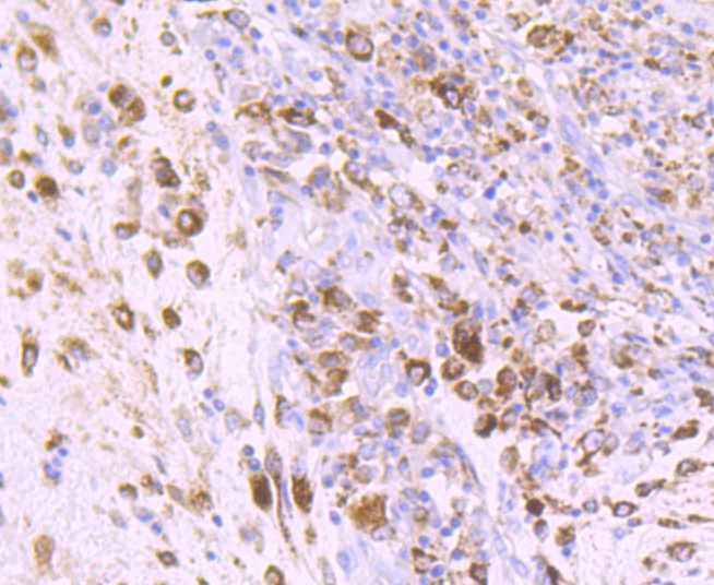 Immunohistochemical analysis of paraffin-embedded human colon carcinoma tissue using anti-IGF2 antibody. The section was pre-treated using heat mediated antigen retrieval with Tris-EDTA buffer (pH 9.0) for 20 minutes.The tissues were blocked in 1% BSA for 30 minutes at room temperature, washed with ddH2O and PBS, and then probed with the primary antibody (SLM-52776R, 1/50) for 30 minutes at room temperature. The detection was performed using an HRP conjugated compact polymer system. DAB was used as the chromogen. Tissues were counterstained with hematoxylin and mounted with DPX.
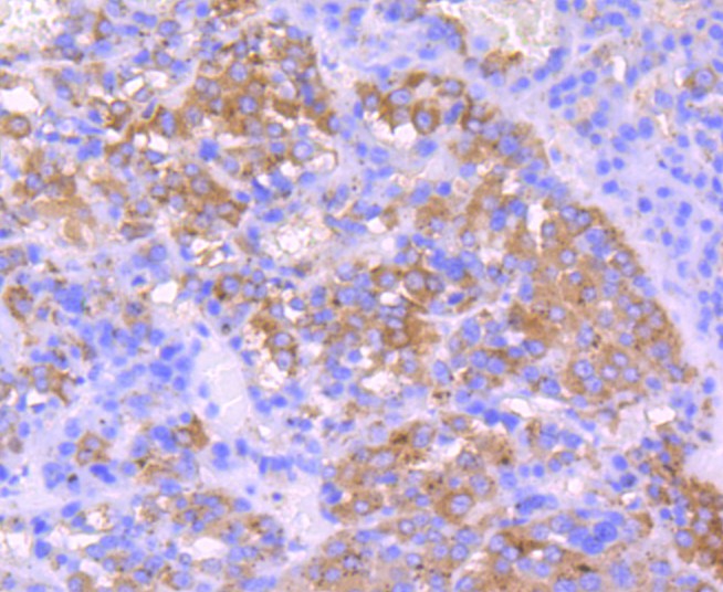 Immunohistochemical analysis of paraffin-embedded human liver carcinoma tissue using anti-IGF2 antibody. The section was pre-treated using heat mediated antigen retrieval with Tris-EDTA buffer (pH 9.0) for 20 minutes.The tissues were blocked in 1% BSA for 30 minutes at room temperature, washed with ddH2O and PBS, and then probed with the primary antibody (SLM-52776R, 1/50) for 30 minutes at room temperature. The detection was performed using an HRP conjugated compact polymer system. DAB was used as the chromogen. Tissues were counterstained with hematoxylin and mounted with DPX.
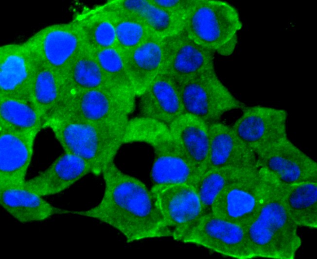 ICC staining of IGF2 in 293 cells (green). Formalin fixed cells were permeabilized with 0.1% Triton X-100 in TBS for 10 minutes at room temperature and blocked with 10% negative goat serum for 15 minutes at room temperature. Cells were probed with the primary antibody (SLM-52776R, 1/50) for 1 hour at room temperature, washed with PBS. Alexa Fluor®488 conjugate-Goat anti-Rabbit IgG was used as the secondary antibody at 1/1,000 dilution. The nuclear counter stain is DAPI (blue).
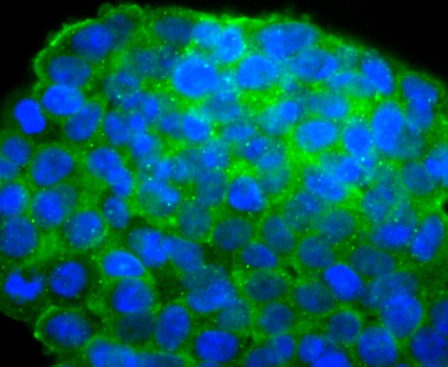 ICC staining of IGF2 in 293T cells (green). Formalin fixed cells were permeabilized with 0.1% Triton X-100 in TBS for 10 minutes at room temperature and blocked with 10% negative goat serum for 15 minutes at room temperature. Cells were probed with the primary antibody (SLM-52776R, 1/50) for 1 hour at room temperature, washed with PBS. Alexa Fluor®488 conjugate-Goat anti-Rabbit IgG was used as the secondary antibody at 1/1,000 dilution. The nuclear counter stain is DAPI (blue).
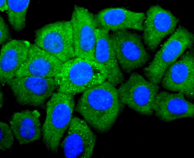 ICC staining of IGF2 in HepG2 cells (green). Formalin fixed cells were permeabilized with 0.1% Triton X-100 in TBS for 10 minutes at room temperature and blocked with 10% negative goat serum for 15 minutes at room temperature. Cells were probed with the primary antibody (SLM-52776R, 1/50) for 1 hour at room temperature, washed with PBS. Alexa Fluor®488 conjugate-Goat anti-Rabbit IgG was used as the secondary antibody at 1/1,000 dilution. The nuclear counter stain is DAPI (blue).
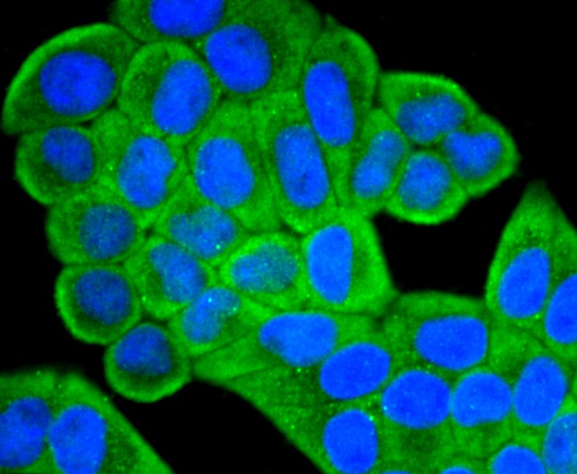 ICC staining of IGF2 in Hela cells (green). Formalin fixed cells were permeabilized with 0.1% Triton X-100 in TBS for 10 minutes at room temperature and blocked with 10% negative goat serum for 15 minutes at room temperature. Cells were probed with the primary antibody (SLM-52776R, 1/50) for 1 hour at room temperature, washed with PBS. Alexa Fluor®488 conjugate-Goat anti-Rabbit IgG was used as the secondary antibody at 1/1,000 dilution. The nuclear counter stain is DAPI (blue).
Cartpieces
Totalgoods,subtotals:¥Checkout
References (0)
No References
Bought notes(bought amounts latest0)
No one bought this product
User Comment(Total0User Comment Num)
- No comment


 +86 571 56623320
 +86 18668110335

