
Rabbit Anti-ADRB2 antibody
Beta 2-adrenergic receptor; beta 2 Adrenergic Receptor; ADRB2R; ADRBR; ADRB2_HUMAN; Adrenergic beta 2 receptor surface; B2AR; BAR; Beta 2 adrenoreceptor; BETA2AR; Catecholamine receptor; beta2-adrenergic receptor.
View History [Clear]
Details
Product Name ADRB2 Chinese Name 肾上腺素能受体β2Recombinant rabbit monoclonal anti Alias Beta 2-adrenergic receptor; beta 2 Adrenergic Receptor; ADRB2R; ADRBR; ADRB2_HUMAN; Adrenergic beta 2 receptor surface; B2AR; BAR; Beta 2 adrenoreceptor; BETA2AR; Catecholamine receptor; beta2-adrenergic receptor. Research Area Tumour Cell biology Neurobiology The cell membrane受体 Immunogen Species Rabbit Clonality Monoclonal Clone NO. 7C1 React Species Human, (predicted: Mouse, Rat, Zebrafish, ) Applications WB=1:500-2000 IHC-P=1:100-500 Flow-Cyt=1:100 ICC=1:100 (Paraffin sections need antigen repair)
not yet tested in other applications.
optimal dilutions/concentrations should be determined by the end user.Theoretical molecular weight 60kDa Form Liquid Concentration 1mg/ml immunogen KLH conjugated synthetic peptide derived from human ADRB2 Lsotype IgG Purification affinity purified by Protein A Buffer Solution 0.01M TBS(pH7.4) with 1% BSA, 0.03% Proclin300 and 50% Glycerol. Storage Shipped at 4℃. Store at -20 °C for one year. Avoid repeated freeze/thaw cycles. Attention This product as supplied is intended for research use only, not for use in human, therapeutic or diagnostic applications. PubMed PubMed Product Detail Beta 2 Adrenergic Receptor is a member of the G protein coupled receptor superfamily. This receptor is directly associated with one of its ultimate effectors, the class C L type calcium channel Ca(V)1.2. This receptor channel complex also contains a G protein, an adenylyl cyclase, cAMP dependent kinase, and the counterbalancing phosphatase, PP2A. The assembly of the signaling complex provides a mechanism that ensures specific and rapid signaling by this G protein coupled receptor. This gene contains no introns in either its coding or untranslated sequences. Different polymorphic forms, point mutations, and/or downregulation of this gene are associated with nocturnal asthma, obesity and type 2 diabetes. Expression of the beta 2 Adrenergic Receptor has been reported in adipose, blood, brain, heart, lung, nose, pancreas, skeletal muscle, skin, and vessel.
Function:
Beta-adrenergic receptors mediate the catecholamine-induced activation of adenylate cyclase through the action of G proteins. The beta-2-adrenergic receptor binds epinephrine with an approximately 30-fold greater affinity than it does norepinephrine.
Subunit:
Binds SLC9A3R1 and GPRASP1. Interacts with ARRB1 and ARRB2. Interacts with SRC, USP20 and USP33. Interacts with VHL; the interaction, which is increased on hydroxylation of ADRB2, ubiquitinates ADRB2 leading to its degradation. Interacts with EGLN3; the interaction hydroxylates ADRB2 facilitating VHL-E3 ligase-mediated ubiquitination.
Subcellular Location:
Cell membrane; Multi-pass membrane protein. Note=Colocalizes with VHL at the cell membrane.
Similarity:
Belongs to the G-protein coupled receptor 1 family.
Adrenergic receptor subfamily. ADRB2 sub-subfamily.
SWISS:
P07550
Gene ID:
154
Product Picture 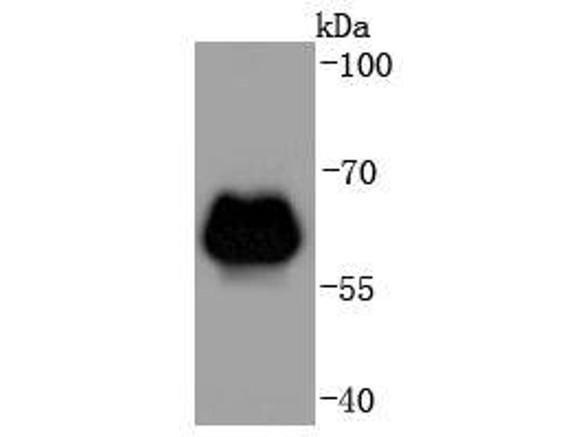 Western blot analysis of beta 2 Adrenergic Receptor on zebrafish tissue lysates. Proteins were transferred to a PVDF membrane and blocked with 5% BSA in PBS for 1 hour at room temperature. The primary antibody (SLM-52880R, 1/500) was used in 5% BSA at room temperature for 2 hours. Goat Anti-Rabbit IgG - HRP Secondary Antibody at 1:5,000 dilution was used for 1 hour at room temperature.
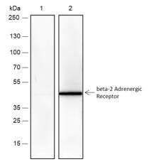 Blocking buffer: 5% NFDM/TBST
Primary Ab dilution: 1:1000
Primary Ab incubation condition: lane 1:
PTM-6016+Competitive peptides, lane 2: PTM-
6016, 2 hours at room temperature
Secondary Ab: Goat Anti-Rabbit IgG H&L
(HRP)
Lysate: Mouse kidney
Protein loading quantity: 20 μg
Exposure time: 60 s
Predicted MW: 46 kDa
Observed MW: 46 kDa
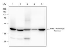 Blocking buffer: 5% NFDM/TBST
Primary Ab dilution: 1:2000
Primary Ab incubation condition: 2 hours at
room temperature
Secondary Ab: Goat Anti-Rabbit IgG H&L
(HRP)
Lysate: 1: A431, 2: MCF-7, 3: MDA-MB-231, 4:
Rat kidney, 5: Mouse kidney
Protein loading quantity: 20 μg
Exposure time: 60 s
Predicted MW: 46 kDa
Observed MW: 46 kDa
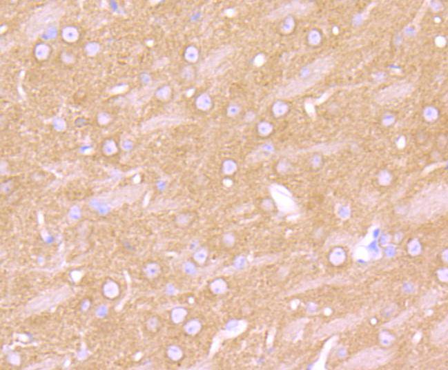 Immunohistochemical analysis of paraffin-embedded mouse brain tissue using anti-beta 2 Adrenergic Receptor antibody. The section was pre-treated using heat mediated antigen retrieval with Tris-EDTA buffer (pH 8.0-8.4) for 20 minutes.The tissues were blocked in 5% BSA for 30 minutes at room temperature, washed with ddH2O and PBS, and then probed with the primary antibody (SLM-52880R, 1/50) for 30 minutes at room temperature. The detection was performed using an HRP conjugated compact polymer system. DAB was used as the chromogen. Tissues were counterstained with hematoxylin and mounted with DPX.
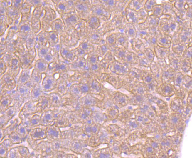 Immunohistochemical analysis of paraffin-embedded mouse liver tissue using anti-beta 2 Adrenergic Receptor antibody. The section was pre-treated using heat mediated antigen retrieval with Tris-EDTA buffer (pH 8.0-8.4) for 20 minutes.The tissues were blocked in 5% BSA for 30 minutes at room temperature, washed with ddH2O and PBS, and then probed with the primary antibody (SLM-52880R, 1/50) for 30 minutes at room temperature. The detection was performed using an HRP conjugated compact polymer system. DAB was used as the chromogen. Tissues were counterstained with hematoxylin and mounted with DPX.
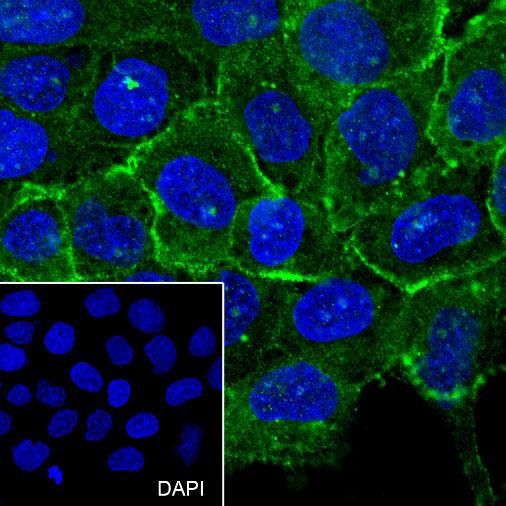 Cell line: A431
Fixation: 100% Ice-cold methanol
Permeabilization: 0.1% TritonX-100
Primary Ab dilution: 1:100
Primary Ab incubation condition: 4°C overnight
Secondary Ab: Goat Anti-Rabbit IgG
Nuclear counter stain: DAPI (Blue)
Comment: Color green is the positive signal for SLM-52880R
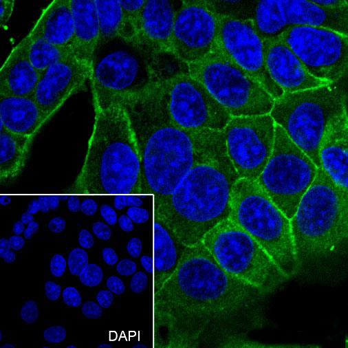 Cell line: MCF7
Fixation: 100% Ice-cold methanol
Permeabilization: 0.1% TritonX-100
Primary Ab dilution: 1:100
Primary Ab incubation condition: 4°C overnight
Secondary Ab: Goat Anti-Rabbit IgG
Nuclear counter stain: DAPI (Blue)
Comment: Color green is the positive signal for SLM-52880R
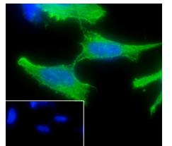 Cell line: SH-SY5Y
Fixation: 100% Ice-cold methanol
Permeabilization: 0.1% TritonX-100
Primary Ab dilution: 1:50
Primary Ab incubation condition: 4℃
overnight
Secondary Ab: Goat Anti-Rabbit IgG
Nuclear counter stain: DAPI (Blue)
Comment: Color green is the positive signal for
PTM-6016
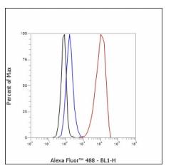 Cell line: SH-SY5Y
Fixation: 4% Paraformaldehyde
Permeabilization: 90% Methanol
Primary Ab dilution: 1:100
Secondary Ab: Goat Anti-Rabbit IgG
Unlabelled control: The cell without incubation
with primary antibody and secondary antibody
(Black line).
Isotype control: Rabbit monoclonal IgG (Blue
line).
Comment: Line red is the positive signal for
SLM-52880R
Cartpieces
Totalgoods,subtotals:¥Checkout
References (0)
No References
Bought notes(bought amounts latest0)
No one bought this product
User Comment(Total0User Comment Num)
- No comment


 +86 571 56623320
 +86 18668110335

