
Rabbit Anti-Heme Oxygenase 1 antibody
Heme oxygenase (decycling) 1; Heme oxygenase 1; Hemox; HMOX 1; Hmox; HMOX1; HO 1; HO-1; HO; HO1; Hsp32; bK286B10; D8Wsu38e; HMOX1_HUMAN.
View History [Clear]
Details
Product Name Heme Oxygenase 1 Chinese Name 血红素氧合酶 1抗体 Alias Heme oxygenase (decycling) 1; Heme oxygenase 1; Hemox; HMOX 1; Hmox; HMOX1; HO 1; HO-1; HO; HO1; Hsp32; bK286B10; D8Wsu38e; HMOX1_HUMAN. Research Area Tumour Cardiovascular immunology Neurobiology Signal transduction Epigenetics Alzheimer's Immunogen Species Rabbit Clonality Monoclonal React Species Human, Mouse, Rat, Applications WB=1:2000-20000 IP=1:25-100 IHC-P=1:100-500 ICC=1:50 IF=1:50 (Paraffin sections need antigen repair)
not yet tested in other applications.
optimal dilutions/concentrations should be determined by the end user.Theoretical molecular weight 32kDa Cellular localization cytoplasmic Form Liquid Concentration 1mg/ml immunogen KLH conjugated synthetic peptide derived from human Heme Oxygenase 1 Lsotype IgG Purification affinity purified by Protein A Buffer Solution PBS, Glycerol, BSA Storage Shipped at 4℃. Store at -20 °C for one year. Avoid repeated freeze/thaw cycles. Attention This product as supplied is intended for research use only, not for use in human, therapeutic or diagnostic applications. PubMed PubMed Product Detail The hemeoxygenase-1 calls that the hemoglobin oxidizes to synthesize the enzyme again-1( hemeoxygenase-1, HO-1) is the catalyst enzyme that a kind of hemoglobin declines the solution, under the NADPH and the cell dye P-450 revivification enzymes and the member oxygen functions, the catalyst HO-1 hemoglobin declines the solution as the courage green vegetable, CO and irons, the former revivification has the very strong anti- to oxidize the ability after become the red vegetable of courage , the latter is a kind of important letter to make the member.
SWISS:
P09601
Gene ID:
3162
Product Picture 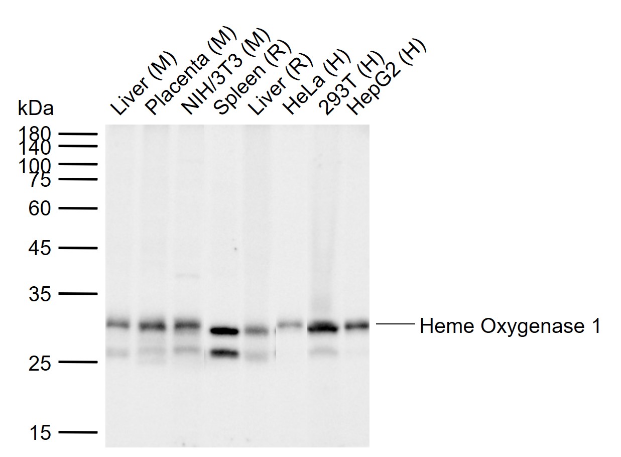 Sample:
Lane 1: Mouse Liver tissue lysates
Lane 2: Mouse Placenta tissue lysates
Lane 3: Mouse NIH/3T3 cell lysates
Lane 4: Rat Spleen tissue lysates
Lane 5: Rat Liver tissue lysates
Lane 6: Human HeLa cell lysates
Lane 7: Human 293T cell lysates
Lane 8: Human HepG2 cell lysates
Primary: Anti-Heme Oxygenase 1 (SLM-60751R) at 1/10000 dilution
Secondary: IRDye800CW Goat Anti-Rabbit IgG at 1/20000 dilution
Predicted band size: 32 kDa
Observed band size: 30 kDa
 Paraformaldehyde-fixed, paraffin embedded (mouse thymus); Antigen retrieval by boiling in sodium citrate buffer (pH6.0) for 15min; Block endogenous peroxidase by 3% hydrogen peroxide for 20 minutes; Blocking buffer (normal goat serum) at 37°C for 30min; Incubation with (Heme Oxygenase 1) Monoclonal Antibody, Unconjugated (SLM-60751R) at 1:200 overnight at 4°C, followed by operating according to SP Kit(Rabbit) (sp-0023) instructionsand DAB staining.
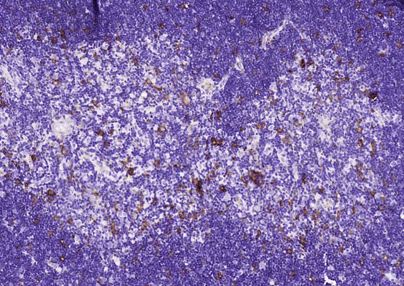
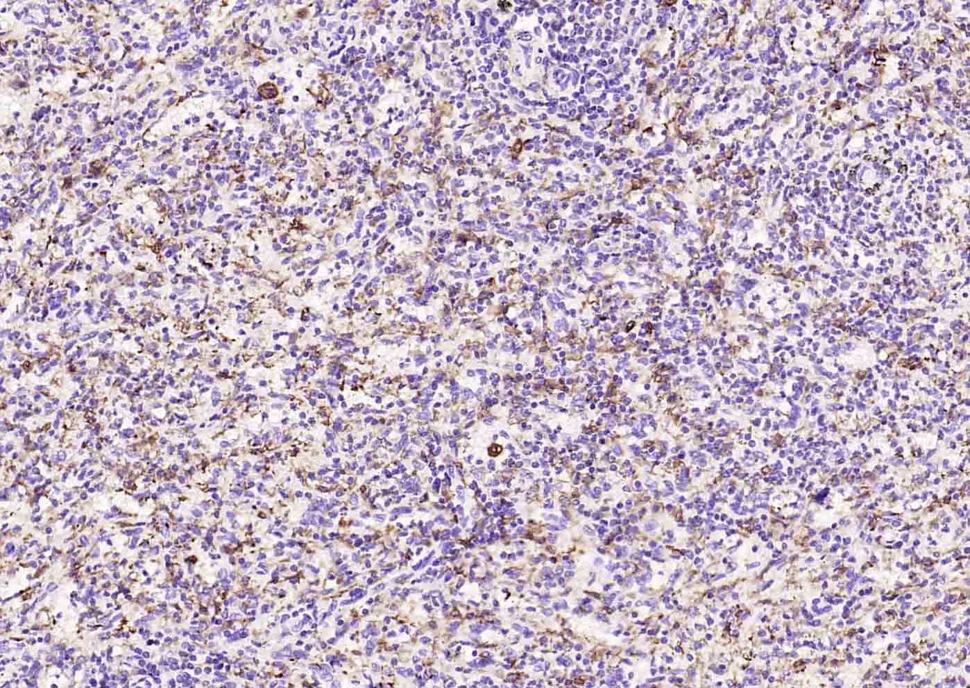 Paraformaldehyde-fixed, paraffin embedded (human spleen); Antigen retrieval by boiling in sodium citrate buffer (pH6.0) for 15min; Block endogenous peroxidase by 3% hydrogen peroxide for 20 minutes; Blocking buffer (normal goat serum) at 37°C for 30min; Incubation with (Heme Oxygenase 1) Monoclonal Antibody, Unconjugated (SLM-60751R) at 1:200 overnight at 4°C, followed by operating according to SP Kit(Rabbit) (sp-0023) instructionsand DAB staining.
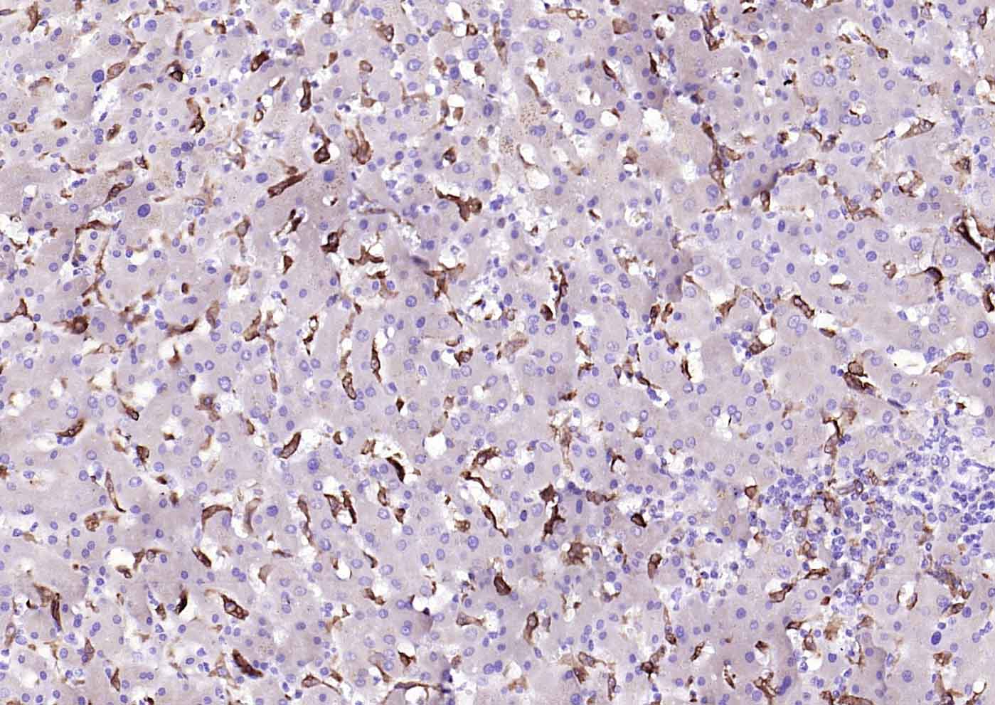 Paraformaldehyde-fixed, paraffin embedded (human liver); Antigen retrieval by boiling in sodium citrate buffer (pH6.0) for 15min; Block endogenous peroxidase by 3% hydrogen peroxide for 20 minutes; Blocking buffer (normal goat serum) at 37°C for 30min; Incubation with (Heme Oxygenase 1) Monoclonal Antibody, Unconjugated (SLM-60751R) at 1:200 overnight at 4°C, followed by operating according to SP Kit(Rabbit) (sp-0023) instructionsand DAB staining.
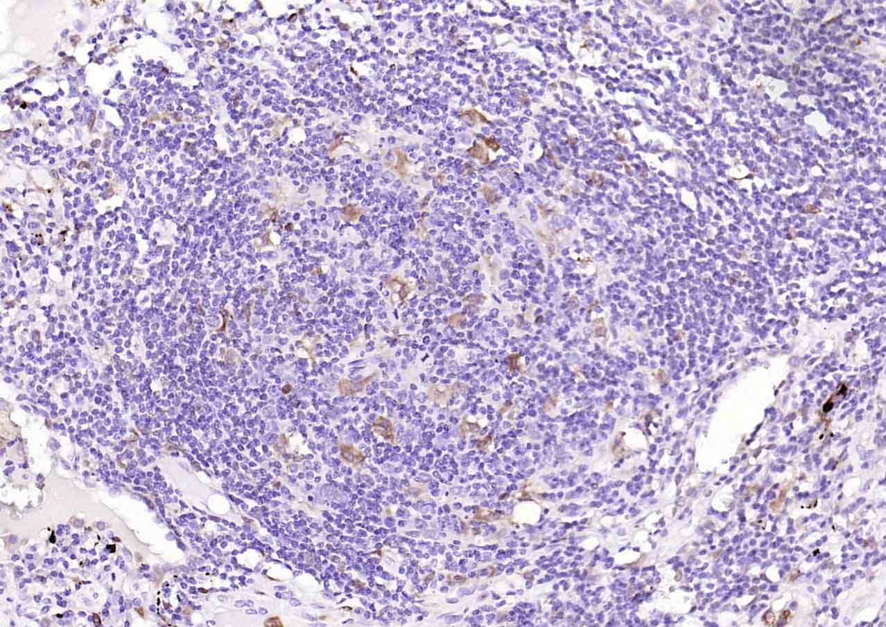 Paraformaldehyde-fixed, paraffin embedded (human lung carcinoma); Antigen retrieval by boiling in sodium citrate buffer (pH6.0) for 15min; Block endogenous peroxidase by 3% hydrogen peroxide for 20 minutes; Blocking buffer (normal goat serum) at 37°C for 30min; Incubation with (Heme Oxygenase 1) Monoclonal Antibody, Unconjugated (SLM-60751R) at 1:200 overnight at 4°C, followed by operating according to SP Kit(Rabbit) (sp-0023) instructionsand DAB staining.
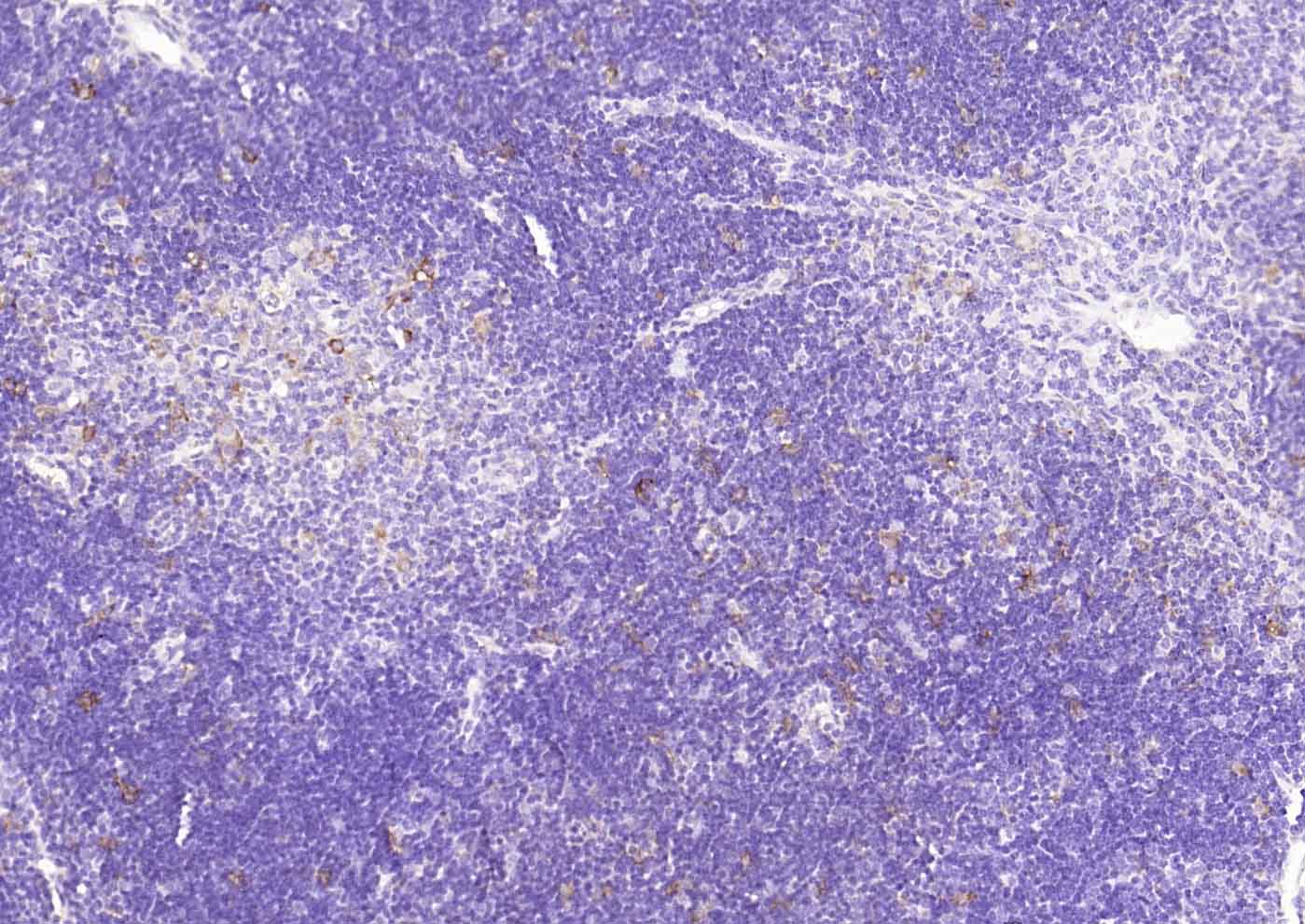 Paraformaldehyde-fixed, paraffin embedded (rat thymus); Antigen retrieval by boiling in sodium citrate buffer (pH6.0) for 15min; Block endogenous peroxidase by 3% hydrogen peroxide for 20 minutes; Blocking buffer (normal goat serum) at 37°C for 30min; Incubation with (Heme Oxygenase 1) Monoclonal Antibody, Unconjugated (SLM-60751R) at 1:200 overnight at 4°C, followed by operating according to SP Kit(Rabbit) (sp-0023) instructionsand DAB staining.
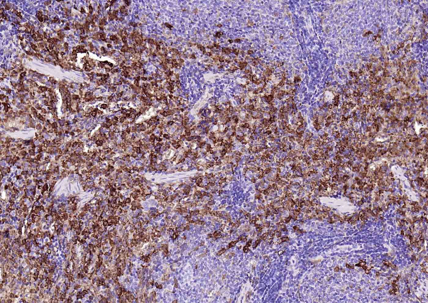 Paraformaldehyde-fixed, paraffin embedded (rat spleen); Antigen retrieval by boiling in sodium citrate buffer (pH6.0) for 15min; Block endogenous peroxidase by 3% hydrogen peroxide for 20 minutes; Blocking buffer (normal goat serum) at 37°C for 30min; Incubation with (Heme Oxygenase 1) Monoclonal Antibody, Unconjugated (SLM-60751R) at 1:200 overnight at 4°C, followed by operating according to SP Kit(Rabbit) (sp-0023) instructionsand DAB staining.
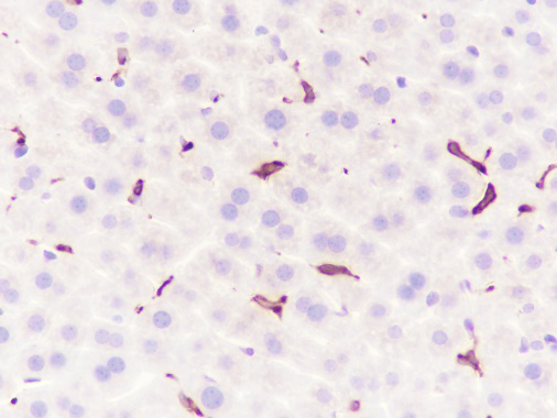 Tissue: mouse liver
Section type: Formalin-fixed & Paraffinembedded
section
Retrieval method: High temperature and high
pressure
Retrieval buffer: Tris/EDTA buffer, pH 9.0
Primary Ab dilution: 1:2000
Primary Ab incubation condition: 1 hour at
room temperature
Secondary Ab:SP Kit(Rabbit) (sp-0023)
Polymer HRP (Ready to use)
Counter stain: Hematoxylin (Blue)
Comment: Color brown is the positive signal
for SLM-60751R
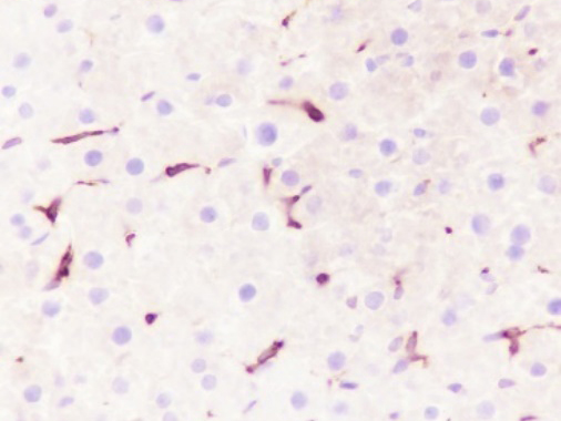 Tissue: rat liver
Section type: Formalin-fixed & Paraffinembedded
section
Retrieval method: High temperature and high
pressure
Retrieval buffer: Tris/EDTA buffer, pH 9.0
Primary Ab dilution: 1:2000
Primary Ab incubation condition: 1 hour at
room temperature
Secondary Ab:SP Kit(Rabbit) (sp-0023)
Polymer HRP (Ready to use)
Counter stain: Hematoxylin (Blue)
Comment: Color brown is the positive signal
for SLM-60751R
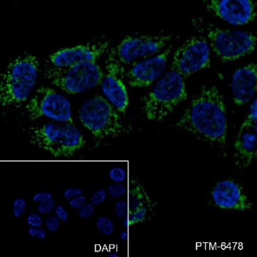 Cell line: MCF7
Fixative: 100% Ice-cold methanol
Permeabilization: 0.1% TritonX-100
Primary ab dilution: 1:50
Primary incubation condition: 4°C overnight
Secondary ab: Goat Anti-Rabbit IgG
Nuclear counter stain: DAPI (Blue)
Comment: Color green is the positive signal for SLM-60751R
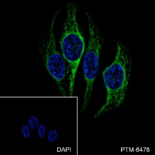 Cell line: HeLa
Fixative: 100% Ice-cold methanol
Permeabilization: 0.1% TritonX-100
Primary ab dilution: 1:50
Primary incubation condition: 4°C overnight
Secondary ab: Goat Anti-Rabbit IgG
Nuclear counter stain: DAPI (Blue)
Comment: Color green is the positive signal for SLM-60751R
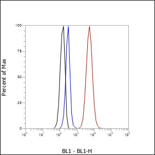
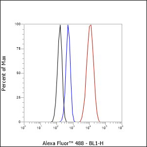 Cell line: MCF-7
Fixative: 4% Paraformaldehyde
Permeabilization: 90% Methanol
Primary ab dilution: 1:100
Secondary ab: Goat Anti-Rabbit IgG
Unlabelled control: The cell without incubation with primary antibody and secondary antibody(Black line)
Isotype control: Rabbit monoclonal IgG (Blueline).
Comment: Line red is the positive signal for SLM-60751R
 Cell line: HeLa
Fixative: 4% Paraformaldehyde
Permeabilization: 90% Methanol
Primary ab dilution: 1:100
Secondary ab: Goat Anti-Rabbit IgG
Unlabelled control: The cell without incubation with primary antibody and secondary antibody(Black line)
Isotype control: Rabbit monoclonal IgG (Blueline).
Comment: Line red is the positive signal for SLM-60751R
Cartpieces
Totalgoods,subtotals:¥Checkout
References (0)
No References
Bought notes(bought amounts latest0)
No one bought this product
User Comment(Total0User Comment Num)
- No comment


 +86 571 56623320
 +86 18668110335

