
Rabbit Anti-SCARB1/Scavenger Receptor BI antibody
HDL-R; High Density Lipoprotein Receptor; CD36 Antigen like 1; CD36L1; CLA 1; CLA1;SR BI; SRB1; SRBI; Scavenger Receptor BI; CD36 AND LIMPII ANALOGOUS 1; CD36 Antigen like 1; CD36L1; CLA 1; CLA1; Collagen type I receptor; MGC138242; SCARB1; Scavebger Rece
View History [Clear]
Details
Product Name SCARB1/Scavenger Receptor BI Chinese Name 高密度Lipoprotein受体/清道夫受体Recombinant rabbit monoclonal anti Alias HDL-R; High Density Lipoprotein Receptor; CD36 Antigen like 1; CD36L1; CLA 1; CLA1;SR BI; SRB1; SRBI; Scavenger Receptor BI; CD36 AND LIMPII ANALOGOUS 1; CD36 Antigen like 1; CD36L1; CLA 1; CLA1; Collagen type I receptor; MGC138242; SCARB1; Scavebger Receptor Class B Member 1; Scavenger Receptor Class B Type 1; SR BI; SRB1; SRBI; Thrombospondin receptor like 1; High density lipoprotein receptor SR-BI. Research Area immunology Growth factors and hormones The cell membrane受体 Diabetes Immunogen Species Rabbit Clonality Monoclonal Clone NO. 1C2 React Species (predicted: Human, Mouse, Rat, ) Applications WB=1:500-2000 IHC-P=1:100-500 IHC-F=1:200-500 ICC=1:50-200 (Paraffin sections need antigen repair)
not yet tested in other applications.
optimal dilutions/concentrations should be determined by the end user.Theoretical molecular weight 61kDa Cellular localization cytoplasmic The cell membrane Form Liquid Concentration 1mg/ml immunogen KLH conjugated synthetic peptide derived from human SCARB1/Scavenger Receptor BI Lsotype IgG Purification affinity purified by Protein A Buffer Solution 0.01M TBS(pH7.4) with 1% BSA, 0.03% Proclin300 and 50% Glycerol. Storage Shipped at 4℃. Store at -20 °C for one year. Avoid repeated freeze/thaw cycles. Attention This product as supplied is intended for research use only, not for use in human, therapeutic or diagnostic applications. PubMed PubMed Product Detail High density lipoproteins (HDLs) play a critical role in cholesterol metabolism and their plasma concentrations are inversely correlated with risk for atherosclerosis. The SR-BI (Scavenger Receptor BI) protein binds HDLs and mediates selective uptake of HDL cholesteryl ester. SR-BI binds HDL with high affinity, is expressed primarily in liver and nonplacental steroidgenic tissues, and mediates selective cholesterol uptake by a distinct mechanism. In mice, it seems that SR-BI plays a key role in determining the levels of plasma lipoprotein cholesterol and the accumulation of cholesterol stores in the adrenal gland. Scavenging Receptor SR-BI plays a critical role in HCV attachment and/or cell entry by interacting with HCV E1/E2 glycoproteins heterodimer.
Function:
Receptor for different ligands such as phospholipids, cholesterol ester, lipoproteins, phosphatidylserine and apoptotic cells. Probable receptor for HDL, located in particular region of the plasma membrane, called caveolae. Facilitates the flux of free and esterified cholesterol between the cell surface and extracellular donors and acceptors, such as HDL and to a lesser extent, apoB-containing lipoproteins and modified lipoproteins. Probably involved in the phagocytosis of apoptotic cells, via its phosphatidylserine binding activity. Receptor for hepatitis C virus glycoprotein E2. Binding between SCARB1 and E2 was found to be independent of the genotype of the viral isolate. Plays an important role in the uptake of HDL cholesteryl ester.
Subunit:
Plays a critical role in HCV attachment and/or cell entry by interacting with HCV E1/E2 glycoproteins heterodimer. The C-terminal region binds to PDZK1.
Subcellular Location:
Cell membrane; Multi-pass membrane protein. Membrane, caveola; Multi-pass membrane protein. Note=Predominantly localized to cholesterol and sphingomyelin-enriched domains within the plasma membrane, called caveolae.
Tissue Specificity:
Widely expressed.
The six cysteines of the extracellular domain are all involved in intramolecular disulfide bonds.
Post-translational modifications:
N-glycosylated.
Similarity:
Belongs to the CD36 family.
SWISS:
Q8WTV0
Gene ID:
949
Product Picture 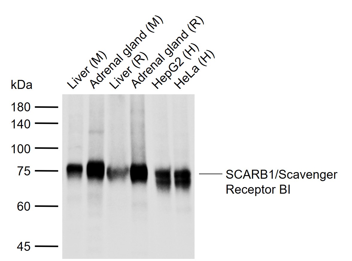 Sample:
Lane 1: Mouse Liver tissue lysates
Lane 2: Mouse Adrenal gland tissue lysates
Lane 3: Rat Liver tissue lysates
Lane 4: Rat Adrenal gland tissue lysates
Lane 5: Human HepG2 cell lysates
Lane 6: Human HeLa cell lysates
Primary: Anti-SCARB1/Scavenger Receptor BI (SLM-52283R) at 1/500 dilution
Secondary: IRDye800CW Goat Anti-Rabbit IgG at 1/20000 dilution
Predicted band size: 61 kDa
Observed band size: 75 kDa
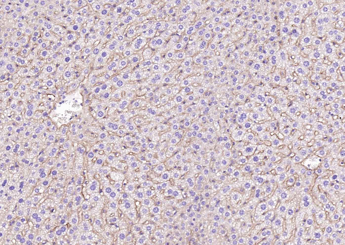 Paraformaldehyde-fixed, paraffin embedded (mouse liver); Antigen retrieval by boiling in sodium citrate buffer (pH6.0) for 15min; Block endogenous peroxidase by 3% hydrogen peroxide for 20 minutes; Blocking buffer (normal goat serum) at 37°C for 30min; Incubation with (SCARB1/Scavenger Receptor BI) Monoclonal Antibody, Unconjugated (SLM-52283R) at 1:200 overnight at 4°C, followed by operating according to SP Kit(Rabbit) (sp-0023) instructionsand DAB staining.
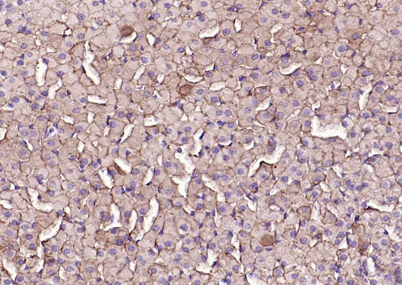 Paraformaldehyde-fixed, paraffin embedded (mouse adrenal gland); Antigen retrieval by boiling in sodium citrate buffer (pH6.0) for 15min; Block endogenous peroxidase by 3% hydrogen peroxide for 20 minutes; Blocking buffer (normal goat serum) at 37°C for 30min; Incubation with (SCARB1/Scavenger Receptor BI) Monoclonal Antibody, Unconjugated (SLM-52283R) at 1:200 overnight at 4°C, followed by operating according to SP Kit(Rabbit) (sp-0023) instructionsand DAB staining.
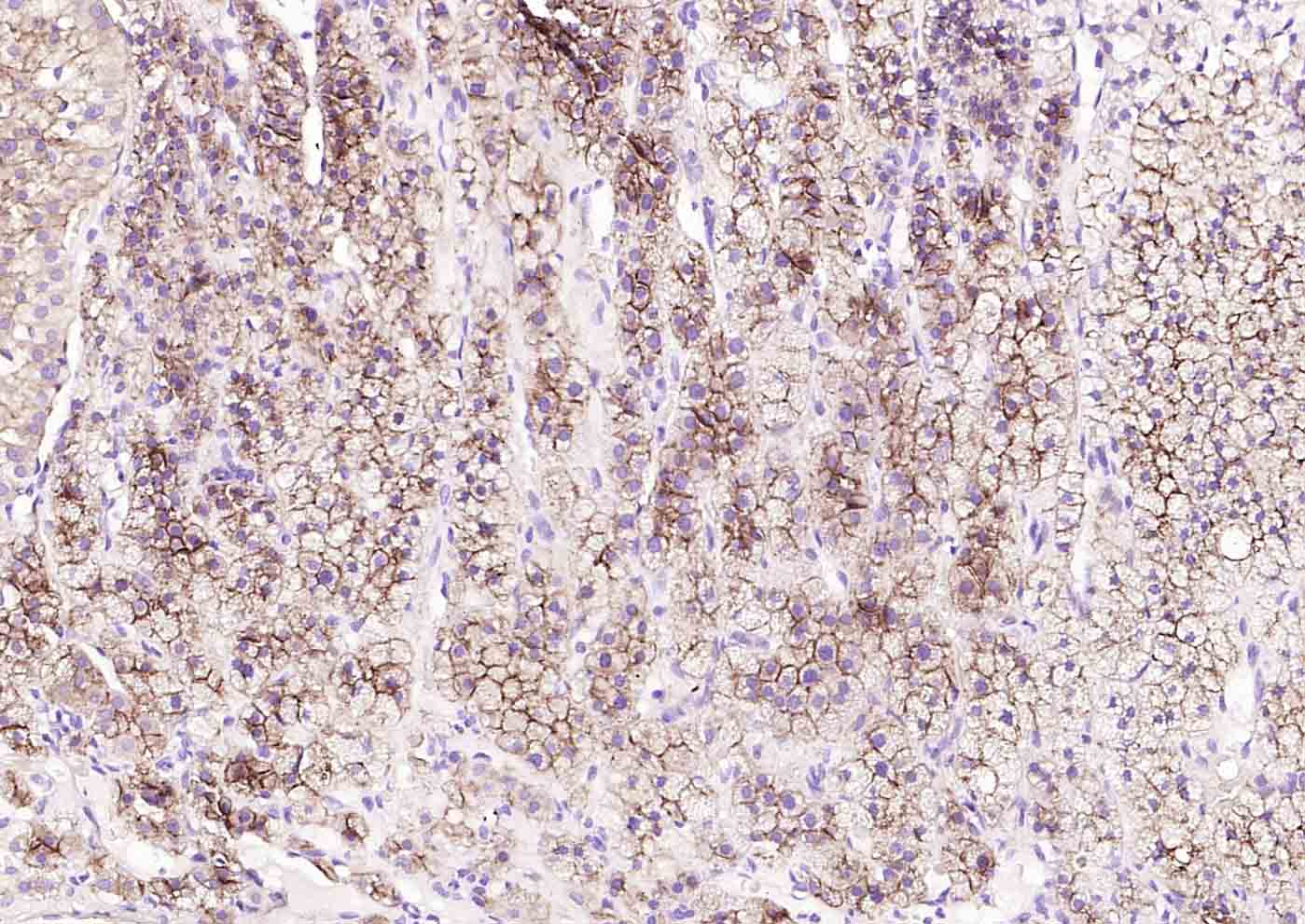 Paraformaldehyde-fixed, paraffin embedded (human adrenal gland); Antigen retrieval by boiling in sodium citrate buffer (pH6.0) for 15min; Block endogenous peroxidase by 3% hydrogen peroxide for 20 minutes; Blocking buffer (normal goat serum) at 37°C for 30min; Incubation with (SCARB1/Scavenger Receptor BI) Monoclonal Antibody, Unconjugated (SLM-52283R) at 1:200 overnight at 4°C, followed by operating according to SP Kit(Rabbit) (sp-0023) instructionsand DAB staining.
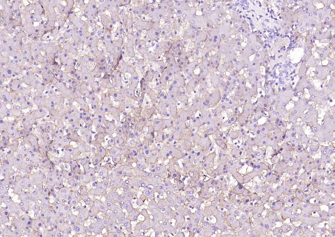 Paraformaldehyde-fixed, paraffin embedded (human liver); Antigen retrieval by boiling in sodium citrate buffer (pH6.0) for 15min; Block endogenous peroxidase by 3% hydrogen peroxide for 20 minutes; Blocking buffer (normal goat serum) at 37°C for 30min; Incubation with (SCARB1/Scavenger Receptor BI) Monoclonal Antibody, Unconjugated (SLM-52283R) at 1:200 overnight at 4°C, followed by operating according to SP Kit(Rabbit) (sp-0023) instructionsand DAB staining.
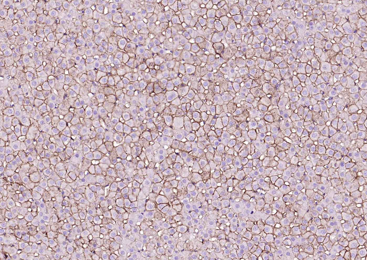 Paraformaldehyde-fixed, paraffin embedded (rat adrenal gland); Antigen retrieval by boiling in sodium citrate buffer (pH6.0) for 15min; Block endogenous peroxidase by 3% hydrogen peroxide for 20 minutes; Blocking buffer (normal goat serum) at 37°C for 30min; Incubation with (SCARB1/Scavenger Receptor BI) Monoclonal Antibody, Unconjugated (SLM-52283R) at 1:200 overnight at 4°C, followed by operating according to SP Kit(Rabbit) (sp-0023) instructionsand DAB staining.
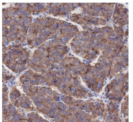 Tissue: Human liver carcinoma
Section type: Formalin-fixed & Paraffin
-embedded section
Retrieval method: High temperature and high
pressure
Retrieval buffer: Tris/EDTA buffer, pH 9.0
Primary Ab dilution: 1:2000
Primary Ab incubation condition: 1 hour at
room temperature
Secondary Ab: Anti-Rabbit and Mouse
Polymer HRP (Ready to use)
Counter stain: Hematoxylin (Blue)
Comment: Color brown is the positive signal for
SLM-52283R
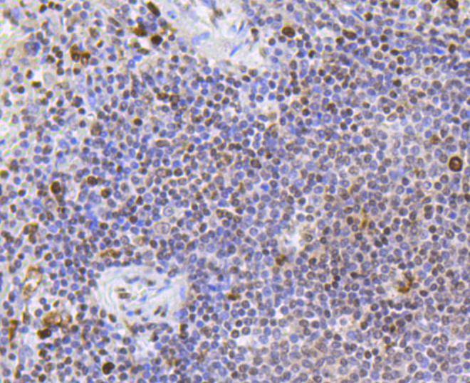 Immunohistochemical analysis of paraffin-embedded human spleen tissue using anti-Scavenging Receptor SR-BI antibody. The section was pre-treated using heat mediated antigen retrieval with Tris-EDTA buffer (pH 8.0-8.4) for 20 minutes.The tissues were blocked in 5% BSA for 30 minutes at room temperature, washed with ddH2O and PBS, and then probed with the primary antibody (SLM-52283R, 1/50) for 30 minutes at room temperature. The detection was performed using an HRP conjugated compact polymer system. DAB was used as the chromogen. Tissues were counterstained with hematoxylin and mounted with DPX.
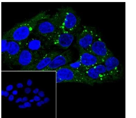 Cell line: HepG2
Fixation: 4% Paraformaldehyde
Permeabilization: 0.1% TritonX-100
Primary Ab dilution: 1:50
Primary Ab incubation condition: 4°C
overnight
Secondary Ab: Goat Anti-Rabbit IgG
Nuclear counter stain: DAPI (Blue)
Comment: Color green is the positive signal for
SLM-52283R
 ICC staining of Scavenging Receptor SR-BI in CRC cells (green). Formalin fixed cells were permeabilized with 0.1% Triton X-100 in TBS for 10 minutes at room temperature and blocked with 1% Blocker BSA for 15 minutes at room temperature. Cells were probed with the primary antibody (SLM-52283R, 1/50) for 1 hour at room temperature, washed with PBS. Alexa Fluor®488 Goat anti-Rabbit IgG was used as the secondary antibody at 1/1,000 dilution. The nuclear counter stain is DAPI (blue).
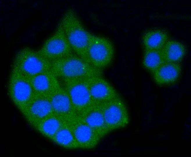 ICC staining of Scavenging Receptor SR-BI in PC-12 cells (green). Formalin fixed cells were permeabilized with 0.1% Triton X-100 in TBS for 10 minutes at room temperature and blocked with 1% Blocker BSA for 15 minutes at room temperature. Cells were probed with the primary antibody (SLM-52283R, 1/50) for 1 hour at room temperature, washed with PBS. Alexa Fluor®488 Goat anti-Rabbit IgG was used as the secondary antibody at 1/1,000 dilution. The nuclear counter stain is DAPI (blue).
Cartpieces
Totalgoods,subtotals:¥Checkout
References (0)
No References
Bought notes(bought amounts latest0)
No one bought this product
User Comment(Total0User Comment Num)
- No comment


 +86 571 56623320
 +86 18668110335

