
Details
Product Name Histone H4 (acetyl K16) Chinese Name 乙酰化组蛋白H4(K16)Recombinant rabbit monoclonal anti Alias Histone H4 (Acetyl Lys16); Acetyl-Histone H4(K16); Acetyl-Histone H4 (Lys16); H4K16ac; H4F2; H4FN; HIST1H4; HIST2H4; HISTH4H4; methyl histone H4; histone H4; H4_HUMAN; Osteogenic growth peptide; OGP. Product Type Acetylated anti Recombinant rabbit monoclonal anti Research Area Cell biology immunology Epigenetics Immunogen Species Rabbit Clonality Monoclonal Clone NO. 2G2 React Species (predicted: Human, Mouse, Rat, ) Applications WB=1:500-2000 IHC-P=1:100-500 ICC=1:200 (Paraffin sections need antigen repair)
not yet tested in other applications.
optimal dilutions/concentrations should be determined by the end user.Theoretical molecular weight 11kDa Cellular localization The nucleus Form Liquid Concentration 1mg/ml immunogen Recombinant protein Lsotype IgG Purification affinity purified by Protein A Buffer Solution 0.01M TBS(pH7.4) with 1% BSA, 0.03% Proclin300 and 50% Glycerol. Storage Shipped at 4℃. Store at -20 °C for one year. Avoid repeated freeze/thaw cycles. Attention This product as supplied is intended for research use only, not for use in human, therapeutic or diagnostic applications. PubMed PubMed Product Detail Histones are basic nuclear proteins that are responsible for the nucleosome structure of the chromosomal fiber in eukaryotes. Nucleosomes consist of approximately 146 bp of DNA wrapped around a histone octamer composed of pairs of each of the four core histones (H2A, H2B, H3, and H4). The chromatin fiber is further compacted through the interaction of a linker histone, H1, with the DNA between the nucleosomes to form higher order chromatin structures. This gene is intronless and encodes a member of the histone H4 family. Transcripts from this gene lack polyA tails; instead, they contain a palindromic termination element. [provided by RefSeq, Jul 2008].
Function:
Core component of nucleosome. Nucleosomes wrap and compact DNA into chromatin, limiting DNA accessibility to the cellular machineries which require DNA as a template. Histones thereby play a central role in transcription regulation, DNA repair, DNA replication and chromosomal stability. DNA accessibility is regulated via a complex set of post-translational modifications of histones, also called histone code, and nucleosome remodeling. Subunit : The nucleosome is a histone octamer containing two molecules each of H2A, H2B, H3 and H4 assembled in one H3-H4 heterotetramer and two H2A-H2B heterodimers. The octamer wraps approximately 147 bp of DNA.
Subunit:
The nucleosome is a histone octamer containing two molecules each of H2A, H2B, H3 and H4 assembled in one H3-H4 heterotetramer and two H2A-H2B heterodimers. The octamer wraps approximately 147 bp of DNA.
Subcellular Location:
Nucleus. Chromosome.
Post-translational modifications:
Acetylation at Lys-6 (H4K5ac), Lys-9 (H4K8ac), Lys-13 (H4K12ac) and Lys-17 (H4K16ac) occurs in coding regions of the genome but not in heterochromatin.
Citrullination at Arg-4 (H4R3ci) by PADI4 impairs methylation.
Monomethylation and asymmetric dimethylation at Arg-4 (H4R3me1 and H4R3me2a, respectively) by PRMT1 favors acetylation at Lys-9 (H4K8ac) and Lys-13 (H4K12ac). Demethylation is performed by JMJD6. Symmetric dimethylation on Arg-4 (H4R3me2s) by the PRDM1/PRMT5 complex may play a crucial role in the germ-cell lineage.
Monomethylated, dimethylated or trimethylated at Lys-21 (H4K20me1, H4K20me2, H4K20me3). Monomethylation is performed by SET8. Trimethylation is performed by SUV420H1 and SUV420H2 and induces gene silencing.
Phosphorylated by PAK2 at Ser-48 (H4S47ph). This phosphorylation increases the association of H3.3-H4 with the histone chaperone HIRA, thus promoting nucleosome assembly of H3.3-H4 and inhibiting nucleosome assembly of H3.1-H4.
Ubiquitinated by the CUL4-DDB-RBX1 complex in response to ultraviolet irradiation. This may weaken the interaction between histones and DNA and facilitate DNA accessibility to repair proteins. Monoubiquitinated at Lys-92 of histone H4 (H4K91ub1) in response to DNA damage. The exact role of H4K91ub1 in DNA damage response is still unclear but it may function as a licensing signal for additional histone H4 post-translational modifications such as H4 Lys-21 methylation (H4K20me).
Sumoylated, which is associated with transcriptional repression.
Crotonylation (Kcr) is specifically present in male germ cells and marks testis-specific genes in post-meiotic cells, including X-linked genes that escape sex chromosome inactivation in haploid cells. Crotonylation marks active promoters and enhancers and confers resistance to transcriptional repressors. It is also associated with post-meiotically activated genes on autosomes.
Similarity:
Belongs to the histone H4 family.
SWISS:
P62805
Gene ID:
8359
Database links:Entrez Gene: 121504 Human
Entrez Gene: 554313 Human
Entrez Gene: 8294 Human
Entrez Gene: 8359 Human
Entrez Gene: 8360 Human
Entrez Gene: 8361 Human
Entrez Gene: 8362 Human
Entrez Gene: 8363 Human
Entrez Gene: 8364 Human
Entrez Gene: 8365 Human
Entrez Gene: 8366 Human
Entrez Gene: 8367 Human
Entrez Gene: 8368 Human
Entrez Gene: 8370 Human
Entrez Gene: 100041230 Mouse
Entrez Gene: 100862646 Mouse
Entrez Gene: 319155 Mouse
Entrez Gene: 319156 Mouse
Entrez Gene: 319157 Mouse
Entrez Gene: 319158 Mouse
Entrez Gene: 319159 Mouse
Entrez Gene: 319160 Mouse
Entrez Gene: 319161 Mouse
Entrez Gene: 320332 Mouse
Entrez Gene: 326619 Mouse
Entrez Gene: 326620 Mouse
Entrez Gene: 69386 Mouse
Entrez Gene: 97122 Mouse
GenBank: NM_003548 Human
Omim: 142750 Human
SwissProt: P84040 Fruit fly (Drosophila melanogaster)
SwissProt: P02304 Human
SwissProt: P62805 Human
SwissProt: P02304 Mouse
SwissProt: P62806 Mouse
SwissProt: P09322 Schizosaccharomyces pombe
Unigene: 21500 Fruit fly (Drosophila melanogaster)
Unigene: 29514 Fruit fly (Drosophila melanogaster)
Unigene: 29527 Fruit fly (Drosophila melanogaster)
Unigene: 30219 Fruit fly (Drosophila melanogaster)
Unigene: 30220 Fruit fly (Drosophila melanogaster)
Unigene: 30221 Fruit fly (Drosophila melanogaster)
Unigene: 30223 Fruit fly (Drosophila melanogaster)
Unigene: 30868 Fruit fly (Drosophila melanogaster)
Unigene: 30869 Fruit fly (Drosophila melanogaster)
Unigene: 30871 Fruit fly (Drosophila melanogaster)
Unigene: 30872 Fruit fly (Drosophila melanogaster)
Unigene: 30873 Fruit fly (Drosophila melanogaster)
Unigene: 30876 Fruit fly (Drosophila melanogaster)
Unigene: 33873 Fruit fly (Drosophila melanogaster)
Unigene: 5747 Fruit fly (Drosophila melanogaster)
Unigene: 143080 Human
Unigene: 247816 Human
Unigene: 248172 Human
Unigene: 248178 Human
Unigene: 248179 Human
Unigene: 278483 Human
Unigene: 352191 Human
Unigene: 46423 Human
Unigene: 528055 Human
Unigene: 533295 Human
Unigene: 55468 Human
Unigene: 591790 Human
Unigene: 655235 Human
Unigene: 662174 Human
Unigene: 706635 Human
Unigene: 742244 Human
Unigene: 14775 Mouse
Unigene: 158272 Mouse
Unigene: 227295 Mouse
Unigene: 228709 Mouse
Unigene: 246720 Mouse
Unigene: 255646 Mouse
Unigene: 260530 Mouse
Unigene: 261642 Mouse
Unigene: 261662 Mouse
Unigene: 261664 Mouse
Unigene: 377875 Mouse
Unigene: 442307 Mouse
Unigene: 486099 Mouse
Unigene: 489077 Mouse
Product Picture 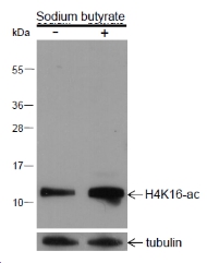 Blocking buffer: 5% NFDM/TBST
Primary ab dilution: 1:2000
Primary ab incubation condition: 2 hours at room temperature
Secondary ab: Goat Anti-Rabbit IgG H&L (HRP)
Lysate: (-) HeLa, (+) HeLa+Sodium butyrate (30mM, 4hr)
Protein loading quantity: 20 μg
Exposure time: 30 s
Predicted MW: 11 kDa
Observed MW: 11 kDa
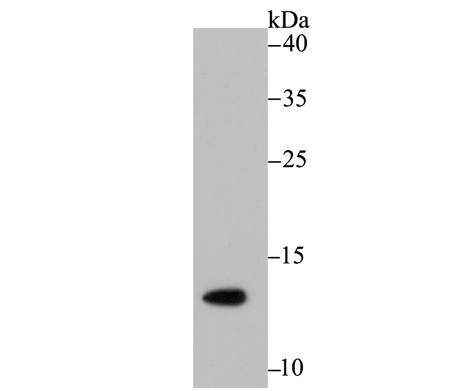 Western blot analysis of Histone H4 (acetyl K16) on SiHa cell lysates. Proteins were transferred to a PVDF membrane and blocked with 5% BSA in PBS for 1 hour at room temperature. The primary antibody (SLM-54330R, 1/500) was used in 5% BSA at room temperature for 2 hours. Goat Anti-Rabbit IgG - HRP Secondary Antibody at 1:200,000 dilution was used for 1 hour at room temperature.
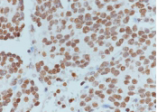 Tissue: Human neuroblastoma
Section type: Formalin fixed & Paraffin-embedded section
Retrieval method: High temperature and high pressure
Retrieval buffer: Tris/EDTA buffer, pH 9.0
Primary ab dilution: 1:200
Primary ab incubation condition: 1 hour at
room temperature
Secondary ab: SP Kit(Rabbit) (sp-0023)
HRP (Ready to use)
Counter stain: Hematoxylin (Blue)
Comment: Color brown is the positive signal for SLM-54330R
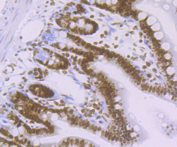 Immunohistochemical analysis of paraffin-embedded mouse colon tissue using anti-Histone H4 (acetyl K16) antibody. The section was pre-treated using heat mediated antigen retrieval with Tris-EDTA buffer (pH 8.0-8.4) for 20 minutes.The tissues were blocked in 5% BSA for 30 minutes at room temperature, washed with ddH2O and PBS, and then probed with the primary antibody (SLM-54330R, 1/50) for 30 minutes at room temperature. The detection was performed using an HRP conjugated compact polymer system. DAB was used as the chromogen. Tissues were counterstained with hematoxylin and mounted with DPX.
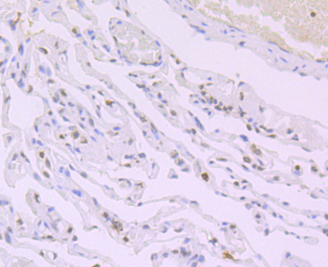 Immunohistochemical analysis of paraffin-embedded human lung carcinoma tissue using anti-Histone H4 (acetyl K16) antibody. The section was pre-treated using heat mediated antigen retrieval with Tris-EDTA buffer (pH 8.0-8.4) for 20 minutes.The tissues were blocked in 5% BSA for 30 minutes at room temperature, washed with ddH2O and PBS, and then probed with the primary antibody (SLM-54330R, 1/50) for 30 minutes at room temperature. The detection was performed using an HRP conjugated compact polymer system. DAB was used as the chromogen. Tissues were counterstained with hematoxylin and mounted with DPX.
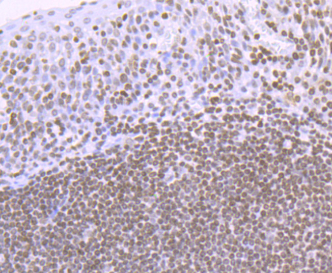 Immunohistochemical analysis of paraffin-embedded human tonsil tissue using anti-Histone H4 (acetyl K16) antibody. The section was pre-treated using heat mediated antigen retrieval with Tris-EDTA buffer (pH 8.0-8.4) for 20 minutes.The tissues were blocked in 5% BSA for 30 minutes at room temperature, washed with ddH2O and PBS, and then probed with the primary antibody (SLM-54330R, 1/50) for 30 minutes at room temperature. The detection was performed using an HRP conjugated compact polymer system. DAB was used as the chromogen. Tissues were counterstained with hematoxylin and mounted with DPX.
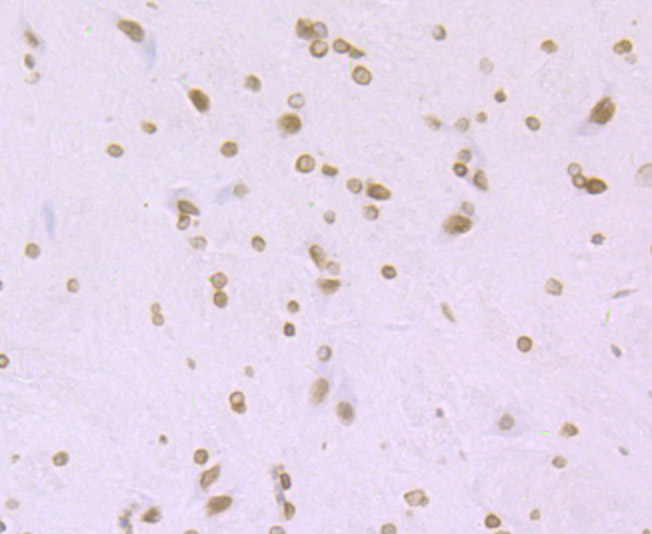 Immunohistochemical analysis of paraffin-embedded rat brain tissue using anti-Histone H4 (acetyl K16) antibody. The section was pre-treated using heat mediated antigen retrieval with Tris-EDTA buffer (pH 8.0-8.4) for 20 minutes.The tissues were blocked in 5% BSA for 30 minutes at room temperature, washed with ddH2O and PBS, and then probed with the primary antibody (SLM-54330R, 1/50) for 30 minutes at room temperature. The detection was performed using an HRP conjugated compact polymer system. DAB was used as the chromogen. Tissues were counterstained with hematoxylin and mounted with DPX.
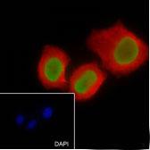 Cell line: HeLa
Fixative: 4% Paraformaldehyde
Permeabilization: 0.1% TritonX-100
Primary ab dilution: 1:200
Primary incubation condition: 4°C overnight
Secondary ab: Goat Anti-Rabbit IgG
Nuclear counter stain: DAPI (Blue)
Counter stain: Tubulin (Red)
Comment: Color green is the positive signal for SLM-54330R
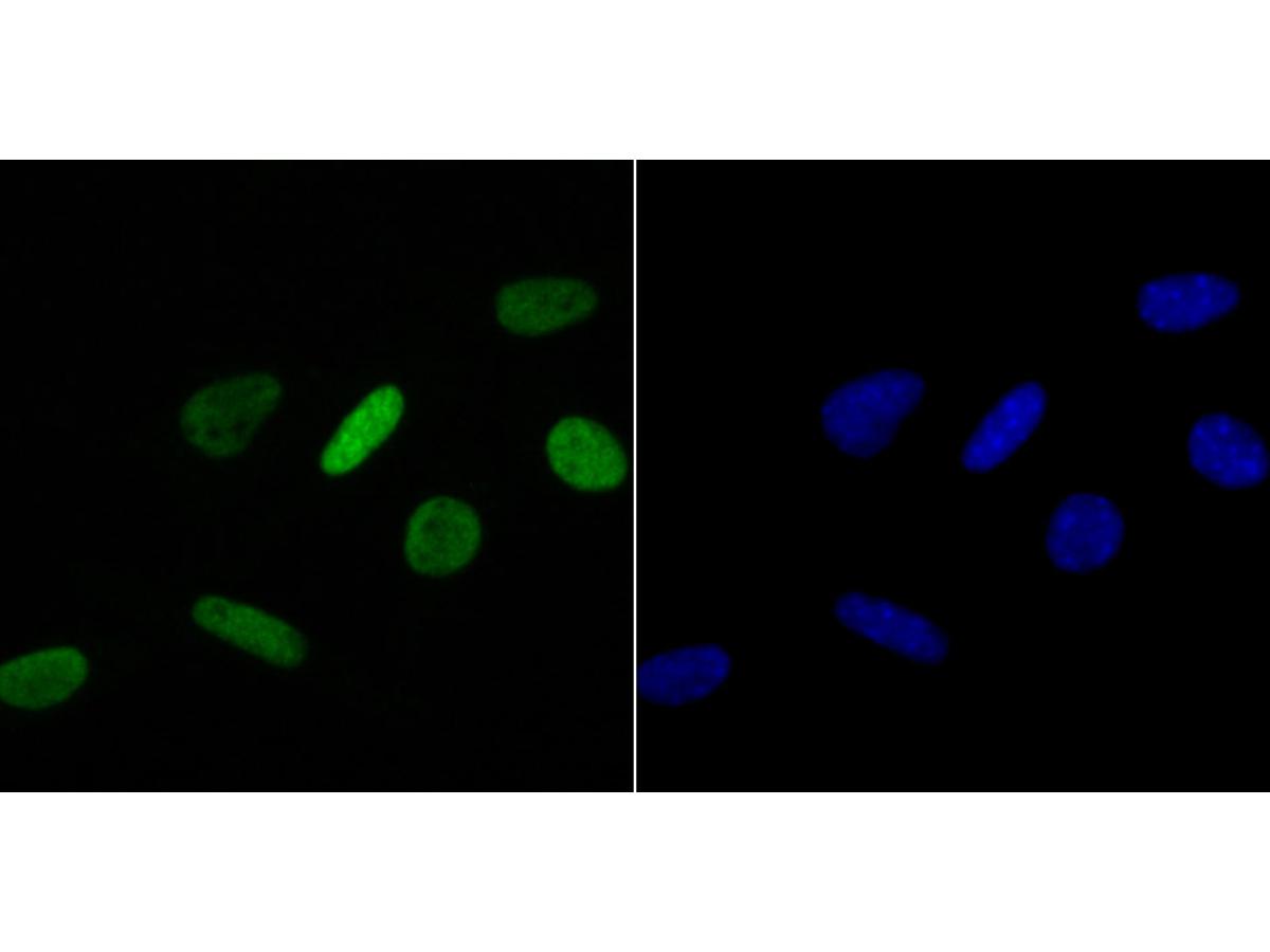 ICC staining of Histone H4 (acetyl K16) in SH-SY5Y cells (green). Formalin fixed cells were permeabilized with 0.1% Triton X-100 in TBS for 10 minutes at room temperature and blocked with 1% Blocker BSA for 15 minutes at room temperature. Cells were probed with the primary antibody (SLM-54330R, 1/50) for 1 hour at room temperature, washed with PBS. Alexa Fluor®488 Goat anti-Rabbit IgG was used as the secondary antibody at 1/1,000 dilution. The nuclear counter stain is DAPI (blue).
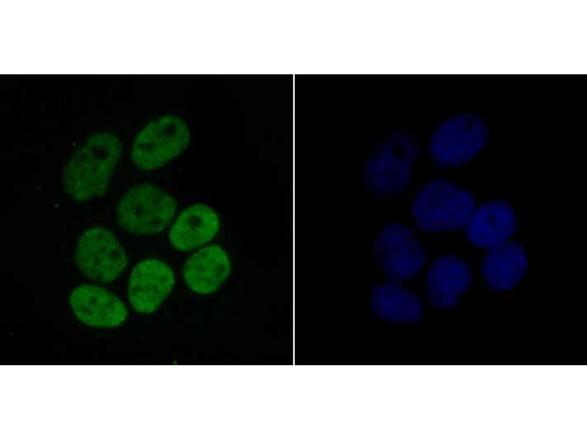 ICC staining of Histone H4 (acetyl K16) in Hela cells (green). Formalin fixed cells were permeabilized with 0.1% Triton X-100 in TBS for 10 minutes at room temperature and blocked with 1% Blocker BSA for 15 minutes at room temperature. Cells were probed with the primary antibody (SLM-54330R, 1/50) for 1 hour at room temperature, washed with PBS. Alexa Fluor®488 Goat anti-Rabbit IgG was used as the secondary antibody at 1/1,000 dilution. The nuclear counter stain is DAPI (blue).
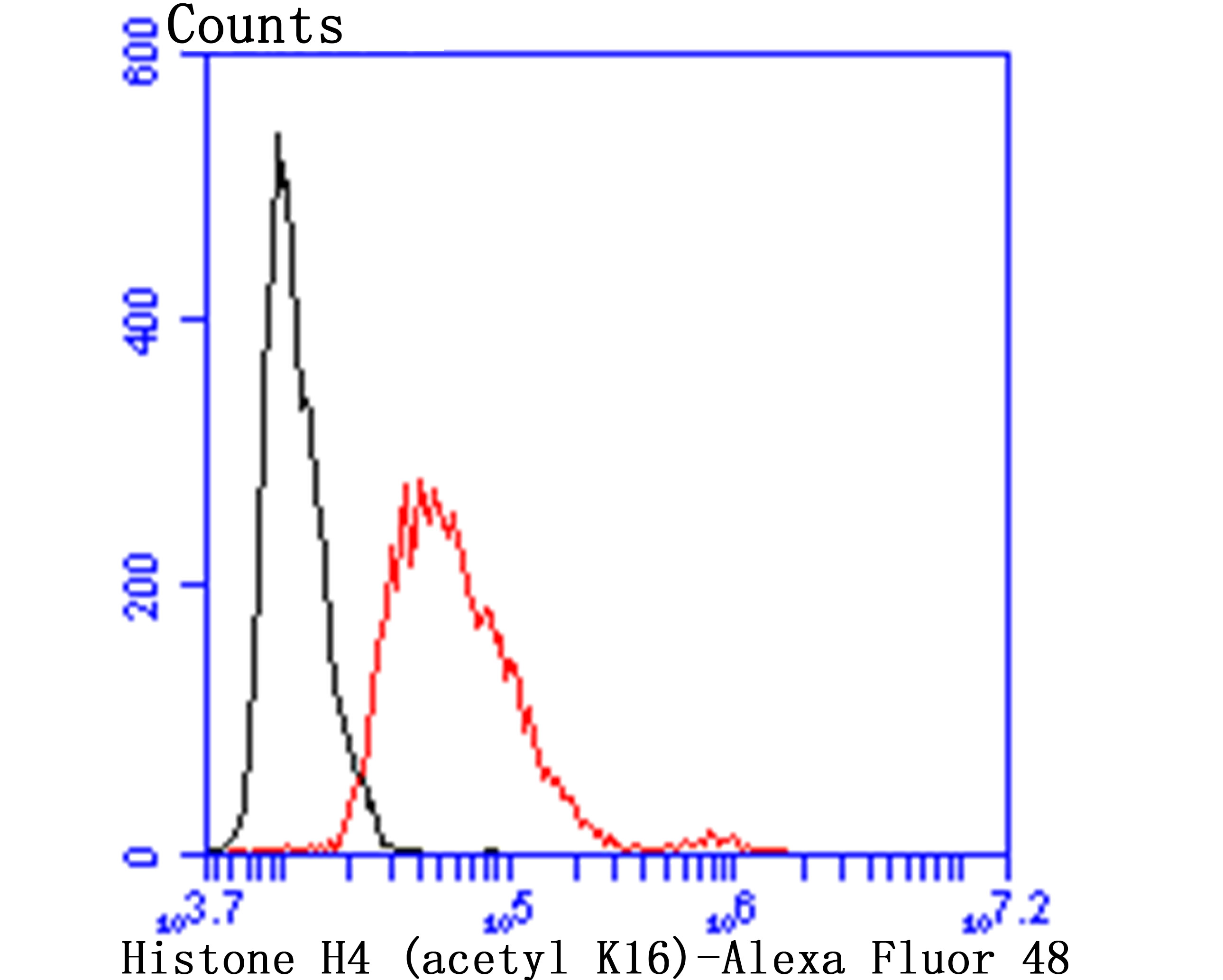 Flow cytometric analysis of Histone H4 (acetyl K16) was done on Hela cells. The cells were fixed, permeabilized and stained with the primary antibody (SLM-54330R, 1/50) (red). After incubation of the primary antibody at room temperature for an hour, the cells were stained with a Alexa Fluor 488-conjugated Goat anti-Rabbit IgG Secondary antibody at 1/1000 dilution for 30 minutes.Unlabelled sample was used as a control (cells without incubation with primary antibody; black).
Cartpieces
Totalgoods,subtotals:¥Checkout
References (0)
Bought notes(bought amounts latest0)
User Comment(Total0User Comment Num)
- No comment


 +86 571 56623320
 +86 18668110335

