
Rabbit Anti-BRD2 antibody
Bromodomain-containing protein 2; KIAA9001; RING3; O27.1.1; Really interesting new gene 3 protein; BRD2_HUMAN;
View History [Clear]
Details
Product Name BRD2 Chinese Name BRD2蛋白Recombinant rabbit monoclonal anti Alias Bromodomain-containing protein 2; KIAA9001; RING3; O27.1.1; Really interesting new gene 3 protein; BRD2_HUMAN; Research Area Signal transduction Kinases and Phosphatases Cell Surface Molecule Immunogen Species Rabbit Clonality Monoclonal Clone NO. R2A5 React Species (predicted: Human, ) Applications WB=1:500 IHC-P=1:100-500 ICC=1:50 (Paraffin sections need antigen repair)
not yet tested in other applications.
optimal dilutions/concentrations should be determined by the end user.Theoretical molecular weight 88kDa Cellular localization The nucleus Form Liquid Concentration 1mg/ml immunogen KLH conjugated synthetic peptide derived from human BRD2: 1-50/801 Lsotype IgG Purification affinity purified by Protein A Buffer Solution 0.01M TBS(pH7.4) with 1% BSA, 0.03% Proclin300 and 50% Glycerol. Storage Shipped at 4℃. Store at -20 °C for one year. Avoid repeated freeze/thaw cycles. Attention This product as supplied is intended for research use only, not for use in human, therapeutic or diagnostic applications. PubMed PubMed Product Detail This gene encodes a transcriptional regulator that belongs to the BET (bromodomains and extra terminal domain) family of proteins. This protein associates with transcription complexes and with acetylated chromatin during mitosis, and it selectively binds to the acetylated lysine-12 residue of histone H4 via its two bromodomains. The gene maps to the major histocompatability complex (MHC) class II region on chromosome 6p21.3, but sequence comparison suggests that the protein is not involved in the immune response. This gene has been implicated in juvenile myoclonic epilepsy, a common form of epilepsy that becomes apparent in adolescence. Multiple alternatively spliced variants have been described for this gene. [provided by RefSeq, Dec 2010]
Function:
May play a role in spermatogenesis or folliculogenesis.
Subunit:
Homodimer. Interacts with E2F1 and with histone H4acetylated at 'Lys-13'.
Subcellular Location:
Nucleus.
Similarity:
Contains 2 bromo domains.
Contains 1 ET domain.
SWISS:
P25440
Gene ID:
6046
Database links:Entrez Gene: 6046 Human
Entrez Gene: 14312 Mouse
SwissProt: P25440 Human
SwissProt: Q7JJ13 Mouse
Product Picture 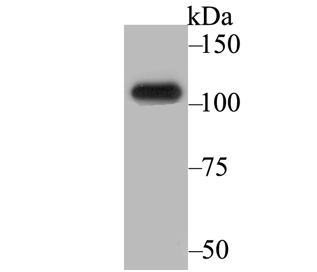 Western blot analysis of BRD2 on SiHa cell lysates. Proteins were transferred to a PVDF membrane and blocked with 5% BSA in PBS for 1 hour at room temperature. The primary antibody (SLM-54434R, 1/500) was used in 5% BSA at room temperature for 2 hours. Goat Anti-Rabbit IgG - HRP Secondary Antibody at 1:200,000 dilution was used for 1 hour at room temperature. Predicted band size: 88 kDa Observed band size: 105 kDa
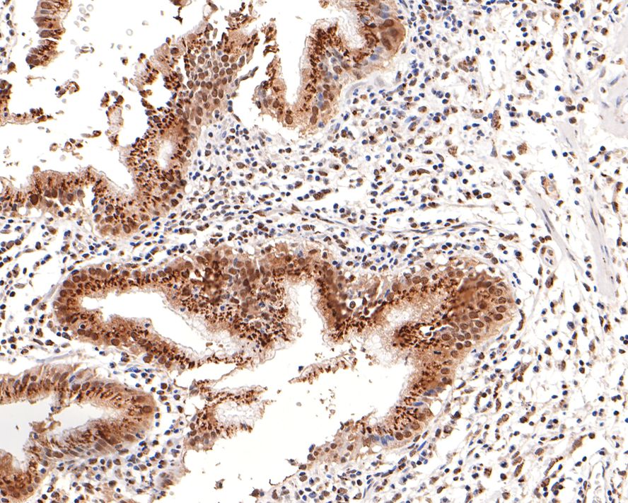 Immunohistochemical analysis of paraffin-embedded human gallbladder tissue using anti-BRD2 antibody. The section was pre-treated using heat mediated antigen retrieval with sodium citrate buffer (pH 6.0) for 20 minutes. The tissues were blocked in 1% BSA for 30 minutes at room temperature, washed with ddH2O and PBS, and then probed with the primary antibody (SLM-54434R, 1/100) for 30 minutes at room temperature. The detection was performed using an HRP conjugated compact polymer system. DAB was used as the chromogen. Tissues were counterstained with hematoxylin and mounted with DPX.
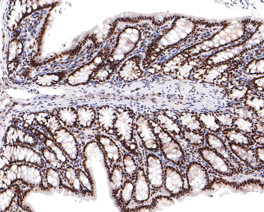 Immunohistochemical analysis of paraffin-embedded rat colon tissue using anti-BRD2 antibody. The section was pre-treated using heat mediated antigen retrieval with sodium citrate buffer (pH 6.0) for 20 minutes. The tissues were blocked in 1% BSA for 30 minutes at room temperature, washed with ddH2O and PBS, and then probed with the primary antibody (SLM-54434R, 1/400) for 30 minutes at room temperature. The detection was performed using an HRP conjugated compact polymer system. DAB was used as the chromogen. Tissues were counterstained with hematoxylin and mounted with DPX.
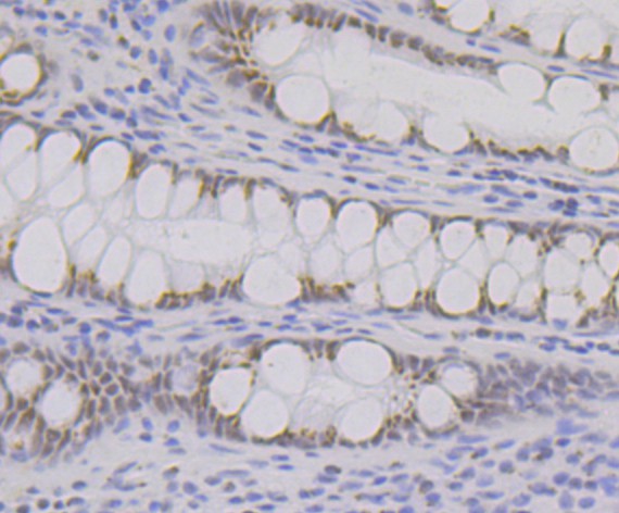 Immunohistochemical analysis of paraffin-embedded human colon tissue using anti-BRD2 antibody. The section was pre-treated using heat mediated antigen retrieval with Tris-EDTA buffer (pH 9.0) for 20 minutes.The tissues were blocked in 1% BSA for 30 minutes at room temperature, washed with ddH2O and PBS, and then probed with the primary antibody (SLM-54434R, 1/50) for 30 minutes at room temperature. The detection was performed using an HRP conjugated compact polymer system. DAB was used as the chromogen. Tissues were counterstained with hematoxylin and mounted with DPX.
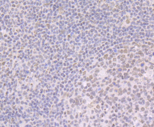 Immunohistochemical analysis of paraffin-embedded human tonsil tissue using anti-BRD2 antibody. The section was pre-treated using heat mediated antigen retrieval with Tris-EDTA buffer (pH 9.0) for 20 minutes.The tissues were blocked in 1% BSA for 30 minutes at room temperature, washed with ddH2O and PBS, and then probed with the primary antibody (SLM-54434R, 1/50) for 30 minutes at room temperature. The detection was performed using an HRP conjugated compact polymer system. DAB was used as the chromogen. Tissues were counterstained with hematoxylin and mounted with DPX.
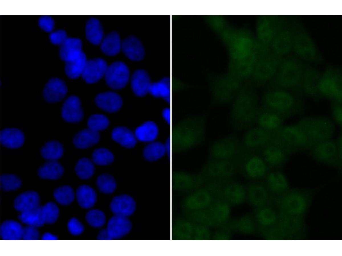 ICC staining of BRD2 in 293T cells (green). Formalin fixed cells were permeabilized with 0.1% Triton X-100 in TBS for 10 minutes at room temperature and blocked with 10% negative goat serum for 15 minutes at room temperature. Cells were probed with the primary antibody (SLM-54434R, 1/50) for 1 hour at room temperature, washed with PBS. Alexa Fluor®488 conjugate-Goat anti-Rabbit IgG was used as the secondary antibody at 1/1,000 dilution. The nuclear counter stain is DAPI (blue).
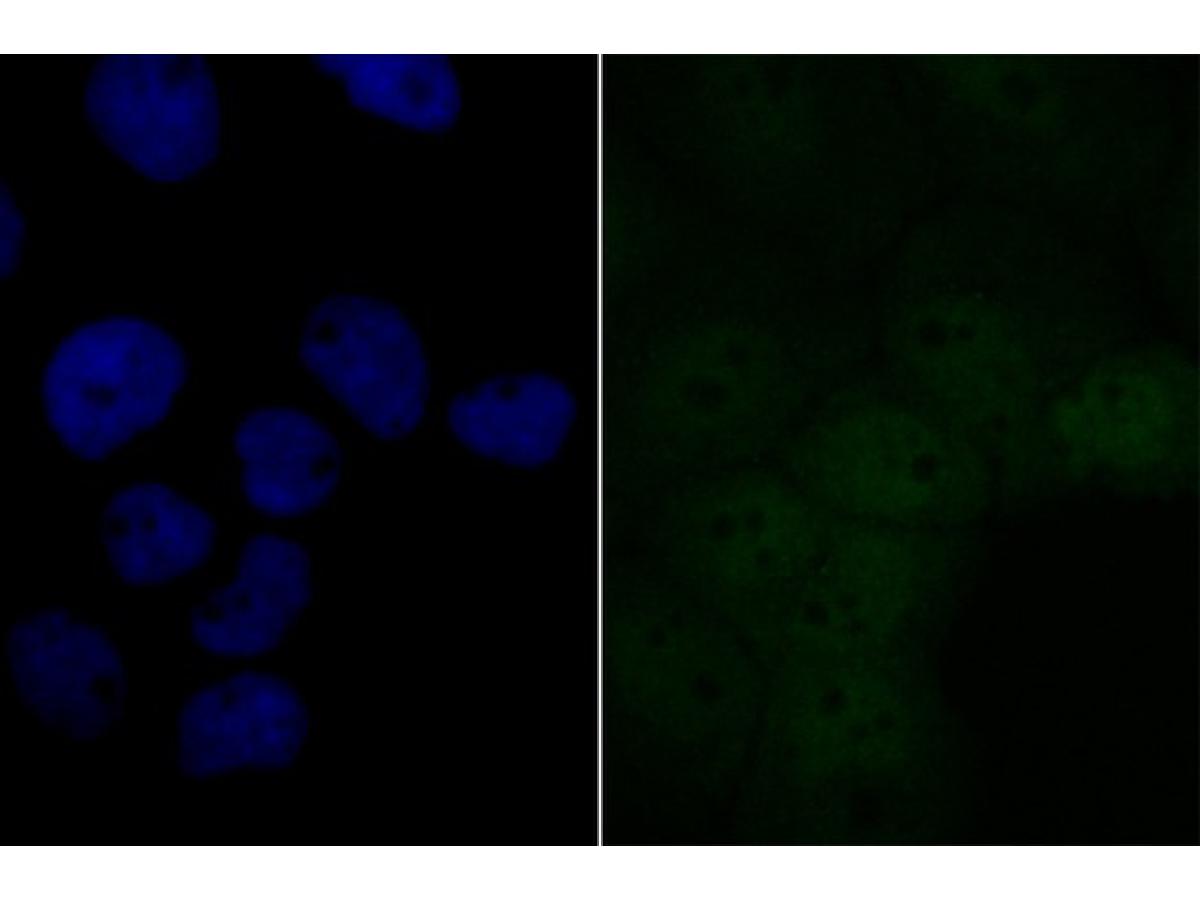 ICC staining of BRD2 in A431 cells (green). Formalin fixed cells were permeabilized with 0.1% Triton X-100 in TBS for 10 minutes at room temperature and blocked with 10% negative goat serum for 15 minutes at room temperature. Cells were probed with the primary antibody (SLM-54434R, 1/50) for 1 hour at room temperature, washed with PBS. Alexa Fluor®488 conjugate-Goat anti-Rabbit IgG was used as the secondary antibody at 1/1,000 dilution. The nuclear counter stain is DAPI (blue).
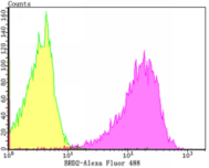 Flow cytometric analysis of BRD2 was done on 293T cells. The cells were fixed, permeabilized and stained with the primary antibody (SLM-54434R, 1/50) (purple). After incubation of the primary antibody at room temperature for an hour, the cells were stained with a Alexa Fluor®488 conjugate-Goat anti-Rabbit IgG Secondary antibody at 1/1,000 dilution for 30 minutes.Unlabelled sample was used as a control (cells without incubation with primary antibody; yellow).
Cartpieces
Totalgoods,subtotals:¥Checkout
References (0)
No References
Bought notes(bought amounts latest0)
No one bought this product
User Comment(Total0User Comment Num)
- No comment


 +86 571 56623320
 +86 18668110335

