
Rabbit Anti-MMP3 antibody
MMP3_HUMAN; Stromelysin-1; EC:3.4.24.17; STMY1; SL-1; SL 1; SL1; Matrix metalloproteinase-3 (MMP-3); MMP-3; MMP 3; Transin-1;
View History [Clear]
Details
Product Name MMP3 Chinese Name 基质金属蛋白酶3Recombinant rabbit monoclonal anti Alias MMP3_HUMAN; Stromelysin-1; EC:3.4.24.17; STMY1; SL-1; SL 1; SL1; Matrix metalloproteinase-3 (MMP-3); MMP-3; MMP 3; Transin-1; Research Area Tumour Immunogen Species Rabbit Clonality Monoclonal React Species Human, (predicted: Mouse, Rat, ) Applications WB=1:500-1000 IHC-P=1:100-500 Flow-Cyt=1:100 ICC=1:50 IF=1:50-100 (Paraffin sections need antigen repair)
not yet tested in other applications.
optimal dilutions/concentrations should be determined by the end user.Theoretical molecular weight 54kDa Cellular localization Extracellular matrix Secretory protein Form Liquid Concentration 1mg/ml immunogen KLH conjugated synthetic peptide derived from human MMP-3 Lsotype IgG Purification affinity purified by Protein A Buffer Solution 0.01M TBS(pH7.4) with 1% BSA, 0.03% Proclin300 and 50% Glycerol. Storage Shipped at 4℃. Store at -20 °C for one year. Avoid repeated freeze/thaw cycles. Attention This product as supplied is intended for research use only, not for use in human, therapeutic or diagnostic applications. PubMed PubMed Product Detail Proteins of the matrix metalloproteinase (MMP) family are involved in the breakdown of extracellular matrix in normal physiological processes, such as embryonic development, reproduction, and tissue remodeling, as well as in disease processes, such as arthritis and metastasis. Most MMP's are secreted as inactive proproteins which are activated when cleaved by extracellular proteinases. This gene encodes an enzyme which degrades fibronectin, laminin, collagens III, IV, IX, and X, and cartilage proteoglycans. The enzyme is thought to be involved in wound repair, progression of atherosclerosis, and tumor initiation. The gene is part of a cluster of MMP genes which localize to chromosome 11q22.3. [provided by RefSeq, Jul 2008].
Function:
Can degrade fibronectin, laminin, gelatins of type I, III, IV, and V; collagens III, IV, X, and IX, and cartilage proteoglycans. Activates procollagenase.
Subcellular Location:
Secreted, extracellular space, extracellular matrix (Probable).
Similarity:
Belongs to the peptidase M10A family. Contains 4 hemopexin-like domains.
SWISS:
P08254
Gene ID:
4314
Synthesis and Degradation(Synthesis and Degradation) 基质金属蛋白酶(matrix metalloproteinases, MMPs)是一族依赖锌离子而降解各种Extracellular matrix的蛋白酶,亦称IV型胶原酶或称明胶酶A,其主要功能为降解IV型胶原,因而它在Tumour细胞突破基底膜屏障和浸润转移中起重要作用。 MMP3目前主要用于各种恶性Tumour(如乳腺癌、胃肠道癌、卵巢癌、膀胱癌等)中的基底膜检测与Tumour转移浸润的研究。Product Picture 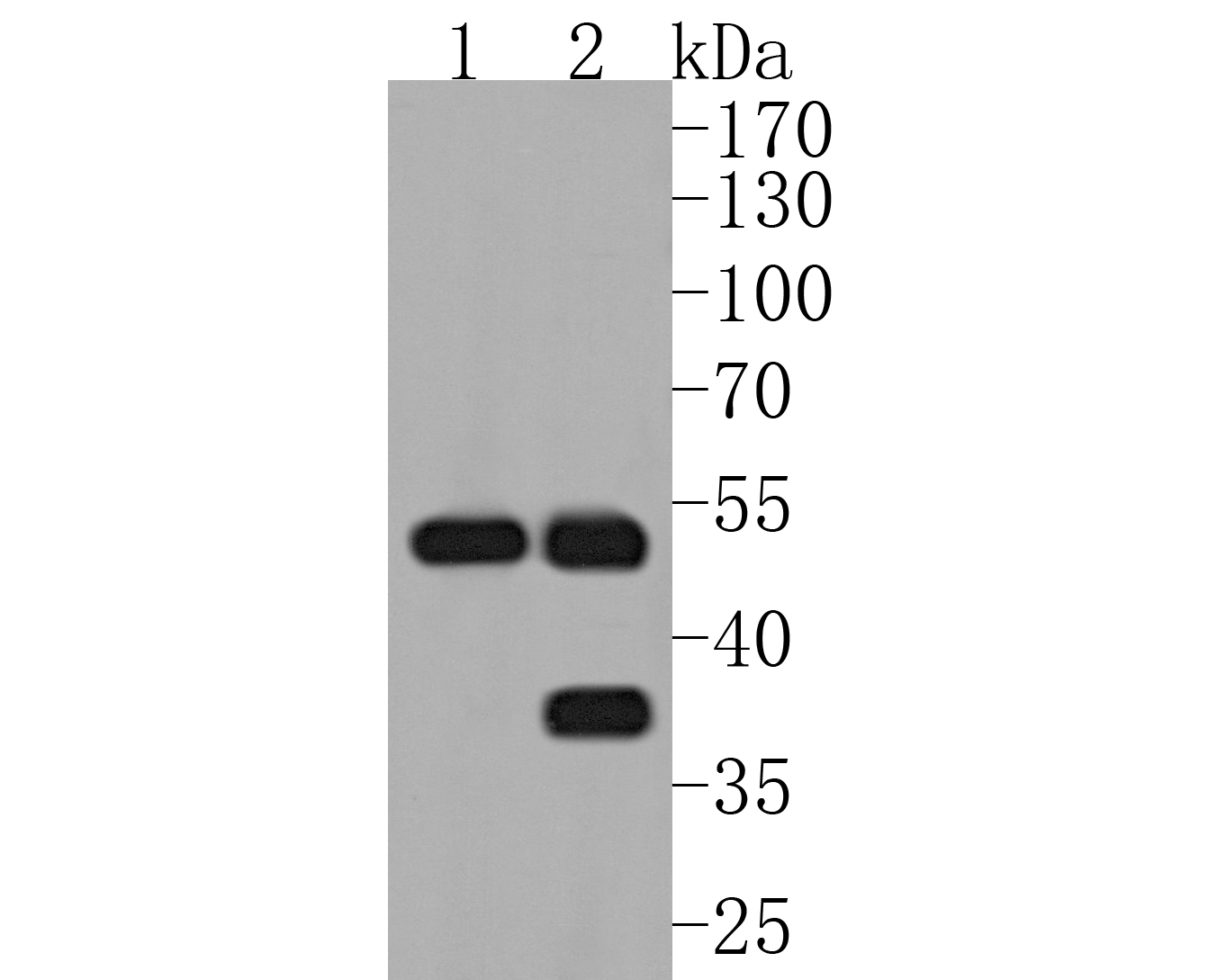 Western blot analysis of MMP3 on different lysates. Proteins were transferred to a PVDF membrane and blocked with 5% BSA in PBS for 1 hour at room temperature. The primary antibody (SLM-54152R, 1/500) was used in 5% BSA at room temperature for 2 hours. Goat Anti-Rabbit IgG - HRP Secondary Antibody (HA1001) at 1:5,000 dilution was used for 1 hour at room temperature. Positive control: Lane 1: human liver tissue lysate Lane 2: rat liver tissue lysate
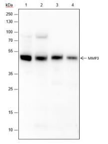 Blocking buffer: 5% NFDM/TBST
Primary ab dilution: 1:1000
Primary ab incubation condition: 2 hours at
room temperature
Secondary ab: Goat Anti-Rabbit IgG H&L
(HRP)
Lysate: 1: Raji, 2: U87-MG, 3: Mouse placenta,
4: Rat placenta
Protein loading quantity: 20 μg
Exposure time: 60 s
Predicted MW: 54 kDa
Observed MW: 54 kDa
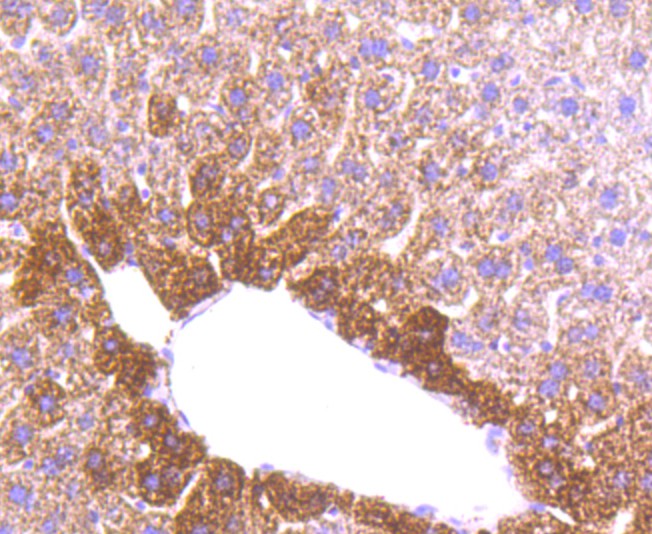 Immunohistochemical analysis of paraffin-embedded mouse liver tissue using anti-MMP3 antibody. The section was pre-treated using heat mediated antigen retrieval with Tris-EDTA buffer (pH 8.0-8.4) for 20 minutes.The tissues were blocked in 5% BSA for 30 minutes at room temperature, washed with ddH2O and PBS, and then probed with the primary antibody (SLM-54152R, 1/50) for 30 minutes at room temperature. The detection was performed using an HRP conjugated compact polymer system. DAB was used as the chromogen. Tissues were counterstained with hematoxylin and mounted with DPX.
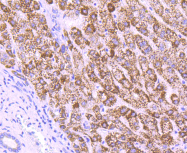 Immunohistochemical analysis of paraffin-embedded human liver tissue using anti-MMP3 antibody. The section was pre-treated using heat mediated antigen retrieval with Tris-EDTA buffer (pH 8.0-8.4) for 20 minutes.The tissues were blocked in 5% BSA for 30 minutes at room temperature, washed with ddH2O and PBS, and then probed with the primary antibody (SLM-54152R, 1/50) for 30 minutes at room temperature. The detection was performed using an HRP conjugated compact polymer system. DAB was used as the chromogen. Tissues were counterstained with hematoxylin and mounted with DPX.
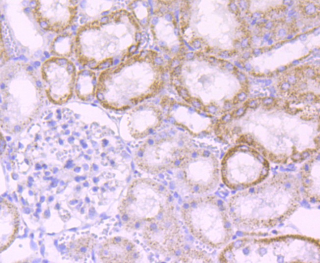 Immunohistochemical analysis of paraffin-embedded rat kidney tissue using anti-MMP3 antibody. The section was pre-treated using heat mediated antigen retrieval with Tris-EDTA buffer (pH 8.0-8.4) for 20 minutes.The tissues were blocked in 5% BSA for 30 minutes at room temperature, washed with ddH2O and PBS, and then probed with the primary antibody (SLM-54152R, 1/50) for 30 minutes at room temperature. The detection was performed using an HRP conjugated compact polymer system. DAB was used as the chromogen. Tissues were counterstained with hematoxylin and mounted with DPX.
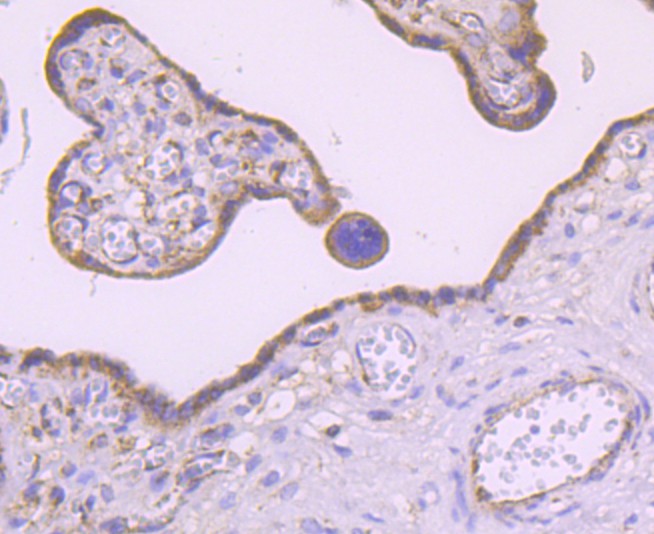 Immunohistochemical analysis of paraffin-embedded human placenta tissue using anti-MMP3 antibody. The section was pre-treated using heat mediated antigen retrieval with Tris-EDTA buffer (pH 8.0-8.4) for 20 minutes.The tissues were blocked in 5% BSA for 30 minutes at room temperature, washed with ddH2O and PBS, and then probed with the primary antibody (SLM-54152R, 1/50) for 30 minutes at room temperature. The detection was performed using an HRP conjugated compact polymer system. DAB was used as the chromogen. Tissues were counterstained with hematoxylin and mounted with DPX.
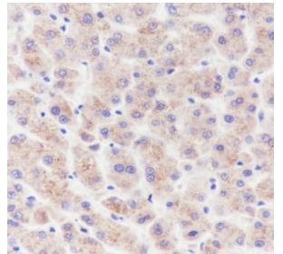 Tissue: Human liver
Section type: Formalin fixed & Paraffin -
embedded section
Retrieval method: High temperature and high
pressure
Retrieval buffer: Tris/EDTA buffer, pH 9.0
Primary ab dilution: 1:100
Primary ab incubation condition: 1 hour at
room temperature
Secondary ab: Anti-Rabbit and Mouse
Polymer HRP (Ready to use)
Counter stain: Hematoxylin (Blue)
Comment:
Color brown is the positive signal
for SLM-54152R
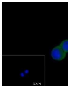
 Cell line: HT-29
Fixative: 4% Paraformaldehyde
Permeabilization: 0.1% TritonX-100
Primary ab dilution: 1:50
Primary incubation condition: 4°C overnight
Secondary ab: Goat Anti-Rabbit IgG
Nuclear counter stain: DAPI (Blue)
Comment: Color green is the positive signal for SLM-54152R
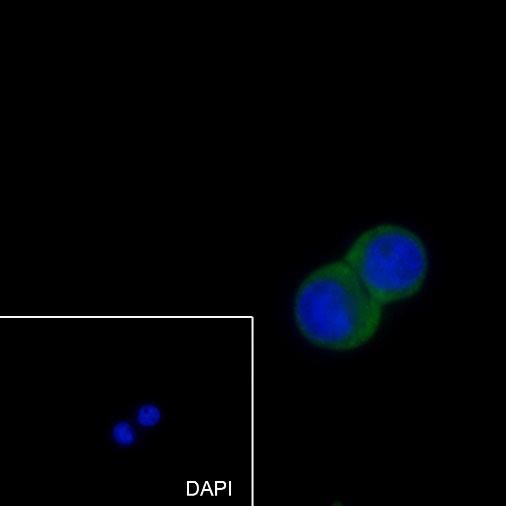
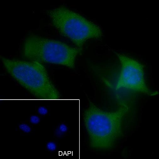 Cell line: NIH/3T3
Fixative: 4% Paraformaldehyde
Permeabilization: 0.1% TritonX-100
Primary ab dilution: 1:50
Primary incubation condition: 4°C overnight
Secondary ab: Goat Anti-Rabbit IgG
Nuclear counter stain: DAPI (Blue)
Comment: Color green is the positive signal for SLM-54152R
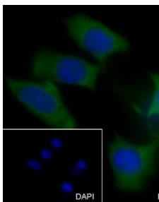
 Cell line: HT-29
Fixative: 4% Paraformaldehyde
Permeabilization: 0.1% TritonX-100
Primary ab dilution: 1:50
Primary incubation condition: 4°C overnight
Secondary ab: Goat Anti-Rabbit IgG
Nuclear counter stain: DAPI (Blue)
Comment: Color green is the positive signal for
SLM-54152R
 Cell line: NIH/3T3
Fixative: 4% Paraformaldehyde
Permeabilization: 0.1% TritonX-100
Primary ab dilution: 1:50
Primary incubation condition: 4°C overnight
Secondary ab: Goat Anti-Rabbit IgG
Nuclear counter stain: DAPI (Blue)
Comment: Color green is the positive signal for
SLM-54152R
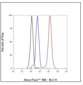
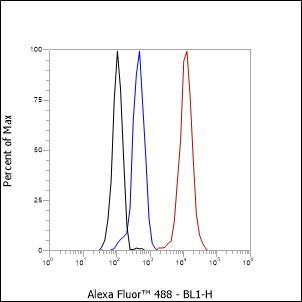 Cell line: HepG2
Fixation: 4% Paraformaldehyde
Permeabilization: 90% Methanol
Primary Ab dilution: 1:100
Secondary Ab: Goat Anti-Rabbit IgG
Unlabelled control: The cell without incubation with primary antibody and secondary antibody (Black line).
Isotype control: Rabbit monoclonal IgG (Blue line).
Comment: Line red is the positive signal for SLM-54152R
 Cell line: HepG2
Fixation: 4% Paraformaldehyde
Permeabilization: 90% Methanol
Primary Ab dilution: 1:100
Secondary Ab: Goat Anti-Rabbit IgG
Unlabelled control: The cell without incubation
with primary antibody and secondary antibody
(Black line).
Isotype control: Rabbit monoclonal IgG (Blue
line).
Comment: Line red is the positive signal for
SLM-54152R
Cartpieces
Totalgoods,subtotals:¥Checkout
References (0)
No References
Bought notes(bought amounts latest0)
No one bought this product
User Comment(Total0User Comment Num)
- No comment


 +86 571 56623320
 +86 18668110335

