
Rabbit Anti-CD90/Thy-1 antibody
CD90 / Thy1; CD7; CD90 antigen; CDw90; FLJ33325; MGC128895; T25; Theta antigen; Thy-1; Thy 1; Thy 1 cell surface antigen; Thy 1 membrane glycoprotein; Thy 1 membrane glycoprotein precursor; Thy 1.2; Thy-1 T-cell antigen; Thy1 antigen; Thy1 T cell antigen;
View History [Clear]
Details
Product Name CD90/Thy-1 Chinese Name CD90Recombinant rabbit monoclonal anti Alias CD90 / Thy1; CD7; CD90 antigen; CDw90; FLJ33325; MGC128895; T25; Theta antigen; Thy-1; Thy 1; Thy 1 cell surface antigen; Thy 1 membrane glycoprotein; Thy 1 membrane glycoprotein precursor; Thy 1.2; Thy-1 T-cell antigen; Thy1 antigen; Thy1 T cell antigen; Thy1.1; Thy1.2; Thymus cell antigen 1, theta; THY1_RAT; THY1_HUMAN. Research Area Cell biology immunology Neurobiology Immunogen Species Rabbit Clonality Monoclonal Clone NO. 4C6 React Species Mouse, Rat, (predicted: Human, ) Applications WB=1:1000-5000 IHC-P=1:100-500 IHC-F=1:20-200 ICC=1:20-200 (Paraffin sections need antigen repair)
not yet tested in other applications.
optimal dilutions/concentrations should be determined by the end user.Theoretical molecular weight 12kDa Cellular localization The cell membrane Form Liquid Concentration 1mg/ml immunogen KLH conjugated synthetic peptide derived from human CD90/Thy-1 Lsotype IgG Purification affinity purified by Protein A Buffer Solution 0.01M TBS(pH7.4) with 1% BSA, 0.03% Proclin300 and 50% Glycerol. Storage Shipped at 4℃. Store at -20 °C for one year. Avoid repeated freeze/thaw cycles. Attention This product as supplied is intended for research use only, not for use in human, therapeutic or diagnostic applications. PubMed PubMed Product Detail Thy-1 or CD90 (Cluster of Differentiation 90) is a 25–37 kDa heavily N-glycosylated, glycophosphatidylinositol (GPI) anchored conserved cell surface protein with a single V-like immunoglobulin domain, originally discovered as a thymocyte antigen. Thy-1can be used as a marker for a variety of stem cells and for the axonal processes of mature neurons. Structural study of Thy-1 lead to the foundation of the Immunoglobulin superfamily, of which it is the smallest member, and led to some of the initial biochemical description and characterization of a vertebrate GPI anchor and also the first demonstration of tissue specific differential glycosylation.
Function:
May play a role in cell-cell or cell-ligand interactions during synaptogenesis and other events in the brain.
Subunit:
Cell membrane; Lipid-anchor, GPI-anchor.
Tissue Specificity:
Abundant in lymphoid tissues.
Post-translational modifications:
Glycosylation is tissue specific. Sialylation of N-glycans at Asn-93 in brain and at Asn-42, Asn-93 and Asn-117 in thymus.
Similarity:
Contains 1 Ig-like V-type (immunoglobulin-like) domain.
SWISS:
P04216
Gene ID:
7070
Database links:Entrez Gene: 7070 Human
Entrez Gene: 21838 Mouse
Omim: 188230 Human
SwissProt: P04216 Human
SwissProt: P01831 Mouse
Unigene: 644697 Human
Unigene: 3951 Mouse
Unigene: 108198 Rat
Product Picture 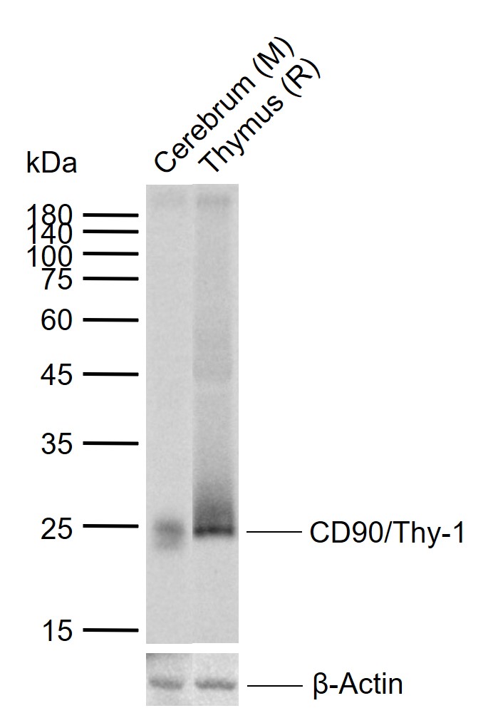 Sample:
Lane 1: Mouse Cerebrum tissue lysates
Lane 2: Rat Thymus tissue lysates
Primary: Anti-CD90/Thy-1 (SLM-52869R) at 1/2000 dilution
Secondary: IRDye800CW Goat Anti-Rabbit IgG at 1/20000 dilution
Predicted band size: 12 kDa
Observed band size: 25 kDa
 Western blot analysis of THY1 on different lysates. Proteins were transferred to a PVDF membrane and blocked with 5% BSA in PBS for 1 hour at room temperature. The primary antibody (SLM-52869R, 1/500) was used in 5% BSA at room temperature for 2 hours. Goat Anti-Rabbit IgG - HRP Secondary Antibody (HA1001) at 1:200,000 dilution was used for 1 hour at room temperature.
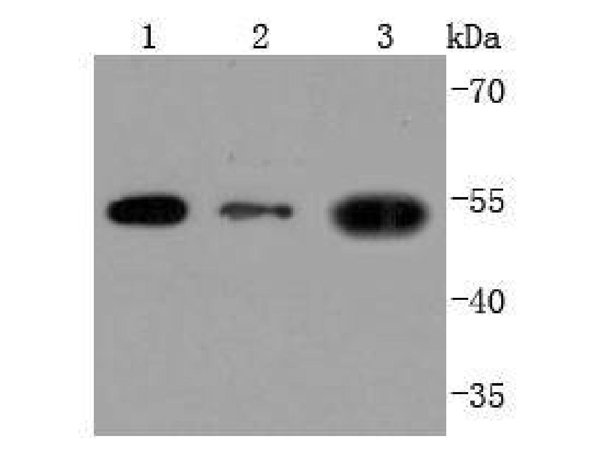
Positive control:
Lane 1: Hela cell lysate
Lane 2: HepG2 cell lysate
Lane 3: HUVEC cell lysate
 All lanes: Western blot analysis of THY1 with anti-THY1 antibody[JF10-09] (SLM-52869R) at 1:500 dilution.
Lane 1: Wild-type Hela whole cell lysate (10 µg).
Lane 2: THY1 knockdown Hela whole cell lysate (10 µg).
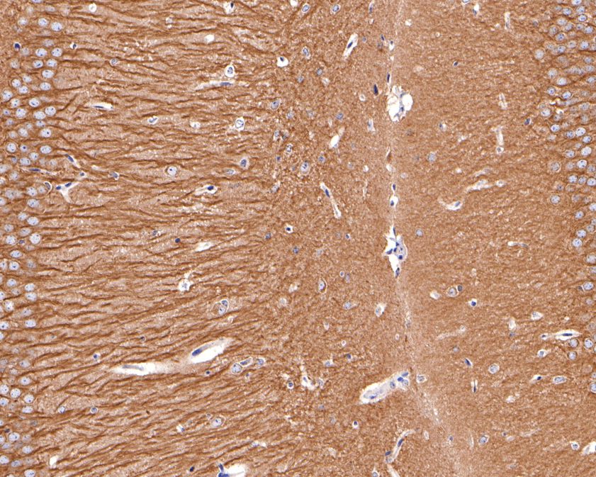 Immunohistochemical analysis of paraffin-embedded mouse hippocampus tissue using anti-THY1 antibody. The section was pre-treated using heat mediated antigen retrieval with Tris-EDTA buffer (pH 9.0) for 20 minutes.The tissues were blocked in 1% BSA for 30 minutes at room temperature, washed with ddH2O and PBS, and then probed with the primary antibody (SLM-52869R, 1/400) for 30 minutes at room temperature. The detection was performed using an HRP conjugated compact polymer system. DAB was used as the chromogen. Tissues were counterstained with hematoxylin and mounted with DPX.
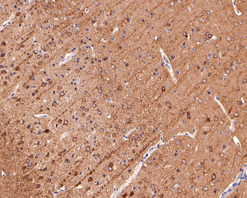 Immunohistochemical analysis of paraffin-embedded mouse hippocampus tissue using anti-THY1 antibody. The section was pre-treated using heat mediated antigen retrieval with Tris-EDTA buffer (pH 9.0) for 20 minutes.The tissues were blocked in 1% BSA for 30 minutes at room temperature, washed with ddH2O and PBS, and then probed with the primary antibody (SLM-52869R, 1/400) for 30 minutes at room temperature. The detection was performed using an HRP conjugated compact polymer system. DAB was used as the chromogen. Tissues were counterstained with hematoxylin and mounted with DPX.
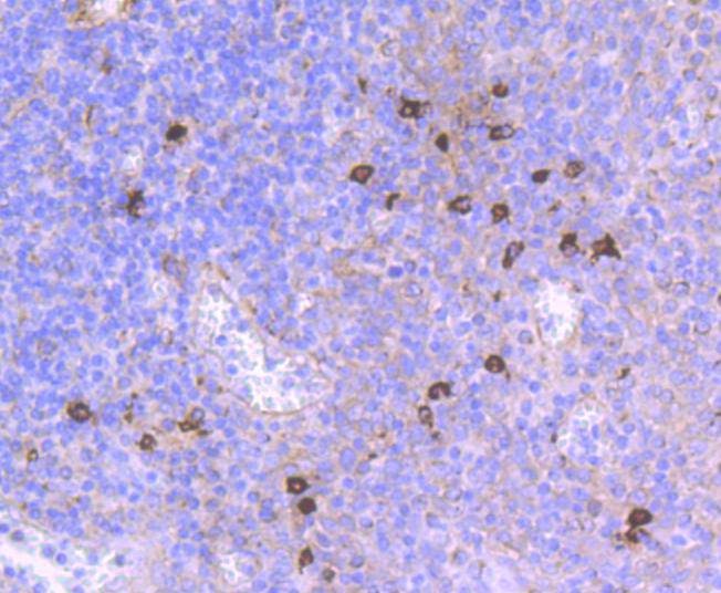 Immunohistochemical analysis of paraffin-embedded human tonsil tissue using anti-THY1 antibody. The section was pre-treated using heat mediated antigen retrieval with Tris-EDTA buffer (pH 9.0) for 20 minutes.The tissues were blocked in 5% BSA for 30 minutes at room temperature, washed with ddH2O and PBS, and then probed with the primary antibody (SLM-52869R, 1/50) for 30 minutes at room temperature. The detection was performed using an HRP conjugated compact polymer system. DAB was used as the chromogen. Tissues were counterstained with hematoxylin and mounted with DPX.
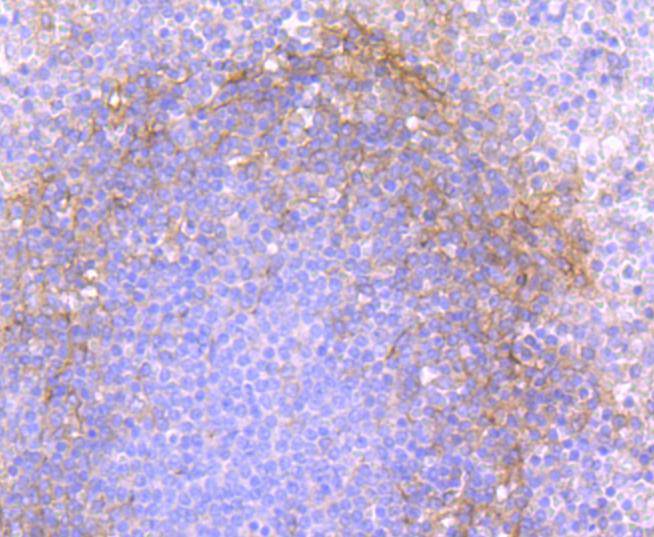 Immunohistochemical analysis of paraffin-embedded human spleen tissue using anti-THY1 antibody. The section was pre-treated using heat mediated antigen retrieval with Tris-EDTA buffer (pH 9.0) for 20 minutes.The tissues were blocked in 5% BSA for 30 minutes at room temperature, washed with ddH2O and PBS, and then probed with the primary antibody (SLM-52869R, 1/50) for 30 minutes at room temperature. The detection was performed using an HRP conjugated compact polymer system. DAB was used as the chromogen. Tissues were counterstained with hematoxylin and mounted with DPX.
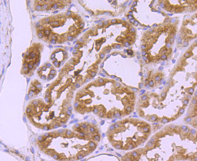 Immunohistochemical analysis of paraffin-embedded human kidney tissue using anti-THY1 antibody. The section was pre-treated using heat mediated antigen retrieval with Tris-EDTA buffer (pH 9.0) for 20 minutes.The tissues were blocked in 5% BSA for 30 minutes at room temperature, washed with ddH2O and PBS, and then probed with the primary antibody (SLM-52869R, 1/50) for 30 minutes at room temperature. The detection was performed using an HRP conjugated compact polymer system. DAB was used as the chromogen. Tissues were counterstained with hematoxylin and mounted with DPX.
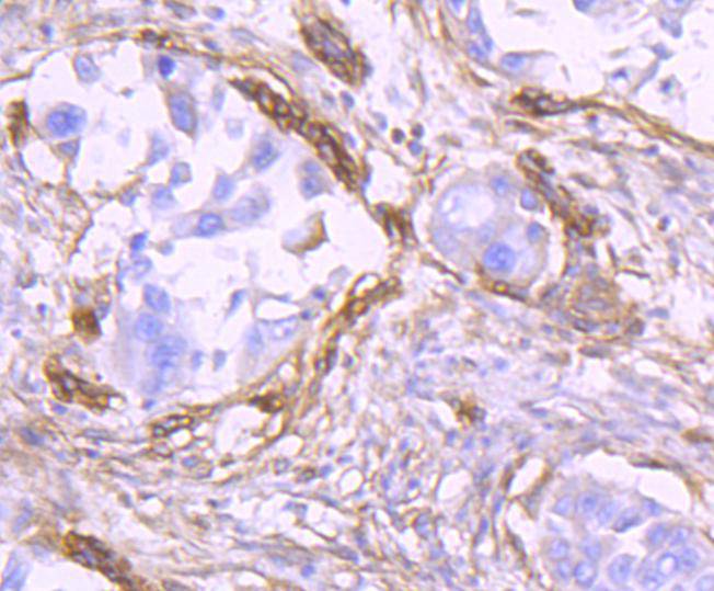 Immunohistochemical analysis of paraffin-embedded human lung carcinoma tissue using anti-THY1 antibody. The section was pre-treated using heat mediated antigen retrieval with Tris-EDTA buffer (pH 9.0) for 20 minutes.The tissues were blocked in 5% BSA for 30 minutes at room temperature, washed with ddH2O and PBS, and then probed with the primary antibody (SLM-52869R, 1/50) for 30 minutes at room temperature. The detection was performed using an HRP conjugated compact polymer system. DAB was used as the chromogen. Tissues were counterstained with hematoxylin and mounted with DPX.
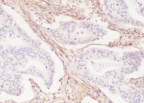 Tissue: Human colon cancer
Section type: Formalin-fixed & Paraffinembedded section
Retrieval method: High temperature and high pressure
Retrieval buffer: Tris/EDTA buffer, pH 9.0
Primary Ab dilution: 1:50
Primary Ab incubation condition: 1 hour at room temperature
Secondary Ab: SP Kit(Rabbit) (sp-0023)
Counter stain: Hematoxylin (Blue)
Comment: Color brown is the positive signal for SLM-52869R
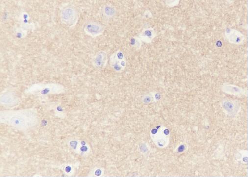 Tissue: Human cerebrum
Section type: Formalin-fixed & Paraffinembedded section
Retrieval method: High temperature and high pressure
Retrieval buffer: Tris/EDTA buffer, pH 9.0
Primary Ab dilution: 1:50
Primary Ab incubation condition: 1 hour at room temperature
Secondary Ab: SP Kit(Rabbit) (sp-0023)
Counter stain: Hematoxylin (Blue)
Comment: Color brown is the positive signal for SLM-52869R
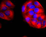
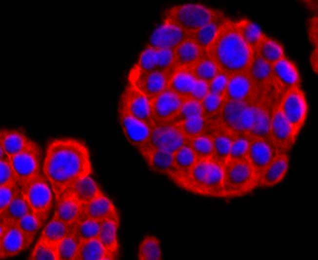 ICC staining of THY1 in PC-12 cells (red). Formalin fixed cells were permeabilized with 0.1% Triton X-100 in TBS for 10 minutes at room temperature and blocked with 10% negative goat serum for 15 minutes at room temperature. Cells were probed with the primary antibody (SLM-52869R, 1/50) for 1 hour at room temperature, washed with PBS. Alexa Fluor®594 conjugate-Goat anti-Rabbit IgG was used as the secondary antibody at 1/1,000 dilution. The nuclear counter stain is DAPI (blue).
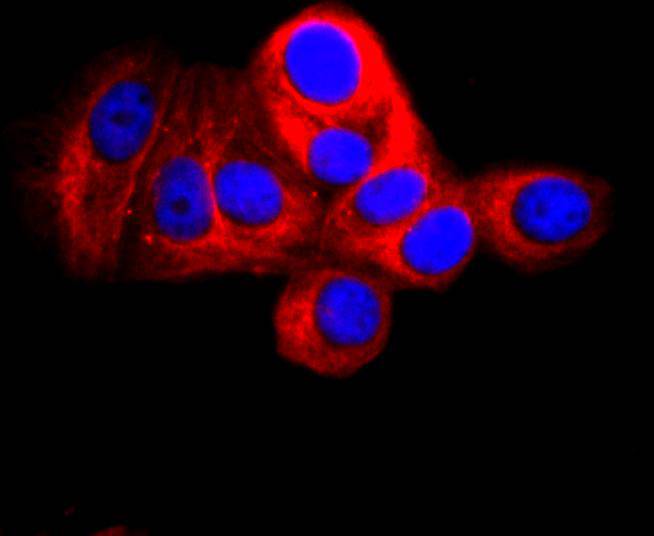 ICC staining of THY1 in MCF-7 cells (red). Formalin fixed cells were permeabilized with 0.1% Triton X-100 in TBS for 10 minutes at room temperature and blocked with 10% negative goat serum for 15 minutes at room temperature. Cells were probed with the primary antibody (SLM-52869R, 1/50) for 1 hour at room temperature, washed with PBS. Alexa Fluor®594 conjugate-Goat anti-Rabbit IgG was used as the secondary antibody at 1/1,000 dilution. The nuclear counter stain is DAPI (blue).ICC staining of THY1 in Hela cells (red). Formalin fixed cells were permeabilized with 0.1% Triton X-100 in TBS for 10 minutes at room temperature and blocked with 10% negative goat serum for 15 minutes at room temperature. Cells were probed with the primary antibody (SLM-52869R, 1/50) for 1 hour at room temperature, washed with PBS. Alexa Fluor®594 conjugate-Goat anti-Rabbit IgG was used as the secondary antibody at 1/1,000 dilution. The nuclear counter stain is DAPI (blue).
Cartpieces
Totalgoods,subtotals:¥Checkout
References (0)
No References
Bought notes(bought amounts latest0)
No one bought this product
User Comment(Total0User Comment Num)
- No comment


 +86 571 56623320
 +86 18668110335

