
Rabbit Anti-ERG antibody
ERG_HUMAN; Transcriptional regulator ERG; ERG; Transforming protein ERG; Avian erythroblastosis virus E-26 (v-ets) oncogene related; V-ets erythroblastosis virus E26 oncogene like (Avian), isoform CRA_e; rCG_53058; Erg-3;
View History [Clear]
Details
Product Name ERG Chinese Name 癌基因ERGRecombinant rabbit monoclonal anti Alias ERG_HUMAN; Transcriptional regulator ERG; ERG; Transforming protein ERG; Avian erythroblastosis virus E-26 (v-ets) oncogene related; V-ets erythroblastosis virus E26 oncogene like (Avian), isoform CRA_e; rCG_53058; Erg-3; Research Area Tumour Cell biology Signal transduction transcriptional regulatory factor Epigenetics Immunogen Species Rabbit Clonality Monoclonal React Species (predicted: Human, Mouse, ) Applications WB=1:500-2000 IHC-P=1:100-500 Flow-Cyt=1:100 ICC=1:100 IF=1:50-200 (Paraffin sections need antigen repair)
not yet tested in other applications.
optimal dilutions/concentrations should be determined by the end user.Theoretical molecular weight 55kDa Cellular localization The nucleus cytoplasmic Form Liquid Concentration 1mg/ml immunogen KLH conjugated synthetic peptide derived from human ERG Lsotype IgG Purification affinity purified by Protein A Buffer Solution 0.01M TBS(pH7.4) with 1% BSA, 0.03% Proclin300 and 50% Glycerol. Storage Shipped at 4℃. Store at -20 °C for one year. Avoid repeated freeze/thaw cycles. Attention This product as supplied is intended for research use only, not for use in human, therapeutic or diagnostic applications. PubMed PubMed Product Detail Ets-1 is the prototype member of a family of genes identified on the basis of homology to the v-Ets oncogene isolated from the E26 erythroblastosis virus. This family of genes currently includes Ets-1, Ets-2, Erg-1–3, Elk-1, Elf-1, Elf-5, NERF, PU.1, PEA3, ERM, FEV, ER8l, Fli-1, TEL, Spi-B, ESE-1, ESE-3A, Net, ABT1 and ERF. Members of the Ets gene family exhibit varied patterns of tissue expression, and share a highly conserved carboxy-terminal domain containing a sequence related to the SV40 large T antigen nuclear localization signal sequence. This conserved domain is essential for Ets-1 binding to DNA and is likely to be responsible for the DNA binding activity of all members of the Ets gene family. Several of these proteins have been shown to recognize similar motifs in DNA that share a centrally located 5'-GGAA-3' element. Erg genes encode for multiple proteins due to alternative splicing and alternative usage of initiation codons.
Function:
Transcriptional regulator. May participate in transcriptional regulation through the recruitment of SETDB1 histone methyltransferase and subsequent modification of local chromatin structure.
Subcellular Location:
Nucleus. Cytoplasm. Localized in cytoplasmic mRNP granules containing untranslated mRNAs.
DISEASE:
Defects in ERG are a cause of Ewing sarcoma (ES) [MIM:612219]. A highly malignant, metastatic, primitive small round cell tumor of bone and soft tissue that affects children and adolescents. It belongs to the Ewing sarcoma family of tumors, a group of morphologically heterogeneous neoplasms that share the same cytogenetic features. They are considered neural tumors derived from cells of the neural crest. Ewing sarcoma represents the less differentiated form of the tumors. Note=A chromosomal aberration involving ERG is found in patients with Erwing sarcoma. Translocation t(21;22)(q22;q12) with EWSR1.
Note=Chromosomal aberrations involving ERG have been found in acute myeloid leukemia (AML). Translocation t(16;21)(p11;q22) with FUS. Translocation t(X;21)(q25-26;q22) with ELF4.
Similarity:
Belongs to the ETS family.
Contains 1 ETS DNA-binding domain.
Contains 1 PNT (pointed) domain.
SWISS:
P11308
Gene ID:
2078
Database links:Entrez Gene: 2078 Human
Entrez Gene: 13876 Mouse
SwissProt: P11308 Human
SwissProt: P81270 Mouse
Product Picture 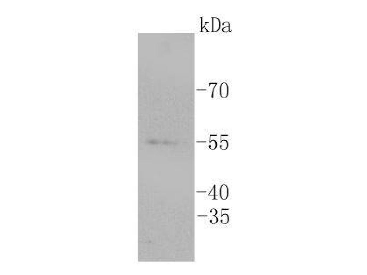 Western blot analysis of ERG on Jurkat cell lysates. Proteins were transferred to a PVDF membrane and blocked with 5% BSA in PBS for 1 hour at room temperature. The primary antibody (SLM-52324R, 1/500) was used in 5% BSA at room temperature for 2 hours. Goat Anti-Rabbit IgG - HRP Secondary Antibody (HA1001) at 1:5,000 dilution was used for 1 hour at room temperature.
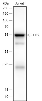 Blocking buffer: 5% NFDM/TBST
Primary Ab dilution: 1:2000
Primary Ab incubation condition: 2 hours at room temperature
Secondary Ab: Goat Anti-Rabbit IgG H&L (HRP)
Lysate: Jurkat
Protein loading quantity: 20 μg
Exposure time: 30 s
Predicted MW: 54 kDa
Observed MW: 54 kDa
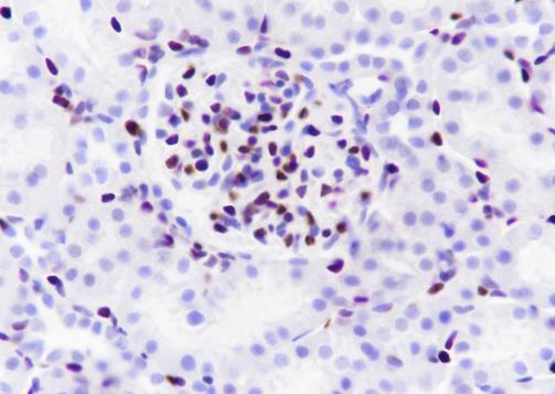
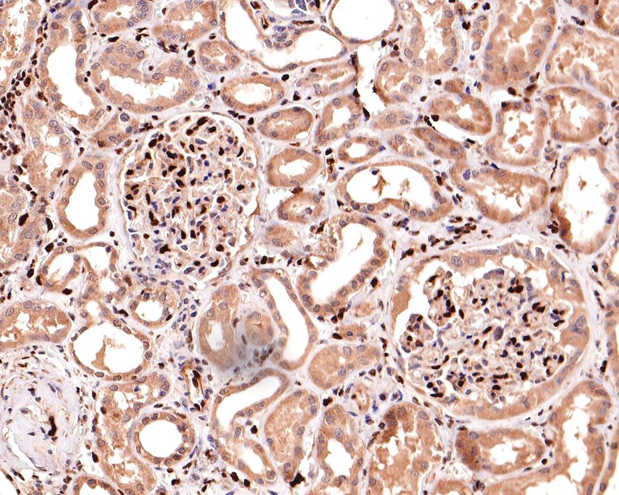 Immunohistochemical analysis of paraffin-embedded human kidney tissue with Rabbit anti-ERG antibody (SLM-52324R) at 1/500 dilution. The section was pre-treated using heat mediated antigen retrieval with sodium citrate buffer (pH 6.0) for 2 minutes. The tissues were blocked in 1% BSA for 20 minutes at room temperature, washed with ddH2O and PBS, and then probed with the primary antibody (SLM-52324R) at 1/500 dilution for 1 hour at room temperature. The detection was performed using an HRP conjugated compact polymer system. DAB was used as the chromogen. Tissues were counterstained with hematoxylin and mounted with DPX.
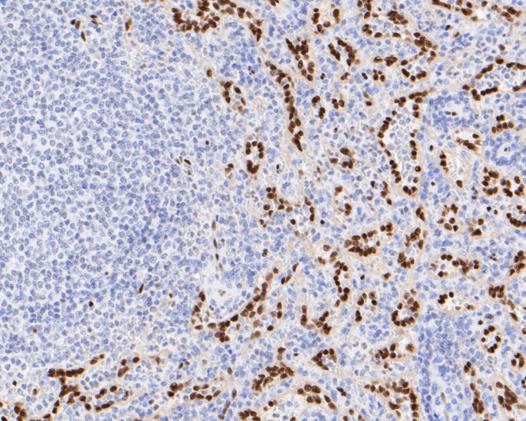 Immunohistochemical analysis of paraffin-embedded human spleen tissue using anti-ERG antibody. The section was pre-treated using heat mediated antigen retrieval with sodium citrate buffer (pH 6.0) for 20 minutes. The tissues were blocked in 5% BSA for 30 minutes at room temperature, washed with ddH2O and PBS, and then probed with the primary antibody (SLM-52324R, 1/100) for 30 minutes at room temperature. The detection was performed using an HRP conjugated compact polymer system. DAB was used as the chromogen. Tissues were counterstained with hematoxylin and mounted with DPX.
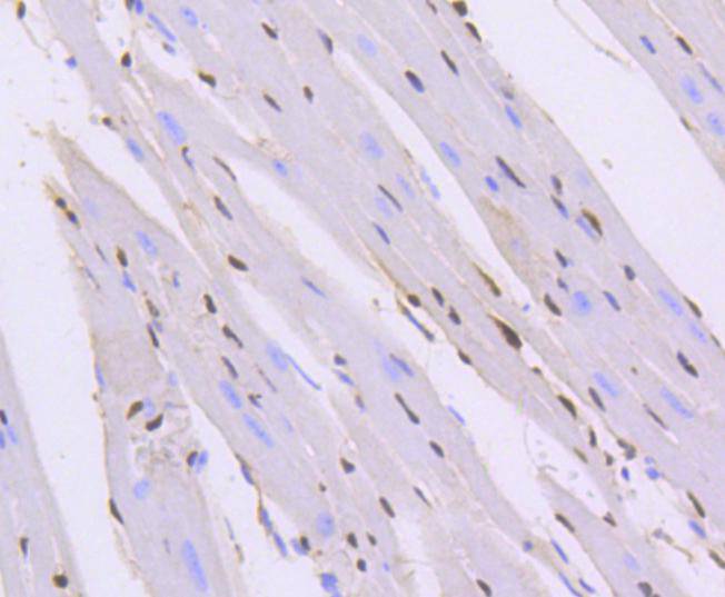 Immunohistochemical analysis of paraffin-embedded mouse heart tissue using anti-ERG antibody. The section was pre-treated using heat mediated antigen retrieval with sodium citrate buffer (pH 6.0) for 20 minutes. The tissues were blocked in 5% BSA for 30 minutes at room temperature, washed with ddH2O and PBS, and then probed with the primary antibody (SLM-52324R, 1/100) for 30 minutes at room temperature. The detection was performed using an HRP conjugated compact polymer system. DAB was used as the chromogen. Tissues were counterstained with hematoxylin and mounted with DPX.
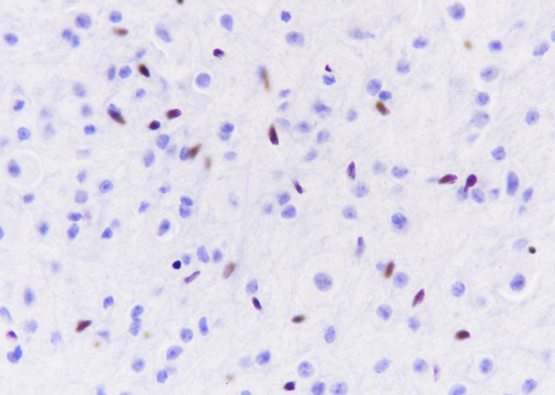
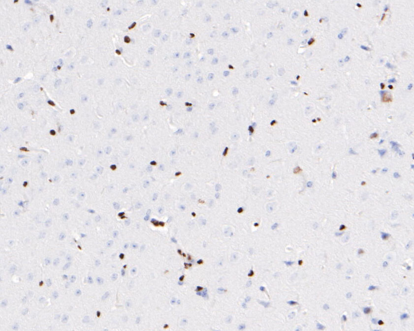 Immunohistochemical analysis of paraffin-embedded mouse brain tissue using anti-ERG antibody. The section was pre-treated using heat mediated antigen retrieval with sodium citrate buffer (pH 6.0) for 20 minutes. The tissues were blocked in 5% BSA for 30 minutes at room temperature, washed with ddH2O and PBS, and then probed with the primary antibody (SLM-52324R, 1/100) for 30 minutes at room temperature. The detection was performed using an HRP conjugated compact polymer system. DAB was used as the chromogen. Tissues were counterstained with hematoxylin and mounted with DPX.
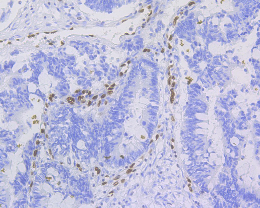 Immunohistochemical analysis of paraffin-embedded human colon carcinoma tissue using anti-ERG antibody. The section was pre-treated using heat mediated antigen retrieval with sodium citrate buffer (pH 6.0) for 20 minutes. The tissues were blocked in 5% BSA for 30 minutes at room temperature, washed with ddH2O and PBS, and then probed with the primary antibody (SLM-52324R, 1/100) for 30 minutes at room temperature. The detection was performed using an HRP conjugated compact polymer system. DAB was used as the chromogen. Tissues were counterstained with hematoxylin and mounted with DPX.
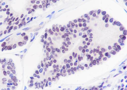 Tissue: Human prostatic cancer
Section type: Formalin-fixed & Paraffin-embedded section
Retrieval method: High temperature and high pressure
Retrieval buffer: Tris/EDTA buffer, pH 9.0 Primary Ab dilution: 1:100
Primary Ab incubation condition: 1 hour at room temperature
Secondary Ab: SP Kit(Rabbit) (sp-0023)
Counter stain: Hematoxylin (Blue)
Comment: Color brown is the positive signal for SLM-52324R
 Tissue: Human kidney
Section type: Formalin-fixed & Paraffin-embedded section
Retrieval method: High temperature and high pressure
Retrieval buffer: Tris/EDTA buffer, pH 9.0 Primary Ab dilution: 1:100
Primary Ab incubation condition: 1 hour at room temperature
Secondary Ab: SP Kit(Rabbit) (sp-0023)
Counter stain: Hematoxylin (Blue)
Comment: Color brown is the positive signal for SLM-52324R
 Tissue: Mouse cerebrum
Section type: Formalin-fixed & Paraffin-embedded section
Retrieval method: High temperature and high pressure
Retrieval buffer: Tris/EDTA buffer, pH 9.0 Primary Ab dilution: 1:100
Primary Ab incubation condition: 1 hour at room temperature
Secondary Ab: SP Kit(Rabbit) (sp-0023)
Counter stain: Hematoxylin (Blue)
Comment: Color brown is the positive signal for SLM-52324R
 Tissue: Rat kidney
Section type: Formalin-fixed & Paraffin-embedded section
Retrieval method: High temperature and high pressure
Retrieval buffer: Tris/EDTA buffer, pH 9.0 Primary Ab dilution: 1:100
Primary Ab incubation condition: 1 hour at room temperature
Secondary Ab: SP Kit(Rabbit) (sp-0023)
Counter stain: Hematoxylin (Blue)
Comment: Color brown is the positive signal for SLM-52324R
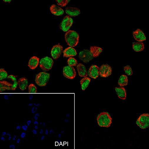 Cell line: THP-1
Fixation: 4% Paraformaldehyde
Permeabilization: 0.1% TritonX-100
Primary Ab dilution: 1:100
Primary Ab incubation condition: 4°C overnight
Secondary Ab: Goat Anti-Rabbit IgG
Nuclear counter stain: DAPI (Blue)
Counter stain: Tubulin (Red)
Comment: Color green is the positive signal for SLM-52324R
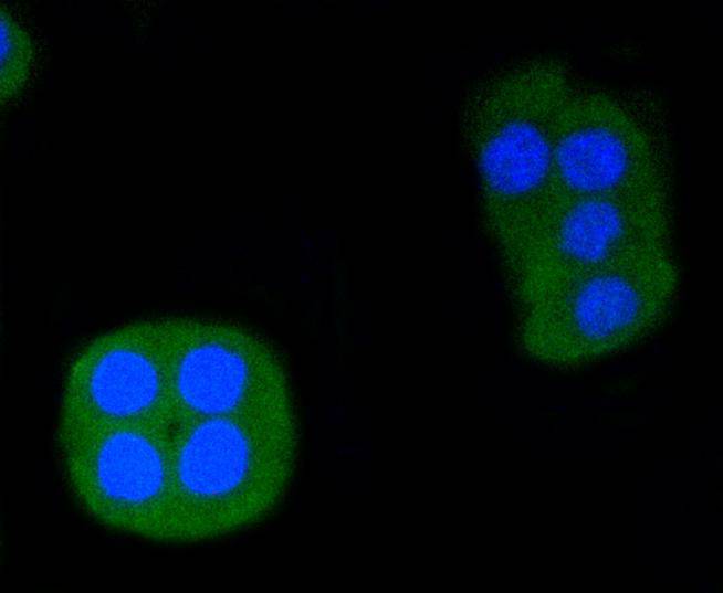 ICC staining of ERG in CRC cells (green). Formalin fixed cells were permeabilized with 0.1% Triton X-100 in TBS for 10 minutes at room temperature and blocked with 1% Blocker BSA for 15 minutes at room temperature. Cells were probed with the primary antibody (SLM-52324R, 1/50) for 1 hour at room temperature, washed with PBS. Alexa Fluor®488 Goat anti-Rabbit IgG was used as the secondary antibody at 1/1,000 dilution. The nuclear counter stain is DAPI (blue). .03
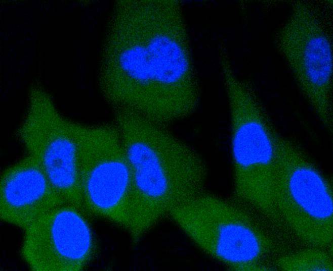 ICC staining of ERG in PC-3M cells (green). Formalin fixed cells were permeabilized with 0.1% Triton X-100 in TBS for 10 minutes at room temperature and blocked with 1% Blocker BSA for 15 minutes at room temperature. Cells were probed with the primary antibody (SLM-52324R, 1/50) for 1 hour at room temperature, washed with PBS. Alexa Fluor®488 Goat anti-Rabbit IgG was used as the secondary antibody at 1/1,000 dilution. The nuclear counter stain is DAPI (blue).
 Cell line: THP-1
Fixation: 4% Paraformaldehyde
Permeabilization: 90% Methanol
Primary Ab dilution: 1:100
Secondary Ab: Goat Anti-Rabbit IgG
Unlabelled control: The cell without incubation with primary antibody and secondary antibody (Black line).
Isotype control: Rabbit monoclonal IgG (Blue line).
Comment: Line red is the positive signal for SLM-52324R
Cartpieces
Totalgoods,subtotals:¥Checkout
References (0)
No References
Bought notes(bought amounts latest0)
No one bought this product
User Comment(Total0User Comment Num)
- No comment


 +86 571 56623320
 +86 18668110335

