
Rabbit Anti-p21 antibody
p21; P21 protein; Activating Fragment 1; CAP20; Cation chloride cotransporter-interacting protein 1; CDK Interacting Protein 1; CDK-interacting protein 1; CDKI; CDKN 1; CDKN1; CDKN1A; CIP1; Cyclin Dependent Kinase Inhibitor 1A; Cyclin-dependent kinase inh
View History [Clear]
Details
Product Name [KO validated anti] p21 Chinese Name p21蛋白抗体 Alias p21; P21 protein; Activating Fragment 1; CAP20; Cation chloride cotransporter-interacting protein 1; CDK Interacting Protein 1; CDK-interacting protein 1; CDKI; CDKN 1; CDKN1; CDKN1A; CIP1; Cyclin Dependent Kinase Inhibitor 1A; Cyclin-dependent kinase inhibitor 1; Cyclin-dependent kinase inhibitor 1A (P21); Cyclin-dependent kinase inhibitor 1A (p21, Cip1); DNA Synthesis Inhibitor; MDA 6; MDA-6; MDA6; Melanoma Differentiation Associated Protein 6; Melanoma differentiation-associated protein 6; Melanoma differentiation-associated protein; p21CIP1; p21WAF; PIC1; SDI1; SLC12A9; WAF1; Wildtype p53 Activating Fragment 1; Wildtype p53-activated fragment 1; CDN1A_HUMAN. literatures Research Area Tumour Cell biology Signal transduction Apoptosis Cyclin Immunogen Species Rabbit Clonality Polyclonal React Species Human, (predicted: Mouse, Rat, ) Applications WB=1:500-2000 IHC-P=1:20-100 IHC-F=1:20-100 (Paraffin sections need antigen repair)
not yet tested in other applications.
optimal dilutions/concentrations should be determined by the end user.Theoretical molecular weight 18kDa Cellular localization The nucleus cytoplasmic Form Liquid Concentration 1mg/ml immunogen Recombinant human p21: 1-164/164 Lsotype IgG Purification affinity purified by Protein A Buffer Solution 0.01M TBS(pH7.4) with 1% BSA, 0.03% Proclin300 and 50% Glycerol. Storage Store at -20 °C for one year. Avoid repeated freeze/thaw cycles. The lyophilized antibody is stable at room temperature for at least one month and for greater than a year when kept at -20°C. When reconstituted in sterile pH 7.4 0.01M PBS or diluent of antibody the antibody is stable for at least two weeks at 2-4 °C. Attention This product as supplied is intended for research use only, not for use in human, therapeutic or diagnostic applications. PubMed PubMed Product Detail This gene encodes a potent cyclin-dependent kinase inhibitor. The encoded protein binds to and inhibits the activity of cyclin-CDK2 or -CDK4 complexes, and thus functions as a regulator of cell cycle progression at G1. The expression of this gene is tightly controlled by the tumor suppressor protein p53, through which this protein mediates the p53-dependent cell cycle G1 phase arrest in response to a variety of stress stimuli. This protein can interact with proliferating cell nuclear antigen (PCNA), a DNA polymerase accessory factor, and plays a regulatory role in S phase DNA replication and DNA damage repair. This protein was reported to be specifically cleaved by CASP3-like caspases, which thus leads to a dramatic activation of CDK2, and may be instrumental in the execution of apoptosis following caspase activation. Two alternatively spliced variants, which encode an identical protein, have been reported. Two families of cyclin dependent kinase inhibitors (CKIs) have been identified. The p21WAF1/Cip1 family inhibits all kinases involved in the G1/S transition. The p16INK4a family inhibits Cdk4 and Cdk6 specifically.
Function:
May be the important intermediate by which p53/TP53 mediates its role as an inhibitor of cellular proliferation in response to DNA damage. Binds to and inhibits cyclin-dependent kinase activity, preventing phosphorylation of critical cyclin-dependent kinase substrates and blocking cell cycle progression. Functions in the nuclear localization and assembly of cyclin D-CDK4 complex and promotes its kinase activity towards RB1. At higher stoichiometric ratios, inhibits the kinase activity of the cyclin D-CDK4 complex.
Subunit:
Interacts with HDAC1; the interaction is prevented by competitive binding of C10orf90/FATS to HDAC1 facilitating acetylation and protein stabilization of CDKN1A/p21. Interacts with MKRN1. Interacts with PSMA3. Interacts with PCNA. Component of the ternary complex, cyclin D-CDK4-CDKN1A. Interacts (via its N-terminal domain) with CDK4; the interaction promotes the assembly of the cyclin D-CDK4 complex, its nuclear translocation and promotes the cyclin D-dependent enzyme activity of CDK4. Binding to CDK2 leads to CDK2/cyclin E inactivation at the G1-S phase DNA damage checkpoint, thereby arresting cells at the G1-S transition during DNA repair. Interacts with PIM1.
Subcellular Location:
Cytoplasmic and Nuclear.
Tissue Specificity:
Expressed in all adult tissues, with 5-fold lower levels observed in the brain.
Post-translational modifications:
Phosphorylation of Thr-145 by Akt or of Ser-146 by PKC impairs binding to PCNA. Phosphorylation at Ser-114 by GSK3-beta enhances ubiquitination by the DCX(DTL) complex. Phosphorylation of Thr-145 by PIM2 enhances CDKN1A stability and inhibits cell proliferation. Phosphorylation of Thr-145 by PIM1 results in the relocation of CDKN1A to the cytoplasm and enhanced CDKN1A protein stability.
Ubiquitinated by MKRN1; leading to polyubiquitination and 26S proteasome-dependent degradation. Ubiquitinated by the DCX(DTL) complex, also named CRL4(CDT2) complex, leading to its degradation during S phase or following UV irradiation. Ubiquitination by the DCX(DTL) complex is essential to control replication licensing and is PCNA-dependent: interacts with PCNA via its PIP-box, while the presence of the containing the 'K+4' motif in the PIP box, recruit the DCX(DTL) complex, leading to its degradation.
Acetylation leads to protein stability. Acetylated in vitro on Lys-141, Lys-154, Lys-161 and Lys-163. Deacetylation by HDAC1 is prevented by competitive binding of C10orf90/FATS to HDAC1.
Similarity:
Belongs to the CDI family.
SWISS:
P39689
Gene ID:
1026
Database links:Entrez Gene: 1026 Human
Entrez Gene: 12575 Mouse
Omim: 116899 Human
SwissProt: P38936 Human
SwissProt: P39689 Mouse
Unigene: 370771 Human
Unigene: 195663 Mouse
Unigene: 10089 Rat
p21蛋白的过度表达与Tumour的类型、恶性度、分期以及病人的预后密切相关。主要用于胃肠道癌肿、乳腺癌、肺癌等恶性Tumour的研究。Product Picture 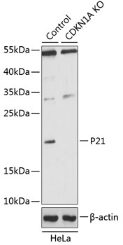 Sample:
Lane 1: Hela (Human) Cell Lysate at 25 ug
Lane 2: NDRG1 knockout (KO) Hela (Human) Cell Lysate at 25 ug
Primary: Anti-CDKN1A/p21CIP1 (SL55160R) at 1/1000 dilution
Secondary: HRP Goat Anti-Rabbit IgG (H+L) at 1:10000 dilution
Predicted band size: 21 kD
Observed band size: 21 kD
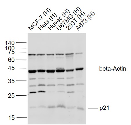 Sample:
Lane 1: MCF-7 (Human) Cell Lysate at 30 ug
Lane 2: Hela (Human) Cell Lysate at 30 ug
Lane 3: Huvec (Human) Cell Lysate at 30 ug
Lane 4: U87MG (Human) Cell Lysate at 30 ug
Lane 5: 293T (Human) Cell Lysate at 30 ug
Lane 6: A673 (Human) Cell Lysate at 30 ug
Primary:
Anti- p21 (SL55160R) at 1/1000 dilution
Anti-beta-Actin (SL0061R) at 1/2000 dilution
Secondary: IRDye800CW Goat Anti-Rabbit IgG at 1/20000 dilution
Predicted band size: 21 kD
Observed band size: 21 kD
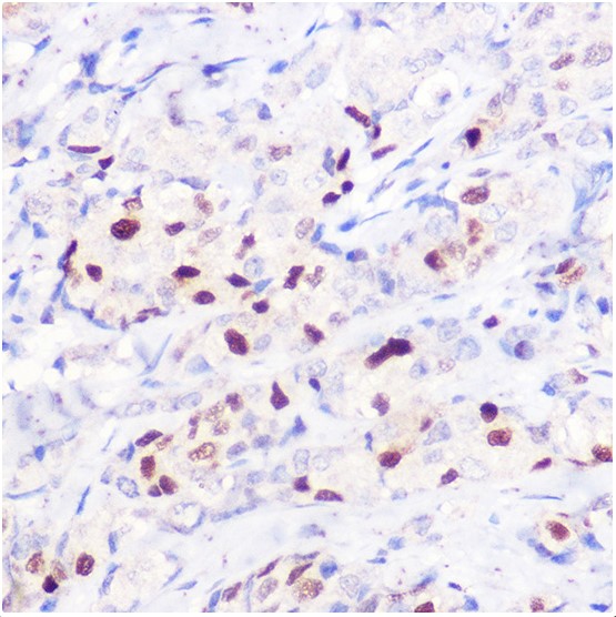 Paraformaldehyde-fixed, paraffin embedded (human breast cancer); Antigen retrieval by boiling in sodium citrate buffer (pH6.0) for 15min; Block endogenous peroxidase by 3% hydrogen peroxide for 20 minutes; Blocking buffer (normal goat serum) at 37°C for 30min; Antibody incubation with (p21) Polyclonal Antibody, Unconjugated (SL55160R) at 1:100 overnight at 4°C, followed by operating according to SP Kit(Rabbit) (sp-0023) instructionsand DAB staining.
Cartpieces
Totalgoods,subtotals:¥Checkout
References (0)
No References
Bought notes(bought amounts latest0)
No one bought this product
User Comment(Total0User Comment Num)
- No comment


 +86 571 56623320
 +86 18668110335

