
Rabbit Anti-ICAM1 antibody
BB2; CD54 antigen; ICAM 1; ICAM-1; Cell surface glycoprotein P3.58; Human rhinovirus receptor; Intercellular adhesion molecule 1; Major group rhinovirus receptor; MALA2; MyD10; P3.58; Surface antigen of activated B cells; ICAM1_HUMAN; MALA-2; MyD10; inter
View History [Clear]
Details
Product Name [KO validated anti] ICAM1 Chinese Name 细胞间粘附分子-1(CD54)抗体 Alias BB2; CD54 antigen; ICAM 1; ICAM-1; Cell surface glycoprotein P3.58; Human rhinovirus receptor; Intercellular adhesion molecule 1; Major group rhinovirus receptor; MALA2; MyD10; P3.58; Surface antigen of activated B cells; ICAM1_HUMAN; MALA-2; MyD10; intercellular adhesion molecule 1 precursor; intercellular adhesion molecule 1 (CD54), human rhinovirus receptor; major group rhinovirus receptor. literatures Research Area Tumour Cell biology immunology Signal transduction Stem cells The cell membrane受体 Cell adhesion molecule Cell Surface Molecule glycoprotein Cell type markers t-lymphocyte b-lymphocyte Immunogen Species Rabbit Clonality Polyclonal React Species Human, Mouse, (predicted: Rat, ) Applications WB=1:500-2000 ELISA=1:5000-10000 IHC-P=1:100-500 IHC-F=1:100-500 Flow-Cyt=1μg/Test ICC=1:100-500 IF=1:100-500 (Paraffin sections need antigen repair)
not yet tested in other applications.
optimal dilutions/concentrations should be determined by the end user.Theoretical molecular weight 56kDa Detection molecular weight 85-110kDa Cellular localization The cell membrane Form Liquid Concentration 1mg/ml immunogen KLH conjugated synthetic peptide derived from human CD54: 201-300/537 <Extracellular> Lsotype IgG Purification affinity purified by Protein A Buffer Solution 0.01M TBS(pH7.4) with 1% BSA, 0.03% Proclin300 and 50% Glycerol. Storage Shipped at 4℃. Store at -20 °C for one year. Avoid repeated freeze/thaw cycles. Attention This product as supplied is intended for research use only, not for use in human, therapeutic or diagnostic applications. PubMed PubMed Product Detail This gene encodes a cell surface glycoprotein which is typically expressed on endothelial cells and cells of the immune system. It binds to integrins of type CD11a / CD18, or CD11b / CD18 and is also exploited by Rhinovirus as a receptor. [provided by RefSeq, Jul 2008]
Function:
ICAM proteins are ligands for the leukocyte adhesion protein LFA-1 (integrin alpha-L/beta-2). During leukocyte trans-endothelial migration, ICAM1 engagement promotes the assembly of endothelial apical cups through ARHGEF26/SGEF and RHOG activation. In case of rhinovirus infection acts as a cellular receptor for the virus.
Subunit:
Homodimer (Probable). Interacts with human herpesvirus 8 MIR2 protein (Probable). Interacts with MUC1 and promotes cell aggregation in epithelial cells. Interacts with ARHGEF26/SGEF. Binds to coxsackievirus A21 capsid proteins and acts as a receptor for this virus.
Subcellular Location:
Membrane; Single-pass type I membrane protein.
Post-translational modifications:
Monoubiquitinated, which is promoted by MARCH9 and leads to endocytosis.
Similarity:
Belongs to the immunoglobulin superfamily. ICAM family.
ontains 5 Ig-like C2-type (immunoglobulin-like) domains.
SWISS:
P05362
Gene ID:
3383
Database links:
Entrez Gene: 3383 Human
Entrez Gene: 15894 Mouse
Omim: 147840 Human
Unigene: 643447 Human
SwissProt: P05362 Human
SwissProt: P13597 Mouse
Unigene: 435508 Mouse
Unigene: 12 Rat
CD54(ICAM-1)是分子量为60-110KD的膜型glycoprotein,表达于单核和endothelial cells。许多细胞如B或Tlymphocyte、胸腺细胞、纤维母细胞和epithelial cells都可诱导表达CD54,在调节免疫和炎症反应中CD54有重要作用。Product Picture 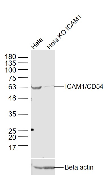 Sample:
Hela(Human) Cell Lysate at 30 ug
Hela KO ICAM1 (Human) Cell Lysate at 30 ug
Primary: Anti-ICAM1/CD54 (SL0608R) at 1/1000 dilution
Secondary: IRDye800CW Goat Anti-Rabbit IgG at 1/20000 dilution
Predicted band size: 56 kD
Observed band size: 56 kD
 Sample:
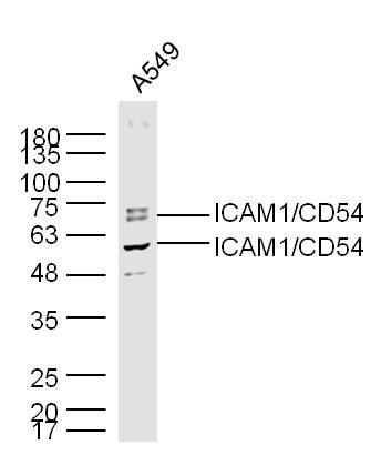
A549(Human) Cell Lysate at 30 ug
Primary: Anti-ICAM1 (SL0608R) at 1/300 dilution
Secondary: IRDye800CW Goat Anti-Rabbit IgG at 1/20000 dilution
Predicted band size: 56 kD
Observed band size: 56/69 kD
 Sample:
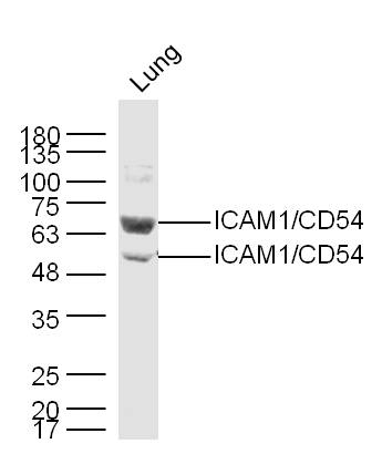
Lung (Mouse) Lysate at 40 ug
Primary: Anti-ICAM1 (SL0608R) at 1/300 dilution
Secondary: IRDye800CW Goat Anti-Rabbit IgG at 1/20000 dilution
Predicted band size: 56 kD
Observed band size: 56/69 kD
 Sample:
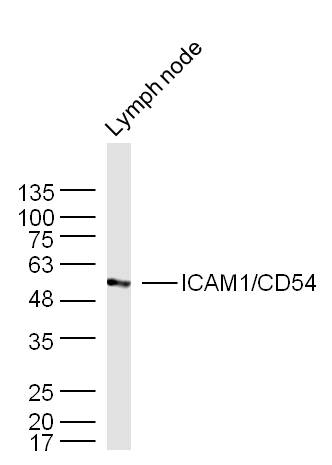
Lymph node (Mouse) Lysate at 40 ug
Primary: Anti-ICAM1 (SL0608R) at 1/300 dilution
Secondary: IRDye800CW Goat Anti-Rabbit IgG at 1/20000 dilution
Predicted band size: 56 kD
Observed band size: 56 kD
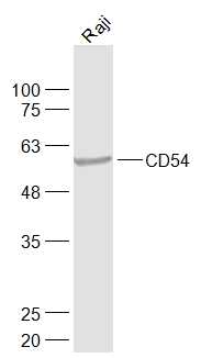 Sample:
Raji(Human) Cell Lysate at 30 ug
Primary: Anti-CD54 (SL0608R) at 1/1000 dilution
Secondary: IRDye800CW Goat Anti-Rabbit IgG at 1/20000 dilution
Predicted band size: 56 kD
Observed band size: 56 kD
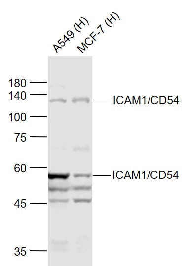 Sample:
Lane 1: A549 (Human) Cell Lysate at 30 ug
Lane 2: MCF-7 (Human) Cell Lysate at 30 ug
Primary: Anti-ICAM1/CD54 (SL0608R) at 1/1000 dilution
Secondary: IRDye800CW Goat Anti-Rabbit IgG at 1/20000 dilution
Predicted band size: 110/58 kD
Observed band size: 110/58 kD
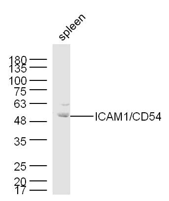 Sample:
Spleen (Mouse) Lysate at 40 ug
Primary: Anti-ICAM1 (SL0608R) at 1/300 dilution
Secondary: IRDye800CW Goat Anti-Rabbit IgG at 1/20000 dilution
Predicted band size: 56 kD
Observed band size: 56/69 kD
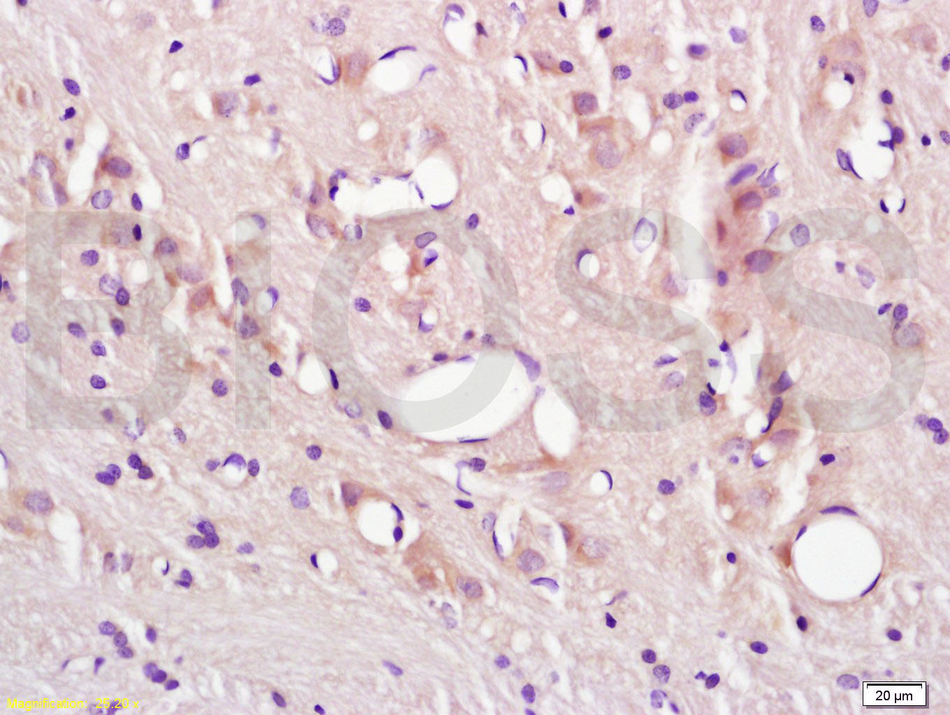 Tissue/cell: rat brain tissue; 4% Paraformaldehyde-fixed and paraffin-embedded;
Antigen retrieval: citrate buffer ( 0.01M, pH 6.0 ), Boiling bathing for 15min; Block endogenous peroxidase by 3% Hydrogen peroxide for 30min; Blocking buffer (normal goat serum,C-0005) at 37℃ for 20 min;
Incubation: Anti-CD54/ICAM-1 Polyclonal Antibody, Unconjugated(SL0608R) 1:200, overnight at 4°C, followed by conjugation to the secondary antibody(SP-0023) and DAB(C-0010) staining
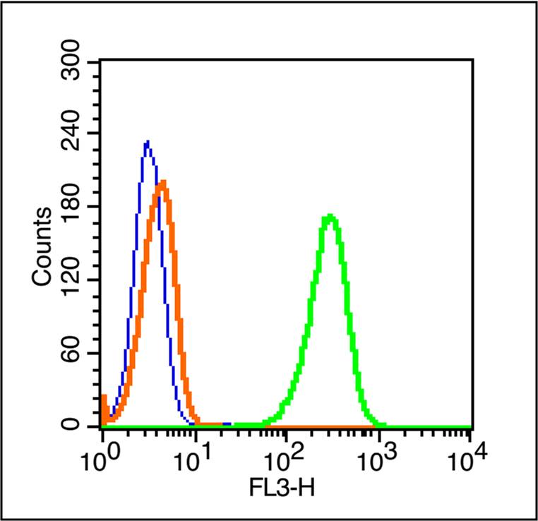 Blank control (blue line): A431 cells(blue).
Primary Antibody (green line): Rabbit Anti-ICAM1/PE-CY7 Conjugated antibody (SL0608R-PE-CY7)
Dilution: 1μg /10^6 cells;
Isotype Control Antibody (orange line): Rabbit IgG-PE-CY7 .
Protocol
The cells were fixed with 70% ice-cold methanol overnight at 4℃ . The cells were then incubated in 1 X PBS/2%BSA/10% goat serum to block non-specific protein-protein interactions followed by the antibody for 15 min at room temperature. Cells stained with Primary Antibody for 30 min at room temperature.Acquisition of 20,000 events was performed.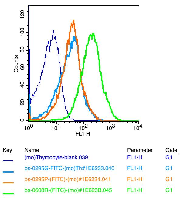 Blank control: mouse thymouses(blue)
Isotype Control Antibody: Rabbit IgG(orange) ; Secondary Antibody: Goat anti-rabbit IgG-FITC(white blue), Dilution: 1:100 in 1 X PBS containing 0.5% BSA ; Primary Antibody Dilution: 1μg in 100 μL 1X PBS containing 0.5% BSA(green).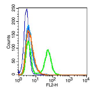 Blank control: HUVEC cells(blue).
Primary Antibody:Rabbit Anti-CD54 antibody(SL0608R), Dilution: 1μg in 100 μL 1X PBS containing 0.5% BSA;
Isotype Control Antibody: Rabbit IgG(orange) ,used under the same conditions );
Secondary Antibody: Goat anti-rabbit IgG-PE(white blue), Dilution: 1:200 in 1 X PBS containing 0.5% BSA.
Protocol
The cells were fixed with 2% paraformaldehyde (10 min) .Primary antibody (SL0608R, 1μg /1x10^6 cells) were incubated for 30 min on the ice, followed by 1 X PBS containing 0.5% BSA + 1 0% goat serum (15 min) to block non-specific protein-protein interactions. Then the Goat Anti-rabbit IgG/PE antibody was added into the blocking buffer mentioned above to react with the primary antibody at 1/200 dilution for 30 min on ice. Acquisition of 20,000 events was performed.
Cartpieces
Totalgoods,subtotals:¥Checkout
References (0)
No References
Bought notes(bought amounts latest0)
No one bought this product
User Comment(Total0User Comment Num)
- No comment


 +86 571 56623320
 +86 18668110335

