
Mouse Anti-ATG5/APG5L antibody
ATG5; APG 5; APG 5L; APG5; APG5 autophagy 5 like; APG5 like; APG5-like; APG5L; Apoptosis specific protein; Apoptosis-specific protein; ASP; ATG 5; ATG5; ATG5 autophagy related 5 homolog; Autophagy protein 5; hAPG5; Homolog of S Cerevisiae autophagy 5; ATG
View History [Clear]
Details
Product Name ATG5/APG5L Chinese Name 自噬蛋白5单克隆抗体 Alias ATG5; APG 5; APG 5L; APG5; APG5 autophagy 5 like; APG5 like; APG5-like; APG5L; Apoptosis specific protein; Apoptosis-specific protein; ASP; ATG 5; ATG5; ATG5 autophagy related 5 homolog; Autophagy protein 5; hAPG5; Homolog of S Cerevisiae autophagy 5; ATG5_HUMAN. Research Area Cell biology Apoptosis Autophagy Immunogen Species Mouse Clonality Monoclonal Clone NO. 10C4 React Species Human, Mouse, Rat, Applications WB=1:500-1000 IHC-P=1:100-500 IHC-F=1:100-500 IF=1:100-500 (Paraffin sections need antigen repair)
not yet tested in other applications.
optimal dilutions/concentrations should be determined by the end user.Theoretical molecular weight 32kDa Cellular localization cytoplasmic Form Liquid Concentration 1mg/ml immunogen Recombinant human ATG5 Protein Lsotype IgG Purification affinity purified by Protein G Buffer Solution 0.01M TBS(pH7.4) with 1% BSA, 0.03% Proclin300 and 50% Glycerol. Storage Shipped at 4℃. Store at -20 °C for one year. Avoid repeated freeze/thaw cycles. Attention This product as supplied is intended for research use only, not for use in human, therapeutic or diagnostic applications. PubMed PubMed Product Detail In yeast, autophagy is an essential process for survival during nutrient starvation and cell differentiation. The process of autophagy is characterized as a non-selective degradation of cytoplasmic proteins into membrane stuctures called autophagosomes, and it is dependent on several proteins, including the autophagy proteins APG5 and APG7. Yeast Apg7 and the human homolog, APG7, share similarities with the ubiquitin-activating enzyme E1 in Saccharomyces cerevisiae and are likewise responsible for enzymatically activating the autophagy conjugation system. Apg5 and the human homolog, APG5 (also designated apoptosis-specific protein or APS), function as substrates for the autophagy protein Apg12. These proteins are covalently bonded together to form Apg12/APG5 conjugates, which are required for the progression of autophagy.
Function:
Required for autophagy. Conjugates to ATG12 and associates with isolation membrane to form cup-shaped isolation membrane and autophagosome. The conjugate detaches from the membrane immediately before or after autophagosome formation is completed (By similarity).
May play an important role in the apoptotic process, possibly within the modified cytoskeleton. Its expression is a relatively late event in the apoptotic process, occurring downstream of caspase activity.
Subunit:
The ATG5-ATG12 conjugate forms a complex with several units of ATG16. Interacts with TECPR1; the interaction is direct and does not take place when ATG16 is associated with the ATG5-ATG12 conjugate.
Subcellular Location:
Cytoplasm. Note=Colocalizes with nonmuscle actin.
Tissue Specificity:
Ubiquitous. The mRNA is present at similar levels in viable and apoptotic cells, whereas the protein is dramatically highly expressed in apoptotic cells.
Post-translational modifications:
Conjugated to ATG12; which is essential for autophagy, but is not required for association with isolation membrane.
Similarity:
Belongs to the ATG5 family.
SWISS:
Q9H1Y0
Gene ID:
9474
Database links:Entrez Gene: 9474 Human
Entrez Gene: 11793 Mouse
Omim: 604261 Human
SwissProt: Q9H1Y0 Human
SwissProt: Q99J83 Mouse
Unigene: 486063 Human
Unigene: 22264 Mouse
Unigene: 98385 Rat
Product Picture 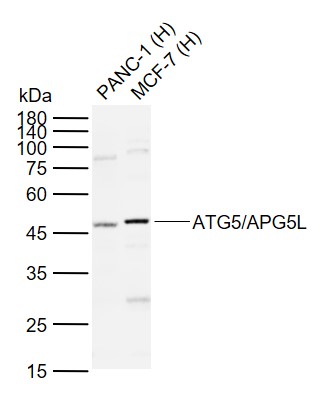 Sample:
Lane 1: Human PANC-1 cell lysates
Lane 2: Human MCF-7 cell lysates
Primary: Anti-ATG5/APG5L (SLM-33386M) at 1/1000 dilution
Secondary: IRDye800CW Goat Anti-Mouse IgG at 1/20000 dilution
Predicted band size: 32 kDa
Observed band size: 47 kDa
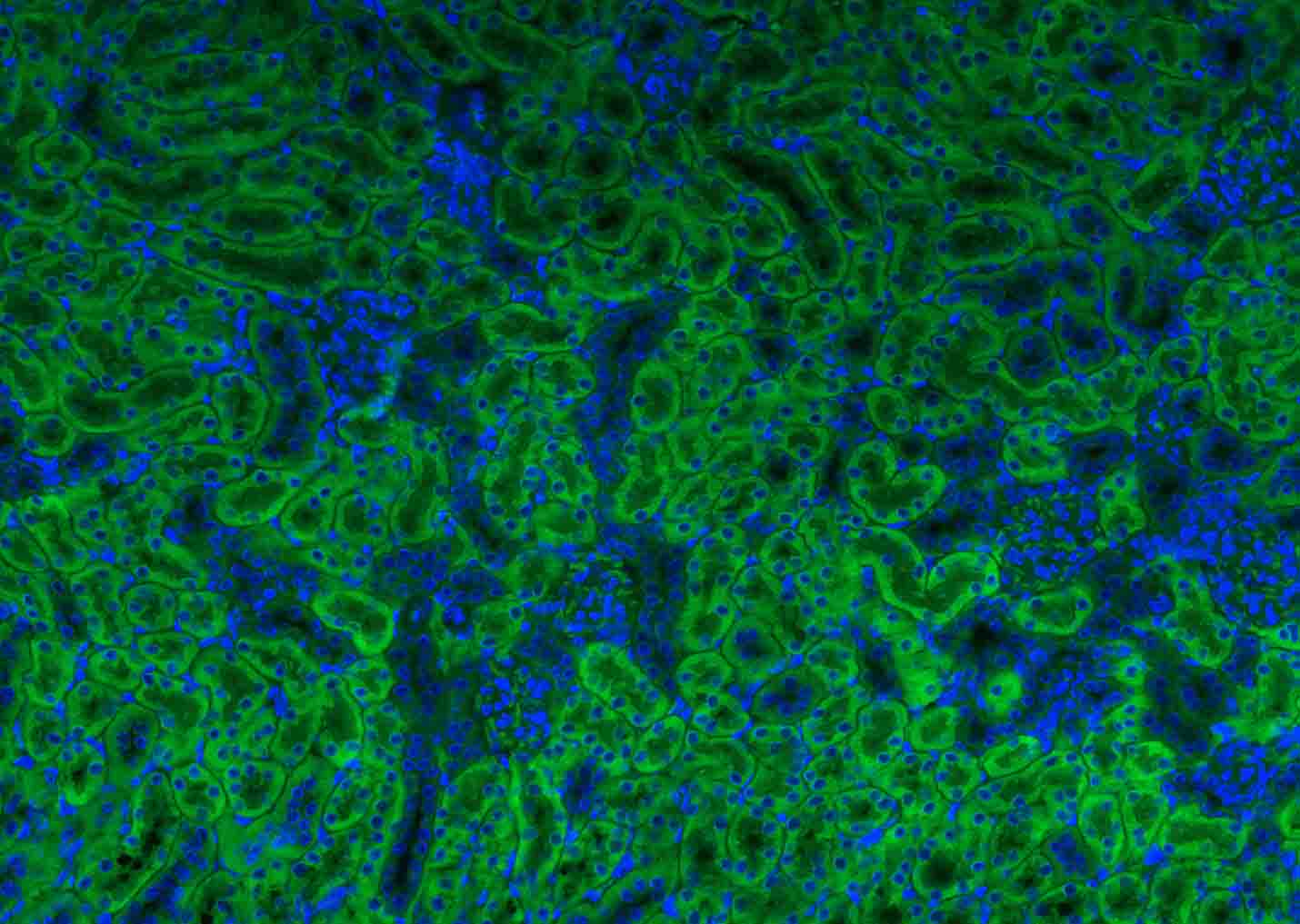 Paraformaldehyde-fixed, paraffin embedded (mouse kidney); Antigen retrieval by boiling in sodium citrate buffer (pH6.0) for 15min; Blocking buffer (normal goat serum) at 37°C for 30min; Antibody incubation with (ATG5/APG5L) Polyclonal Antibody, Unconjugated (SLM-33386M) at 1:200 overnight at 4°C, followed by a conjugated Goat Anti-mouse IgG antibody (SL0296G-AF488) for 90 minutes, and DAPI for nuclei staining.
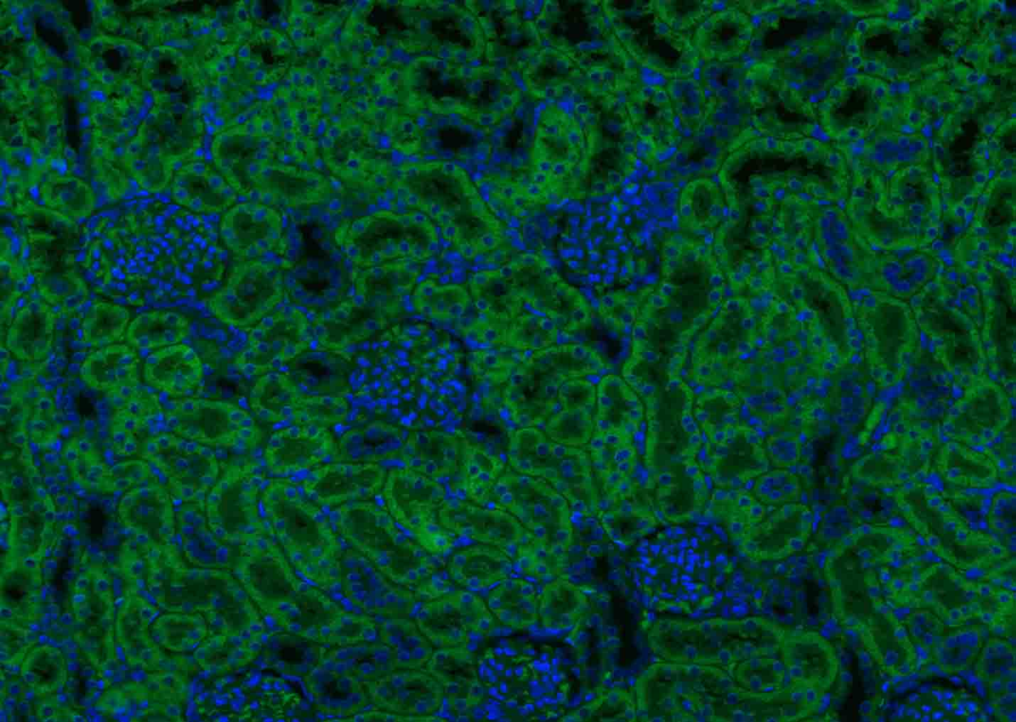 Paraformaldehyde-fixed, paraffin embedded (rat kidney); Antigen retrieval by boiling in sodium citrate buffer (pH6.0) for 15min; Blocking buffer (normal goat serum) at 37°C for 30min; Antibody incubation with (ATG5/APG5L) Polyclonal Antibody, Unconjugated (SLM-33386M) at 1:200 overnight at 4°C, followed by a conjugated Goat Anti-mouse IgG antibody (SL0296G-AF488) for 90 minutes, and DAPI for nuclei staining.
Cartpieces
Totalgoods,subtotals:¥Checkout
References (0)
No References
Bought notes(bought amounts latest0)
No one bought this product
User Comment(Total0User Comment Num)
- No comment


 +86 571 56623320
 +86 18668110335

