
Rabbit Anti-CD3 gamma antibody
CD3-GAMMA; CD3g; CD3g antigen gamma polypeptide; CD3g molecule gamma(CD3 TCRcomplex); CD3gmoleculegamma; CD3G_HUMAN; FLJ17620; FLJ17664 ; FLJ79544; FLJ94613; MGC138597; T cell antigen receptor complex gamma subunit of T3; T-cell receptor T3 gamma chain; T
View History [Clear]
Details
Product Name CD3 gamma Chinese Name CD3GRecombinant rabbit monoclonal anti Alias CD3-GAMMA; CD3g; CD3g antigen gamma polypeptide; CD3g molecule gamma(CD3 TCRcomplex); CD3gmoleculegamma; CD3G_HUMAN; FLJ17620; FLJ17664 ; FLJ79544; FLJ94613; MGC138597; T cell antigen receptor complex gamma subunit of T3; T-cell receptor T3 gamma chain; T-cell surface glycoprotein CD3 gamma chain; T3G. Research Area immunology lymphocyte Immunogen Species Rabbit Clonality Monoclonal Clone NO. 2B11 React Species Human, Mouse, Applications WB=1:500-1000 IP=1:10-50 IHC-P=1:100-500 IHC-F=1:50-200 Flow-Cyt=1:50 (Paraffin sections need antigen repair)
not yet tested in other applications.
optimal dilutions/concentrations should be determined by the end user.Theoretical molecular weight 20kDa Cellular localization The cell membrane Form Liquid Concentration 1mg/ml immunogen KLH conjugated synthetic peptide derived from human CD3 gamma Lsotype IgG Purification affinity purified by Protein A Buffer Solution 0.01M TBS(pH7.4) with 1% BSA, 0.03% Proclin300 and 50% Glycerol. Storage Shipped at 4℃. Store at -20 °C for one year. Avoid repeated freeze/thaw cycles. Attention This product as supplied is intended for research use only, not for use in human, therapeutic or diagnostic applications. PubMed PubMed Product Detail The protein encoded by this gene is the CD3-gamma polypeptide, which together with CD3-epsilon, -delta and -zeta, and the T-cell receptor alpha/beta and gamma/delta heterodimers, forms the T-cell receptor-CD3 complex. This complex plays an important role in coupling antigen recognition to several intracellular signal-transduction pathways. The genes encoding the epsilon, gamma and delta polypeptides are located in the same cluster on chromosome 11. Defects in this gene are associated with T cell immunodeficiency. [provided by RefSeq, Jul 2008]
Function:
Part of the TCR-CD3 complex present on T-lymphocyte cell surface that plays an essential role in adaptive immune response. When antigen presenting cells (APCs) activate T-cell receptor (TCR), TCR-mediated signals are transmitted across the cell membrane by the CD3 chains CD3D, CD3E, CD3G and CD3Z. All CD3 chains contain immunoreceptor tyrosine-based activation motifs (ITAMs) in their cytoplasmic domain. Upon TCR engagement, these motifs become phosphorylated by Src family protein tyrosine kinases LCK and FYN, resulting in the activation of downstream signaling pathways (PubMed:2470098). In addition to this role of signal transduction in T-cell activation, CD3G plays an essential role in the dynamic regulation of TCR expression at the cell surface (PubMed:8187769). Indeed, constitutive TCR cycling is dependent on the di-leucine-based (diL) receptor-sorting motif present in CD3G.
Subcellular Location:
membrane
Post-translational modifications:
Phosphorylated on Tyr residues after T-cell receptor triggering by LCK in association with CD4/CD8 (PubMed:2470098). Phosphorylated also by PKC; leading to the TCR complex down-regulation (PubMed:8187769).
Similarity:
Contains 1 Ig-like (immunoglobulin-like) domain.
Contains 1 ITAM domain.
SWISS:
P09693
Gene ID:
917
Database links:
Entrez Gene: 917 Human
Entrez Gene: 12502 Mouse
Entrez Gene: 443398 Sheep
Omim: 186740 Human
SwissProt: P09693 Human
SwissProt: P11942 Mouse
SwissProt: P18439 Sheep
Unigene: 2259 Human
Unigene: 335106 Mouse
Unigene: 234374 Rat
Product Picture 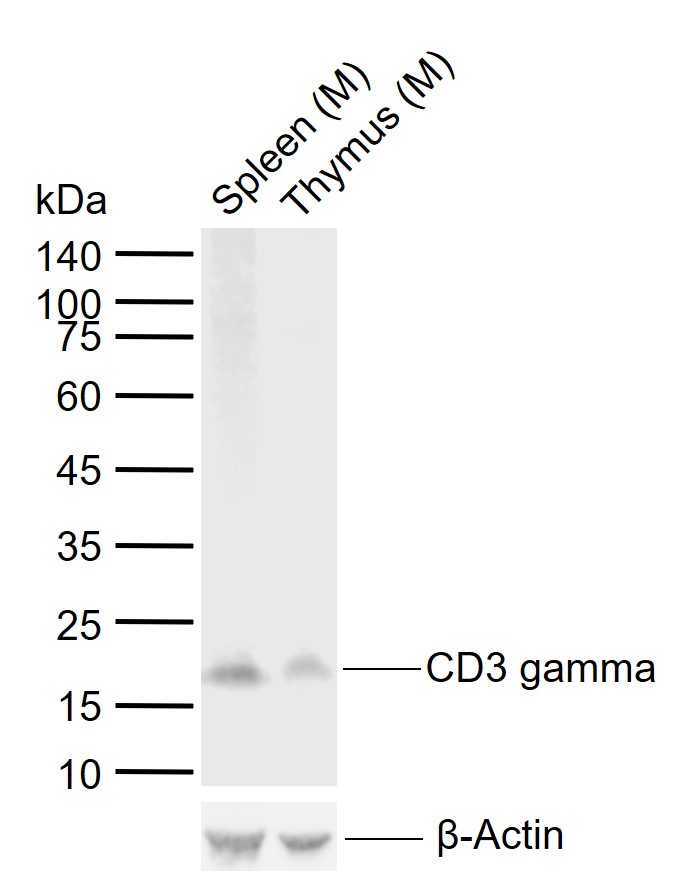 Sample:
Lane 1: Mouse Spleen tissue lysates
Lane 2: Mouse Thymus tissue lysates
Primary: Anti-CD3 gamma (SLM-54300R) at 1/1000 dilution
Secondary: IRDye800CW Goat Anti-Rabbit IgG at 1/20000 dilution
Predicted band size: 20 kDa
Observed band size: 17 kDa
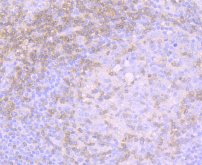 Paraformaldehyde-fixed, paraffin embedded (human tonsil); Antigen retrieval by boiling in sodium citrate buffer (pH6.0) for 15min; Block endogenous peroxidase by 3% hydrogen peroxide for 20 minutes; Blocking buffer (normal goat serum) at 37°C for 30min; Antibody incubation with (CD3 gamma) Monoclonal Antibody, Unconjugated (SLM-54300R) at 1:50 overnight at 4°C, followed by operating according to SP Kit(Rabbit) (sp-0023) instructionsand DAB staining.
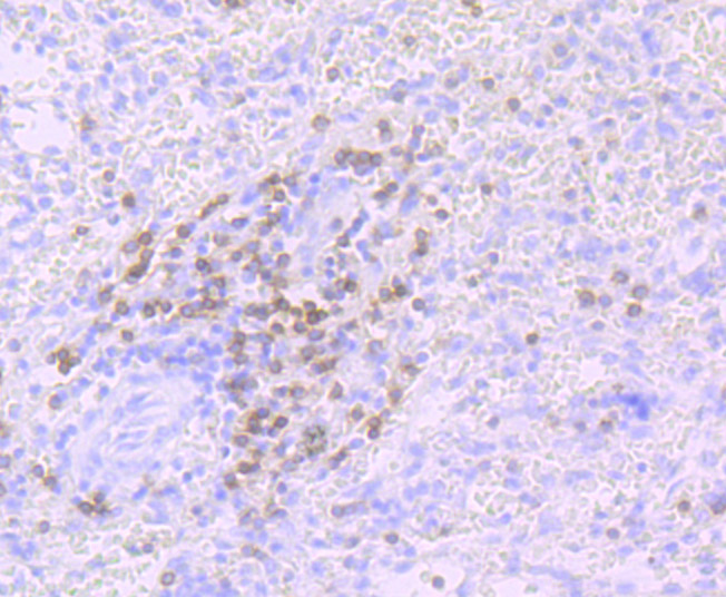 Paraformaldehyde-fixed, paraffin embedded (human spleen); Antigen retrieval by boiling in sodium citrate buffer (pH6.0) for 15min; Block endogenous peroxidase by 3% hydrogen peroxide for 20 minutes; Blocking buffer (normal goat serum) at 37°C for 30min; Antibody incubation with (CD3 gamma) Monoclonal Antibody, Unconjugated (SLM-54300R) at 1:50 overnight at 4°C, followed by operating according to SP Kit(Rabbit) (sp-0023) instructionsand DAB staining.
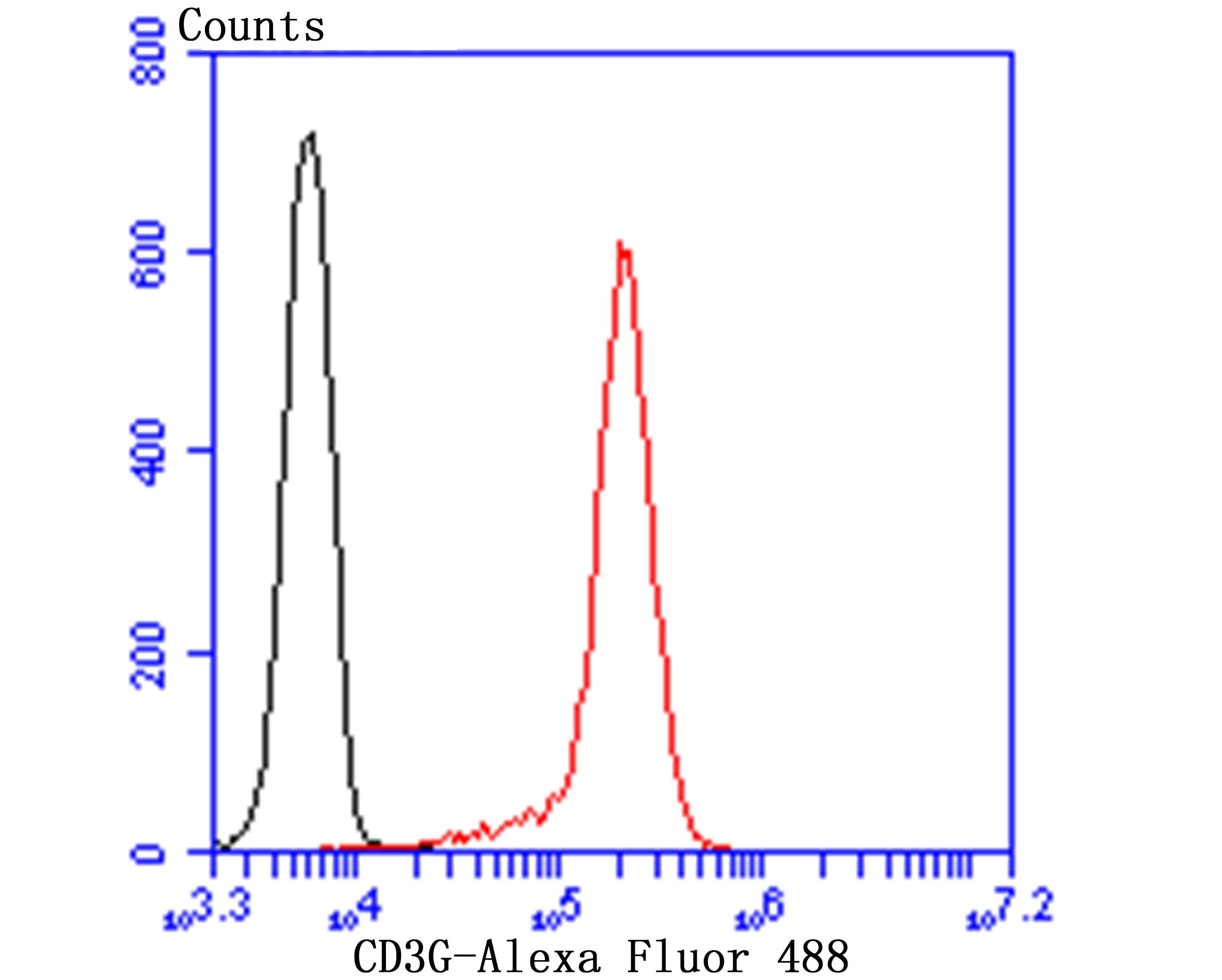 Blank control:Jurkat.
Primary Antibody (green line): Rabbit Anti-CD3G antibody (SLM-54300R)
Dilution: 1:50;
Isotype Control Antibody (orange line): Rabbit IgG .
Secondary Antibody : Goat anti-rabbit IgG-AF488
Dilution: 1:1000.
Protocol
The cells were incubated in 5%BSA to block non-specific protein-protein interactions for 30 min at room temperature .Cells stained with Primary Antibody for 30 min at room temperature. The secondary antibody used for 40 min at room temperature. Acquisition of 20,000 events was performed.
Cartpieces
Totalgoods,subtotals:¥Checkout
References (0)
No References
Bought notes(bought amounts latest0)
No one bought this product
User Comment(Total0User Comment Num)
- No comment


 +86 571 56623320
 +86 18668110335

