
Rabbit Anti-AIF1 (9A3) antibody
AIF 1; AIF1; AIF1 protein; IBa1; iba-1; IBa 1; allograft inflammatory factor 1; Allograft inflammatory factor 1 splice variant G; balloon angioplasty responsive transcription; BART 1; G1; G1 putative splice variant of allograft inflamatory factor 1; IBA 1
View History [Clear]
Details
Product Name AIF1 (9A3) Chinese Name 离子钙接头蛋白单克隆抗体 Alias AIF 1; AIF1; AIF1 protein; IBa1; iba-1; IBa 1; allograft inflammatory factor 1; Allograft inflammatory factor 1 splice variant G; balloon angioplasty responsive transcription; BART 1; G1; G1 putative splice variant of allograft inflamatory factor 1; IBA 1; IBA1; interferon gamma responsive transcript; Ionized calcium binding adapter molecule 1; ionized calcium-binding adapter molecule; IRT 1; IRT1; Microglia response factor; MRF1; Protein G1. Research Area Cell biology immunology Neurobiology Immunogen Species Rabbit Clonality Monoclonal Clone NO. 9A3 React Species Human, Mouse, Rat, Applications WB=1:500-1000 IP=1:10-50 IHC-P=1:50-100 Flow-Cyt=1:50 ICC=1:50-100 IF=1:50-100 (Paraffin sections need antigen repair)
not yet tested in other applications.
optimal dilutions/concentrations should be determined by the end user.Theoretical molecular weight 17kDa Cellular localization cytoplasmic The cell membrane Form Liquid Concentration 1mg/ml immunogen KLH conjugated synthetic peptide derived from human AIF1: 100-147/147 Lsotype IgG Purification affinity purified by Protein A Buffer Solution 0.01M TBS(pH7.4) with 1% BSA, 0.03% Proclin300 and 50% Glycerol. Storage Shipped at 4℃. Store at -20 °C for one year. Avoid repeated freeze/thaw cycles. Attention This product as supplied is intended for research use only, not for use in human, therapeutic or diagnostic applications. PubMed PubMed Product Detail Allograft Inflammatory Factor-1 (AIF1)or ionized calcium-binding adaptor molecule 1 (Iba1) is expressed selectively in microglia/macrophages and is a Ca2+-binding peptide produced by activated monocytes and microglial cells. It has been suggested that AIF1 expression is associated with chronic inflammatory processes. AIF1 is expressed by activated monocytes and might participate in a variety of pathogenic processes in the mammalian brain and in chronic transplant rejection. It has been shown to be expressed early and persistently in chronically rejecting cardiac allografts but not in cardiac syngrafts and host hearts.
Function:
Actin-binding protein that enhances membrane ruffling and RAC activation. Enhances the actin-bundling activity of LCP1. Binds calcium. Plays a role in RAC signaling and in phagocytosis. May play a role in macrophage activation and function. Promotes the proliferation of vascular smooth muscle cells and of T-lymphocytes. Enhances lymphocyte migration. Plays a role in vascular inflammation.
Subunit:
Homodimer (Potential). Monomer. Interacts with LCP1.
Subcellular Location:
Cytoplasm, cytoskeleton. Cell projection, ruffle membrane; Peripheral membrane protein; Cytoplasmic side. Note=Associated with the actin cytoskeleton at membrane ruffles and at sites of phagocytosis.
Tissue Specificity:
Detected in T-lymphocytes and peripheral blood mononuclear cells.
Similarity:
Contains 2 EF-hand domains.
SWISS:
P55008
Gene ID:
199
Product Picture 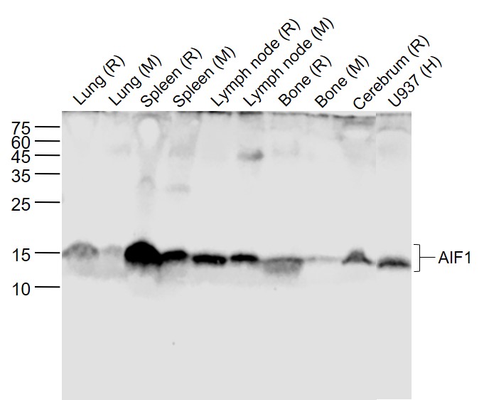 Sample:
Lane 1: Lung (Rat) Lysate at 40 ug
Lane 2: Lung (Mouse) Lysate at 40 ug
Lane 3: Spleen (Rat) Lysate at 40 ug
Lane 4: Spleen (Mouse) Lysate at 40 ug
Lane 5: Lymph node (Rat) Lysate at 40 ug
Lane 6: Lymph node (Mouse) Lysate at 40 ug
Lane 7: Bone (Rat) Lysate at 40 ug
Lane 8: Bone (Mouse) Lysate at 40 ug
Lane 9: Cerebrum (Rat) Lysate at 40 ug
Lane 10: U937 (Human) Cell Lysate at 30 ug
Primary: Anti-AIF1 (SLM-54132R) at 1/1000 dilution
Secondary: IRDye800CW Goat Anti-Rabbit IgG at 1/20000 dilution
Predicted band size: 17 kD
Observed band size: 17 kD
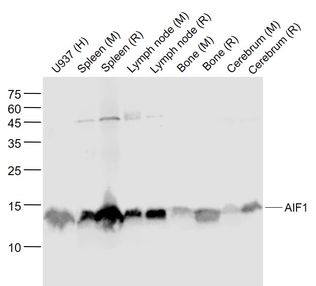 Sample:
Lane 1: U937 (Human) Cell Lysate at 30 ug
Lane 2: Spleen (Mouse) Lysate at 40 ug
Lane 3: Spleen (Rat) Lysate at 40 ug
Lane 4: Lymph node (Mouse) Lysate at 40 ug
Lane 5: Lymph node (Rat) Lysate at 40 ug
Lane 6: Bone (Mouse) Lysate at 40 ug
Lane 7: Bone (Rat) Lysate at 40 ug
Lane 8: Cerebrum (Mouse) Lysate at 40 ug
Lane 9: Cerebrum (Rat) Lysate at 40 ug
Primary: Anti-AIF1 (SLM-54132R) at 1/1000 dilution
Secondary: IRDye800CW Goat Anti-Rabbit IgG at 1/20000 dilution
Predicted band size: 17 kD
Observed band size: 15 kD
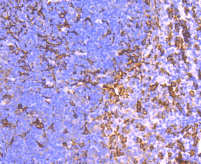 Paraformaldehyde-fixed, paraffin embedded (mouse spleen); Antigen retrieval by boiling in sodium citrate buffer (pH6.0) for 15min; Block endogenous peroxidase by 3% hydrogen peroxide for 20 minutes; Blocking buffer (normal goat serum) at 37°C for 30min; Antibody incubation with (AIF1 (9A3) ) Monoclonal Antibody, Unconjugated (SLM-54132R) at 1:50 overnight at 4°C, followed by operating according to SP Kit(Rabbit) (sp-0023) instructionsand DAB staining.
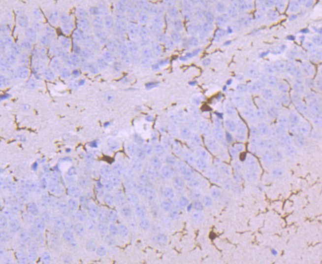 Paraformaldehyde-fixed, paraffin embedded (mouse brain); Antigen retrieval by boiling in sodium citrate buffer (pH6.0) for 15min; Block endogenous peroxidase by 3% hydrogen peroxide for 20 minutes; Blocking buffer (normal goat serum) at 37°C for 30min; Antibody incubation with (AIF1 (9A3) ) Monoclonal Antibody, Unconjugated (SLM-54132R) at 1:50 overnight at 4°C, followed by operating according to SP Kit(Rabbit) (sp-0023) instructionsand DAB staining.
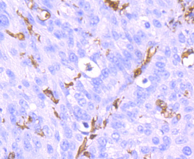 Paraformaldehyde-fixed, paraffin embedded (human lung carcinoma); Antigen retrieval by boiling in sodium citrate buffer (pH6.0) for 15min; Block endogenous peroxidase by 3% hydrogen peroxide for 20 minutes; Blocking buffer (normal goat serum) at 37°C for 30min; Antibody incubation with (AIF1 (9A3) ) Monoclonal Antibody, Unconjugated (SLM-54132R) at 1:50 overnight at 4°C, followed by operating according to SP Kit(Rabbit) (sp-0023) instructionsand DAB staining.
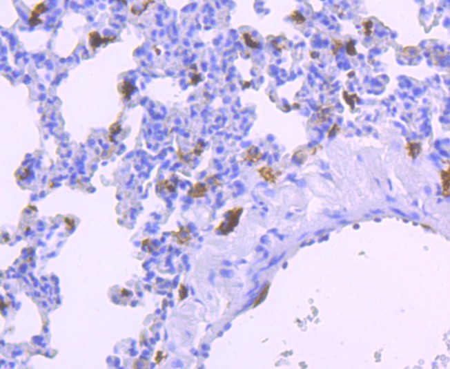 Paraformaldehyde-fixed, paraffin embedded (rat lung); Antigen retrieval by boiling in sodium citrate buffer (pH6.0) for 15min; Block endogenous peroxidase by 3% hydrogen peroxide for 20 minutes; Blocking buffer (normal goat serum) at 37°C for 30min; Antibody incubation with (AIF1 (9A3) ) Monoclonal Antibody, Unconjugated (SLM-54132R) at 1:50 overnight at 4°C, followed by operating according to SP Kit(Rabbit) (sp-0023) instructionsand DAB staining.
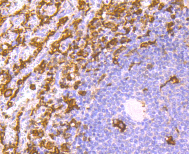 Paraformaldehyde-fixed, paraffin embedded (human spleen); Antigen retrieval by boiling in sodium citrate buffer (pH6.0) for 15min; Block endogenous peroxidase by 3% hydrogen peroxide for 20 minutes; Blocking buffer (normal goat serum) at 37°C for 30min; Antibody incubation with (AIF1 (9A3) ) Monoclonal Antibody, Unconjugated (SLM-54132R) at 1:50 overnight at 4°C, followed by operating according to SP Kit(Rabbit) (sp-0023) instructionsand DAB staining.
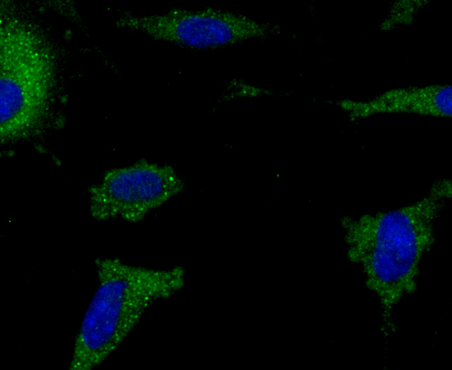 SH-SY5Y cell; 4% Paraformaldehyde-fixed; Triton X-100 at room temperature for 20 min; Blocking buffer (normal goat serum, C-0005) at 37°C for 20 min; Antibody incubation with (Iba1) monoclonal Antibody, Unconjugated (SLM-54132R) 1:50, 90 minutes at 37°C; followed by a conjugated Goat Anti-Rabbit IgG antibody at 37°C for 90 minutes, DAPI (blue, C02-04002) was used to stain the cell nuclei.
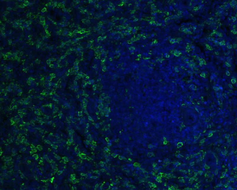 Paraformaldehyde-fixed, paraffin embedded (human spleen); Antigen retrieval by boiling in sodium citrate buffer (pH6.0) for 15min; Blocking buffer (normal goat serum) at 37°C for 30min; Antibody incubation with (AIF1 (9A3)) Monoclonal Antibody, Unconjugated (SLM-54132R) at 1:50 overnight at 4°C, followed by a conjugated Goat Anti-Rabbit IgG antibody (Alexa Fluor® 488 ) for 90 minutes, and DAPI for nuclei staining.
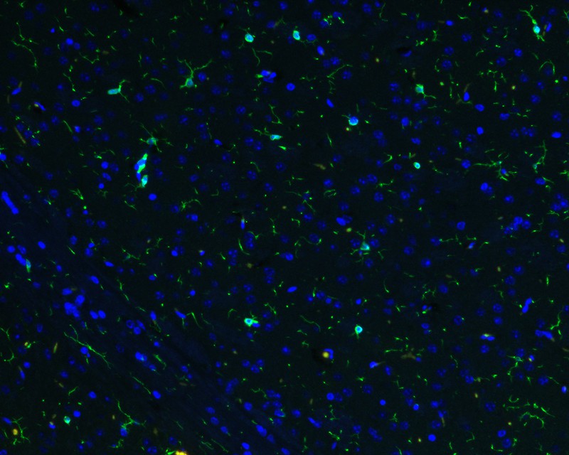 Paraformaldehyde-fixed, paraffin embedded (mouse brain); Antigen retrieval by boiling in sodium citrate buffer (pH6.0) for 15min; Blocking buffer (normal goat serum) at 37°C for 30min; Antibody incubation with (AIF1 (9A3)) Monoclonal Antibody, Unconjugated (SLM-54132R) at 1:50 overnight at 4°C, followed by a conjugated Goat Anti-Rabbit IgG antibody (Alexa Fluor® 488 ) for 90 minutes, and DAPI for nuclei staining.
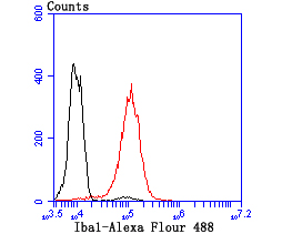 Blank control:THP-1.
Primary Antibody (green line): Rabbit Anti-Iba1 antibody (SLM-54132R)
Dilution: 1:50;
Secondary Antibody : Goat anti-rabbit IgG-AF488
Dilution: 1:1000.
Protocol
The cells were fixed with 4% PFA (10min at room temperature)and then permeabilized with 0.1% PBST for 20 min at room temperature. The cells were then incubated in 5%BSA to block non-specific protein-protein interactions for 30 min at room temperature .Cells stained with Primary Antibody for 30 min at room temperature. The secondary antibody used for 40 min at room temperature. Acquisition of 20,000 events was performed.
Cartpieces
Totalgoods,subtotals:¥Checkout
References (0)
No References
Bought notes(bought amounts latest0)
No one bought this product
User Comment(Total0User Comment Num)
- No comment


 +86 571 56623320
 +86 18668110335

