
Rabbit Anti-phospho-ATM (Ser1981)antibody
ATM(Phospho-Ser1981); ATM (phospho S1981); AT complementation group A; AT complementation group C; AT complementation group D; AT complementation group E; AT mutated; AT protein;AT1;ATA;Ataxia telangiectasia gene mutated in human beings; Ataxia telangiect
View History [Clear]
Details
Product Name phospho-ATM (Ser1981) Chinese Name 磷酸化ATM(Ser1981)Recombinant rabbit monoclonal anti Alias ATM(Phospho-Ser1981); ATM (phospho S1981); AT complementation group A; AT complementation group C; AT complementation group D; AT complementation group E; AT mutated; AT protein;AT1;ATA;Ataxia telangiectasia gene mutated in human beings; Ataxia telangiectasia mutated; ATC; ATDC; ATE; ATM; Human phosphatidylinositol 3 kinase homolog; Serine protein kinase ATM; T cell prolymphocytic leukemia; TEL1; TPLL. Research Area Signal transduction Apoptosis Cyclin Kinases and Phosphatases Immunogen Species Rabbit Clonality Monoclonal Clone NO. 3F10 React Species Human, (predicted: Mouse, ) Applications WB=1:300-1000 IHC-P=1:50-100 IHC-F=1:50-100 ICC=1:50-100 IF=1:50-100 (Paraffin sections need antigen repair)
not yet tested in other applications.
optimal dilutions/concentrations should be determined by the end user.Theoretical molecular weight 370kDa Cellular localization The nucleus cytoplasmic Form Liquid Concentration 1mg/ml immunogen KLH conjugated Synthesised phosphopeptide derived from human ATM around the phosphorylation site of Ser1981: EG(p-S)Q Lsotype IgG Purification affinity purified by Protein A Buffer Solution 0.01M TBS(pH7.4) with 1% BSA, 0.03% Proclin300 and 50% Glycerol. Storage Shipped at 4℃. Store at -20 °C for one year. Avoid repeated freeze/thaw cycles. Attention This product as supplied is intended for research use only, not for use in human, therapeutic or diagnostic applications. PubMed PubMed Product Detail ATM is a 370 kDa nuclear phosphoprotein involved in the autosomal recessive disease Ataxia Telangiectasia (AT). ATM belongs to a novel family of proteins associated with cell cycle regulation, apoptosis, and response to DNA damage repair (DNA damage caused by such things as ionizing irradiation activates ATM kinase). The C terminal region has extensive homology to the catalytic domains of Phosphatidylinositol 3 kinases (PI3 kinases).
Subcellular Location:
Nucleus. Cytoplasmic vesicle. Primarily nuclear. Found also in endocytic vesicles in association with beta-adaptin.
Tissue Specificity:
Found in pancreas, kidney, skeletal muscle, liver, lung, placenta, brain, heart, spleen, thymus, testis, ovary, small intestine, colon and leukocytes.
Post-translational modifications:
Phosphorylated by NUAK1/ARK5. Autophosphorylation on Ser-367, Ser-1893, Ser-1981 correlates with DNA damage-mediated activation of the kinase.
Acetylation, on DNA damage, is required for activation of the kinase activity, dimer-monomer transition, and subsequent autophosphorylation on Ser-1981. Acetylated in vitro by KAT5/TIP60.
DISEASE:
Defects in ATM are the cause of ataxia telangiectasia (AT) [MIM:208900]; also known as Louis-Bar syndrome, which includes four complementation groups: A, C, D and E. This rare recessive disorder is characterized by progressive cerebellar ataxia, dilation of the blood vessels in the conjunctiva and eyeballs, immunodeficiency, growth retardation and sexual immaturity. AT patients have a strong predisposition to cancer; about 30% of patients develop tumors, particularly lymphomas and leukemias. Cells from affected individuals are highly sensitive to damage by ionizing radiation and resistant to inhibition of DNA synthesis following irradiation.
Note=Defects in ATM contribute to T-cell acute lymphoblastic leukemia (TALL) and T-prolymphocytic leukemia (TPLL). TPLL is characterized by a high white blood cell count, with a predominance of prolymphocytes, marked splenomegaly, lymphadenopathy, skin lesions and serous effusion. The clinical course is highly aggressive, with poor response to chemotherapy and short survival time. TPLL occurs both in adults as a sporadic disease and in younger AT patients.
Note=Defects in ATM contribute to B-cell non-Hodgkin lymphomas (BNHL), including mantle cell lymphoma (MCL).
Note=Defects in ATM contribute to B-cell chronic lymphocytic leukemia (BCLL). BCLL is the commonest form of leukemia in the elderly. It is characterized by the accumulation of mature CD5+ B lymphocytes, lymphadenopathy, immunodeficiency and bone marrow failure.
Similarity:
Belongs to the PI3/PI4-kinase family. ATM subfamily.
Contains 1 FAT domain.
Contains 1 FATC domain.
Contains 1 PI3K/PI4K domain.
SWISS:
Q13315
Gene ID:
472
Database links:Entrez Gene: 472 Human
Entrez Gene: 11920 Mouse
Omim: 607585 Human
SwissProt: Q13315 Human
SwissProt: Q62388 Mouse
Unigene: 367437 Human
Unigene: 5088 Mouse
Unigene: 214048 Rat
Product Picture 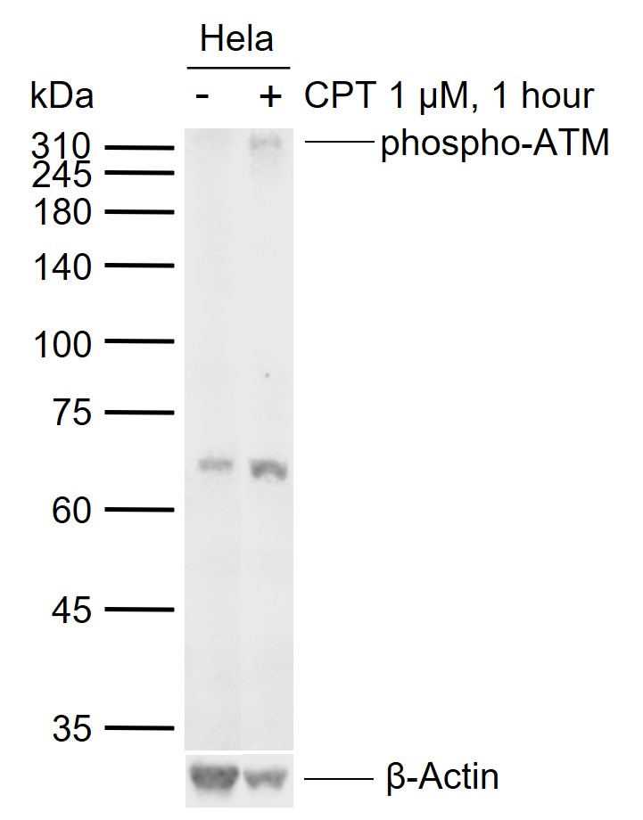 Sample:
Lane 1: Normal Human HeLa cell lysates
Lane 2: HeLa cell treated with 1 µM camptothecin (CPT) for 1 hour
Primary: Anti-phospho-ATM (Ser1981) (SLM-54103R) at 1/1000 dilution
Secondary: IRDye800CW Goat Anti-Rabbit IgG at 1/20000 dilution
Predicted band size: 370 kDa
Observed band size: 65 kDa
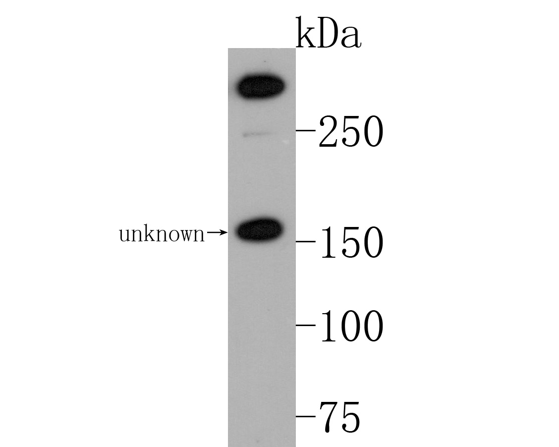 Sample:
Lane 1: HepG2 cell lysates
Primary: Anti-phospho-ATM (Ser1981) (SLM-54103R) at 1:1000 dilution
Secondary: Goat Anti-Rabbit IgG - HRP at 1:20000 dilution
Predicted band size: 370 kD
Observed band size: 350 kD
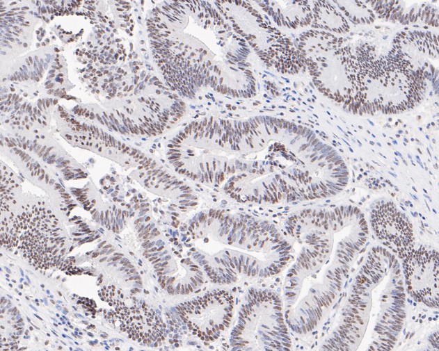 Paraformaldehyde-fixed, paraffin embedded (human colon carcinoma); Antigen retrieval by boiling in sodium citrate buffer (pH6.0) for 15min; Block endogenous peroxidase by 3% hydrogen peroxide for 20 minutes; Blocking buffer (normal goat serum) at 37°C for 30min; Antibody incubation with (phospho-ATM (Ser1981)) Monoclonal Antibody, Unconjugated (SLM-54103R) at 1:50 overnight at 4°C, followed by operating according to SP Kit(Rabbit) (sp-0023) instructionsand DAB staining.
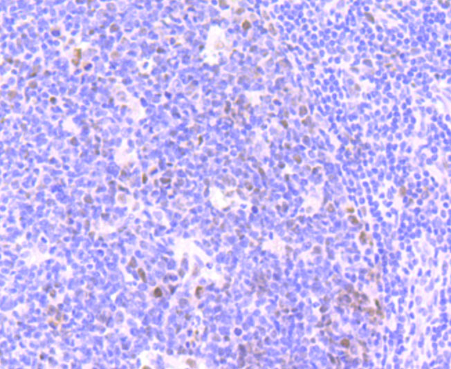 Paraformaldehyde-fixed, paraffin embedded (human tonsil); Antigen retrieval by boiling in sodium citrate buffer (pH6.0) for 15min; Block endogenous peroxidase by 3% hydrogen peroxide for 20 minutes; Blocking buffer (normal goat serum) at 37°C for 30min; Antibody incubation with (phospho-ATM (Ser1981)) Monoclonal Antibody, Unconjugated (SLM-54103R) at 1:50 overnight at 4°C, followed by operating according to SP Kit(Rabbit) (sp-0023) instructionsand DAB staining.
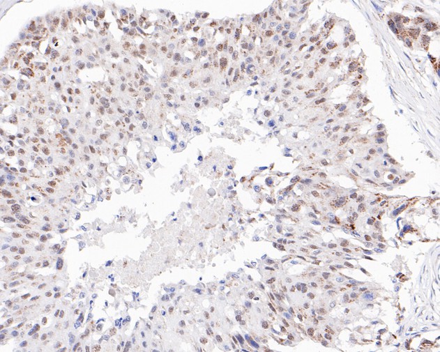 Paraformaldehyde-fixed, paraffin embedded (human breast carcinoma); Antigen retrieval by boiling in sodium citrate buffer (pH6.0) for 15min; Block endogenous peroxidase by 3% hydrogen peroxide for 20 minutes; Blocking buffer (normal goat serum) at 37°C for 30min; Antibody incubation with (phospho-ATM (Ser1981)) Monoclonal Antibody, Unconjugated (SLM-54103R) at 1:50 overnight at 4°C, followed by operating according to SP Kit(Rabbit) (sp-0023) instructionsand DAB staining.
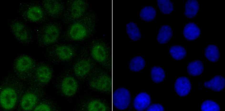 HepG2 cell; 4% Paraformaldehyde-fixed; Triton X-100 at room temperature for 20 min; Blocking buffer (normal goat serum, C-0005) at 37°C for 20 min; Antibody incubation with (ATM(phospho S1981)) monoclonal Antibody, Unconjugated (SLM-54103R) 1:50, 90 minutes at 37°C; followed by a conjugated Goat Anti-Rabbit IgG antibody at 37°C for 90 minutes, DAPI (blue, C02-04002) was used to stain the cell nuclei.
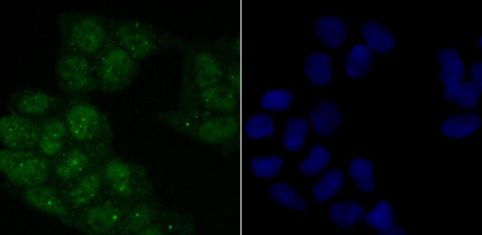 Hela cell; 4% Paraformaldehyde-fixed; Triton X-100 at room temperature for 20 min; Blocking buffer (normal goat serum, C-0005) at 37°C for 20 min; Antibody incubation with (ATM(phospho S1981)) monoclonal Antibody, Unconjugated (SLM-54103R) 1:50, 90 minutes at 37°C; followed by a conjugated Goat Anti-Rabbit IgG antibody at 37°C for 90 minutes, DAPI (blue, C02-04002) was used to stain the cell nuclei.
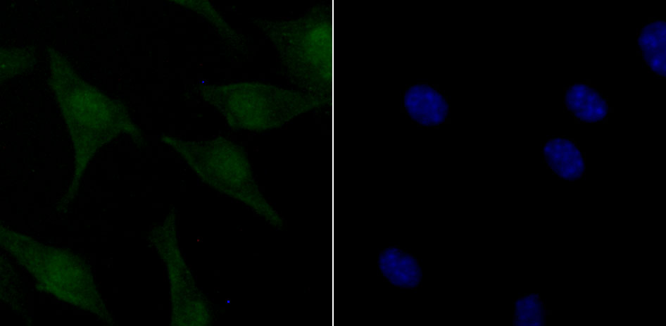 SH-SY5Y cell; 4% Paraformaldehyde-fixed; Triton X-100 at room temperature for 20 min; Blocking buffer (normal goat serum, C-0005) at 37°C for 20 min; Antibody incubation with (ATM(phospho S1981)) monoclonal Antibody, Unconjugated (SLM-54103R) 1:50, 90 minutes at 37°C; followed by a conjugated Goat Anti-Rabbit IgG antibody at 37°C for 90 minutes, DAPI (blue, C02-04002) was used to stain the cell nuclei.
Cartpieces
Totalgoods,subtotals:¥Checkout
Bought notes(bought amounts latest0)
No one bought this product
User Comment(Total0User Comment Num)
- No comment


 +86 571 56623320
 +86 18668110335

