
Rabbit Anti-PDPK1 antibody
3 Phosphoinositide Dependent Protein Kinase 1; hPDK 1; hPDK 1; PDK 1; PkB kinase; PkB kinase like gene 1; PDK1; PkB like 1; PDPK1_HUMAN.3磷酸肌醇依赖性蛋白激酶1
View History [Clear]
Details
Product Name PDPK1 Chinese Name PDK1Recombinant rabbit monoclonal anti Alias 3 Phosphoinositide Dependent Protein Kinase 1; hPDK 1; hPDK 1; PDK 1; PkB kinase; PkB kinase like gene 1; PDK1; PkB like 1; PDPK1_HUMAN. 3磷酸肌醇依赖性蛋白激酶1 Research Area Cell biology Neurobiology Signal transduction Apoptosis Kinases and Phosphatases Immunogen Species Rabbit Clonality Monoclonal Clone NO. 2F9 React Species Human, Mouse, Rat, Applications WB=1:500-2000 IP=1:20-100 IHC-P=1:50-200 Flow-Cyt=1:50 ICC=1:50 (Paraffin sections need antigen repair)
not yet tested in other applications.
optimal dilutions/concentrations should be determined by the end user.Theoretical molecular weight 61kDa Cellular localization cytoplasmic The cell membrane Form Liquid Concentration 1mg/ml immunogen KLH conjugated synthetic peptide derived from human PDK1 Lsotype IgG Purification affinity purified by Protein A Buffer Solution 0.01M TBS(pH7.4) with 1% BSA, 0.03% Proclin300 and 50% Glycerol. Storage Shipped at 4℃. Store at -20 °C for one year. Avoid repeated freeze/thaw cycles. Attention This product as supplied is intended for research use only, not for use in human, therapeutic or diagnostic applications. PubMed PubMed Product Detail PDK1 (3 Phosphoinositide Dependent Protein Kinase 1) phosphorylates AGC kinases.
PDK1 activates conventional PKC and PKC zeta through phosphorylation of critical threonine residues in the activation loop. PDK1 also phosphorylates Protein Kinase B (PKB) at threonine 308 in the presence of phosphatidylinositol-3,4,5-trisphosphate. Active Akt inactivates Glycogen Synthase Kinase 3 (GSK3), eventually leading to the dephosphorylation and activation of glycogen synthase and the stimulation of glycogen synthesis. Because of the role that PDK plays in insulin-induced glycogen synthesis and PKC activation it is a potentially important target for metabolic drug research. There are three named isoforms.
Function:
Phosphorylates and activates not only PKB/AKT, but also PKA, PKC-zeta, RPS6KA1 and RPS6KB1. May play a general role in signaling processes and in development (By similarity). Isoform 3 is catalytically inactive.
Subunit:
Interacts with NPRL2.
Subcellular Location:
Cytoplasm. Membrane; Peripheral membrane protein. Note=Membrane-associated after cell stimulation leading to its translocation. Tyrosine phosphorylation seems to occur only at the plasma membrane.
Tissue Specificity:
Appears to be expressed ubiquitously.
Post-translational modifications:
Phosphorylated on tyrosine and serine/threonine. Phosphorylation on Ser-241 in the activation loop is required for full activity. PDK1 itself can autophosphorylate Ser-241, leading to its own activation.
Similarity:
Belongs to the protein kinase superfamily. AGC Ser/Thr protein kinase family. PDK1 subfamily.
Contains 1 PH domain.
Contains 1 protein kinase domain.
SWISS:
O15530
Gene ID:
5170
Database links:Entrez Gene: 5170 Human
Entrez Gene: 18607 Mouse
Omim: 605213 Human
SwissProt: O15530 Human
SwissProt: Q9Z2A0 Mouse
Unigene: 459691 Human
Unigene: 10504 Mouse
Unigene: 10905 Rat
Product Picture 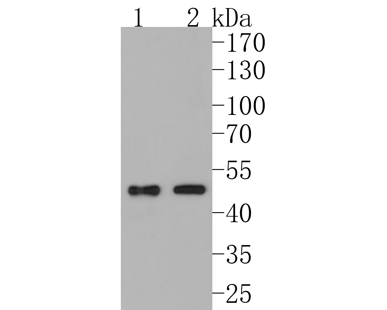 Sample:
Lane 1: rat heart tissue lysate
Lane 2: mouse heart tissue lysate
Primary: Anti-PDK1 (SLM-54037R) at 1:500 dilution
Secondary: Goat Anti-Rabbit IgG - HRP at 1:5000 dilution
Predicted band size: 61 kD
Observed band size: 50 kD
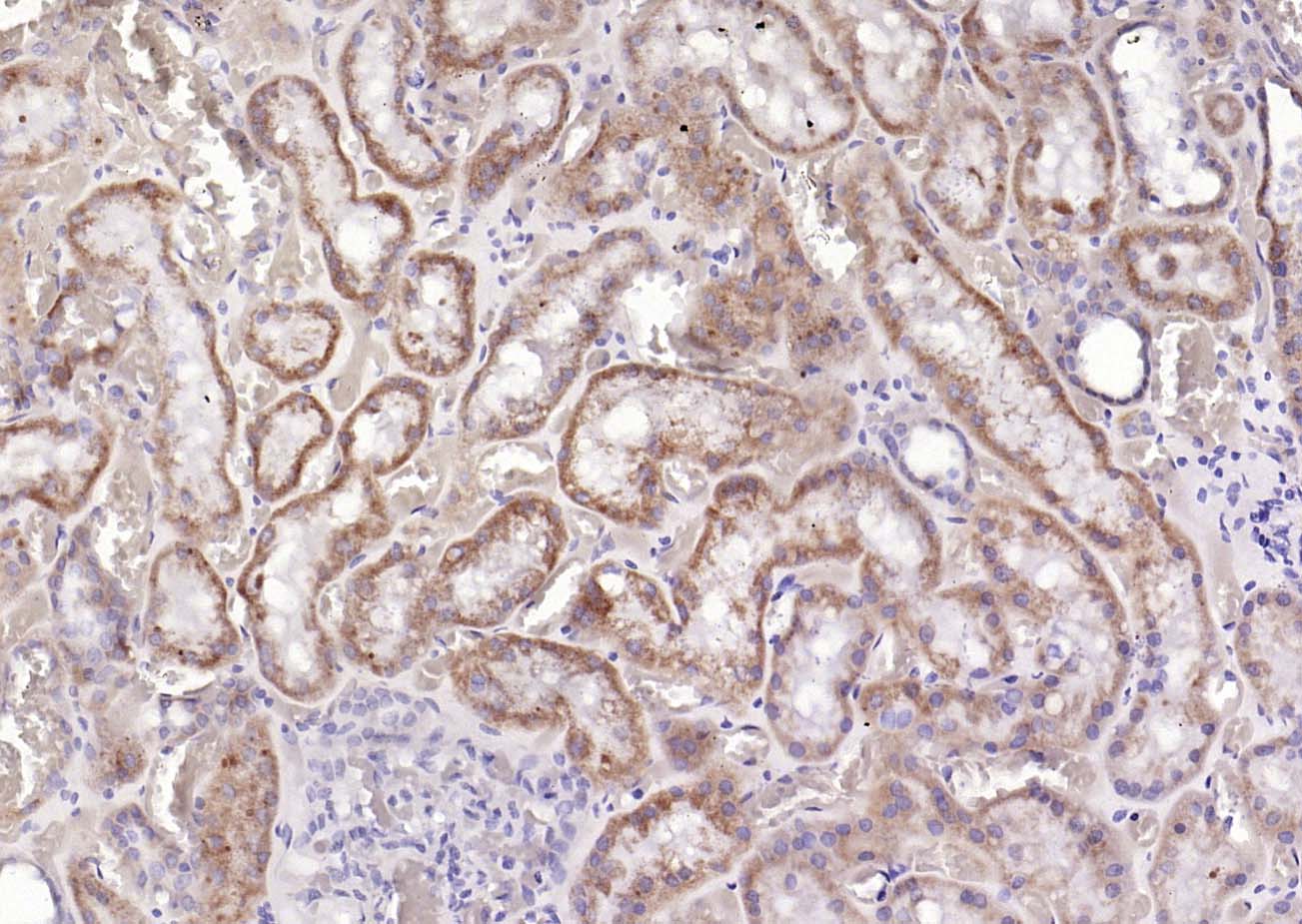 Paraformaldehyde-fixed, paraffin embedded (Human kidney); Antigen retrieval by boiling in sodium citrate buffer (pH6.0) for 15min; Block endogenous peroxidase by 3% hydrogen peroxide for 20 minutes; Blocking buffer (normal goat serum) at 37°C for 30min; Antibody incubation with (PDK1) Monoclonal Antibody, Unconjugated (SLM-54037R) at 1:200 overnight at 4°C, followed by operating according to SP Kit(Rabbit) (sp-0023) instructionsand DAB staining.
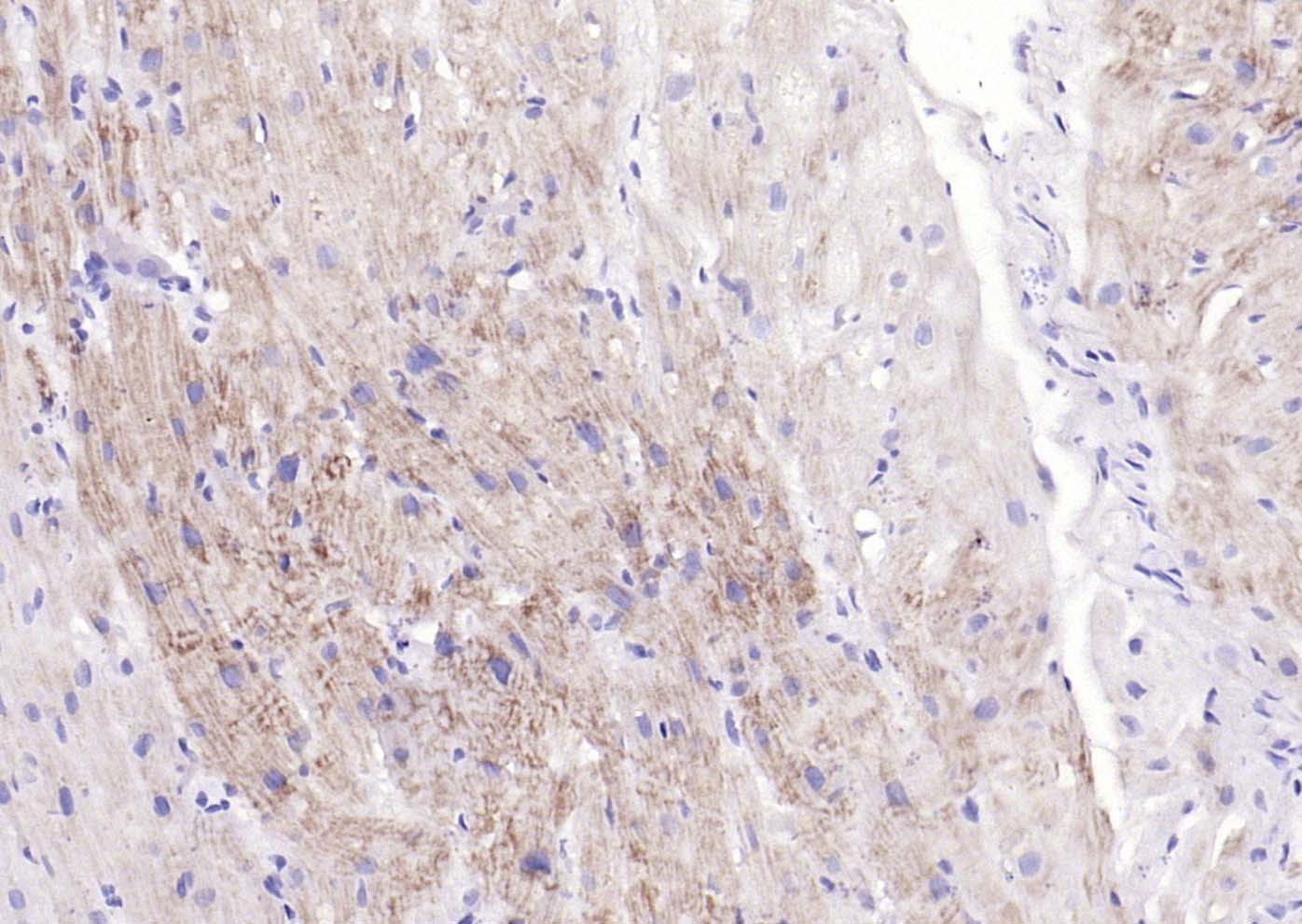 Paraformaldehyde-fixed, paraffin embedded (human myocardium); Antigen retrieval by boiling in sodium citrate buffer (pH6.0) for 15min; Block endogenous peroxidase by 3% hydrogen peroxide for 20 minutes; Blocking buffer (normal goat serum) at 37°C for 30min; Antibody incubation with (PDK1) Monoclonal Antibody, Unconjugated (SLM-54037R) at 1:200 overnight at 4°C, followed by operating according to SP Kit(Rabbit) (sp-0023) instructionsand DAB staining.
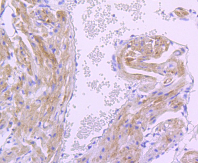 Paraformaldehyde-fixed, paraffin embedded (mouse heart tissue); Antigen retrieval by boiling in sodium citrate buffer (pH6.0) for 15min; Block endogenous peroxidase by 3% hydrogen peroxide for 20 minutes; Blocking buffer (normal goat serum) at 37°C for 30min; Antibody incubation with (PDK1) Monoclonal Antibody, Unconjugated (SLM-54037R) at 1:50 overnight at 4°C, followed by operating according to SP Kit(Rabbit) (sp-0023) instructionsand DAB staining.
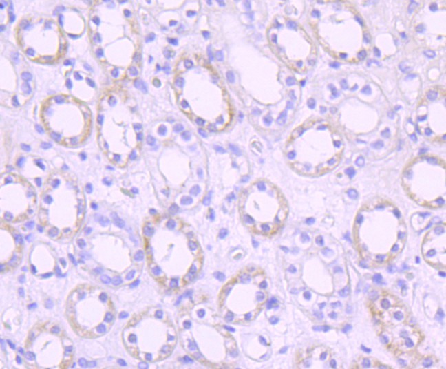 Paraformaldehyde-fixed, paraffin embedded (human kidney tissue); Antigen retrieval by boiling in sodium citrate buffer (pH6.0) for 15min; Block endogenous peroxidase by 3% hydrogen peroxide for 20 minutes; Blocking buffer (normal goat serum) at 37°C for 30min; Antibody incubation with (PDK1) Monoclonal Antibody, Unconjugated (SLM-54037R) at 1:50 overnight at 4°C, followed by operating according to SP Kit(Rabbit) (sp-0023) instructionsand DAB staining.
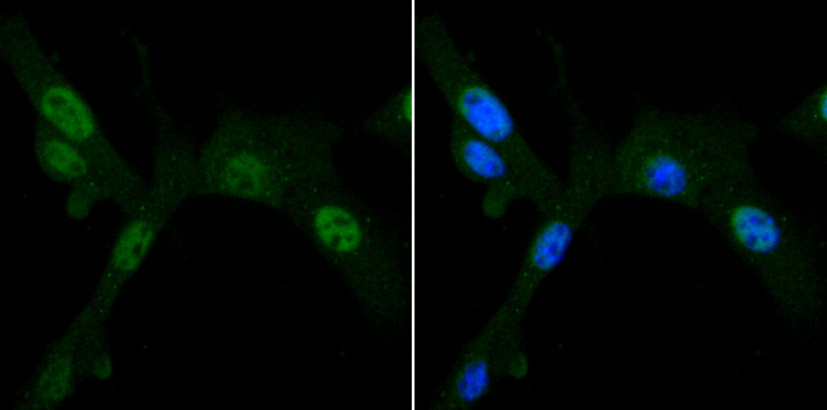 NIH/3T3 cell; 4% Paraformaldehyde-fixed; Triton X-100 at room temperature for 20 min; Blocking buffer (normal goat serum, C-0005) at 37°C for 20 min; Antibody incubation with (PDK1) monoclonal Antibody, Unconjugated (SLM-54037R) 1:50, 90 minutes at 37°C; followed by a conjugated Goat Anti-Rabbit IgG antibody at 37°C for 90 minutes, DAPI (blue, C02-04002) was used to stain the cell nuclei.
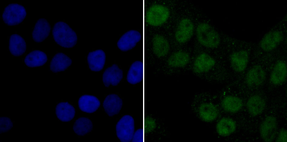 Hela cell; 4% Paraformaldehyde-fixed; Triton X-100 at room temperature for 20 min; Blocking buffer (normal goat serum, C-0005) at 37°C for 20 min; Antibody incubation with (PDK1) monoclonal Antibody, Unconjugated (SLM-54037R) 1:50, 90 minutes at 37°C; followed by a conjugated Goat Anti-Rabbit IgG antibody at 37°C for 90 minutes, DAPI (blue, C02-04002) was used to stain the cell nuclei.
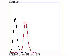 Blank control:NIH/3T3.
Primary Antibody (green line): Rabbit Anti-PDK1 antibody (SLM-54037R)
Dilution: 1:50;
Isotype Control Antibody (orange line): Rabbit IgG .
Secondary Antibody : Goat anti-rabbit IgG-AF488
Dilution: 1:1000.
Protocol
The cells were fixed with 4% PFA (10min at room temperature)and then permeabilized with 90% ice-cold methanol for 20 min at -20℃. The cells were then incubated in 5%BSA to block non-specific protein-protein interactions for 30 min at room temperature .Cells stained with Primary Antibody for 30 min at room temperature. The secondary antibody used for 40 min at room temperature. Acquisition of 20,000 events was performed.
Cartpieces
Totalgoods,subtotals:¥Checkout
References (0)
No References
Bought notes(bought amounts latest0)
No one bought this product
User Comment(Total0User Comment Num)
- No comment


 +86 571 56623320
 +86 18668110335

