
Rabbit Anti-LXR alpha antibody
Liver X receptor alpha; LXR a; LXRa; LXRA; NR1H3; NR1H3_HUMAN; Nuclear orphan receptor LXR alpha; Nuclear receptor subfamily 1 group H member 3; Oxysterols receptor LXR alpha; Oxysterols receptor LXR-alpha; RLD 1; RLD1.
View History [Clear]
Details
Product Name LXR alpha Chinese Name 肝脏X受体兔单克隆抗体 Alias Liver X receptor alpha; LXR a; LXRa; LXRA; NR1H3; NR1H3_HUMAN; Nuclear orphan receptor LXR alpha; Nuclear receptor subfamily 1 group H member 3; Oxysterols receptor LXR alpha; Oxysterols receptor LXR-alpha; RLD 1; RLD1. Research Area Cell biology immunology Bacteria and viruses Immunogen Species Rabbit Clonality Monoclonal Clone NO. 4A8 React Species Human, Mouse, (predicted: Rat, ) Applications WB=1:500-1000 IHC-P=1:50-200 Flow-Cyt=1:50 ICC=1:50 (Paraffin sections need antigen repair)
not yet tested in other applications.
optimal dilutions/concentrations should be determined by the end user.Theoretical molecular weight 50kDa Cellular localization The nucleus Form Liquid Concentration 1mg/ml immunogen KLH conjugated synthetic peptide derived from human LXR alpha: 60-110/447 Lsotype IgG Purification affinity purified by Protein A Buffer Solution 0.01M TBS(pH7.4) with 1% BSA, 0.03% Proclin300 and 50% Glycerol. Storage Shipped at 4℃. Store at -20 °C for one year. Avoid repeated freeze/thaw cycles. Attention This product as supplied is intended for research use only, not for use in human, therapeutic or diagnostic applications. PubMed PubMed Product Detail Peroxisome proliferators include hypolipidemic drugs, herbicides, leukotriene antagonists, and plasticizers; this term arises because they induce an increase in the size and number of peroxisomes. Peroxisomes are subcellular organelles found in plants and animals that contain enzymes for respiration and for cholesterol and lipid metabolism. The action of peroxisome proliferators is thought to be mediated via specific receptors, called PPARs, which belong to the steroid hormone receptor superfamily. PPARs affect the expression of target genes involved in cell proliferation, cell differentiation and in immune and inflammation responses. Three closely related subtypes (alpha, beta/delta, and gamma) have been identified. This gene encodes the subtype PPAR-alpha, which is a nuclear transcription factor. Multiple alternatively spliced transcript variants have been described for this gene, although the full-length nature of only two has been determined. [provided by RefSeq, Jul 2008].
Function:
Orphan receptor. Interaction with RXR shifts RXR from its role as a silent DNA-binding partner to an active ligand-binding subunit in mediating retinoid responses through target genes defined by LXRES. LXRES are DR4-type response elements characterized by direct repeats of two similar hexanuclotide half-sites spaced by four nucleotides. Plays an important role in the regulation of cholesterol homeostasis, regulating cholesterol uptake through MYLIP-dependent ubiquitination of LDLR, VLDLR and LRP8.
Subunit:
Heterodimer of LXRA and RXR.
Subcellular Location:
Nucleus (Potential).
Tissue Specificity:
Visceral organs specific expression. Strong expression was found in liver, kidney and intestine followed by spleen and to a lesser extent the adrenals.
Similarity:
Belongs to the nuclear hormone receptor family. NR1 subfamily.
Contains 1 nuclear receptor DNA-binding domain.
SWISS:
Q13133
Gene ID:
10062
Database links:Entrez Gene: 10062 Human
Entrez Gene: 22259 Mouse
Omim: 602423 Human
SwissProt: Q13133 Human
SwissProt: Q9Z0Y9 Mouse
Unigene: 438863 Human
Unigene: 22690 Mouse
Unigene: 11209 Rat
肝脏X受体LXRa(Liver X Receptor a)属孤核受体家族,是一种与脂类代谢有关的核受体,该蛋白具有调节脂类的吸收、运输、转化和生物合成的功能,并且在在糖类的代谢等方面也有重要的调控作用Product Picture 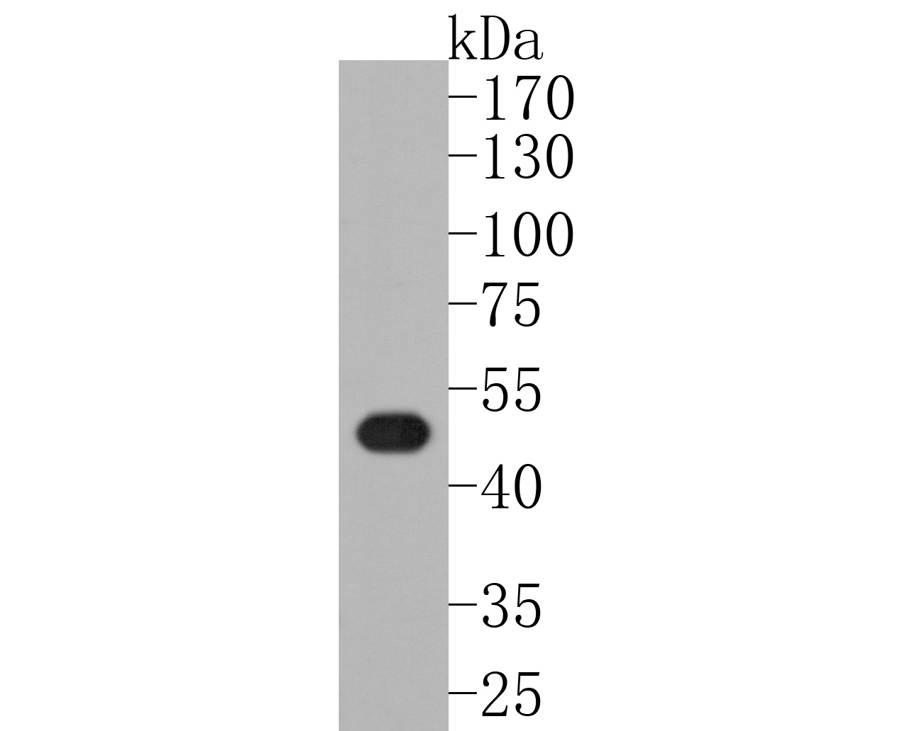 Sample:
Lane 1: mouse colon tissue lysates
Primary: Anti-LXR alpha (SLM-54023R) at 1:500 dilution
Secondary: Goat Anti-Rabbit IgG - HRP at 1:5000 dilution
Predicted band size: 50 kD
Observed band size: 50 kD
 Sample:
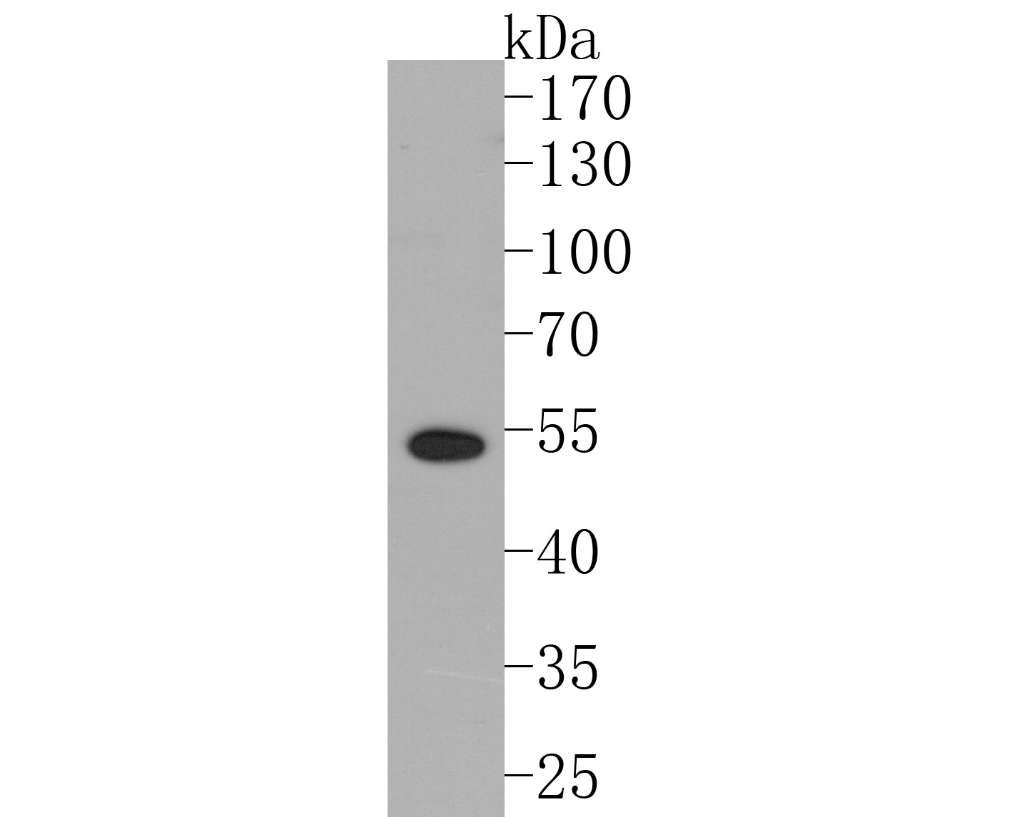
Lane 1: human liver tissue lysates
Primary: Anti-LXR alpha (SLM-54023R) at 1:500 dilution
Secondary: Goat Anti-Rabbit IgG - HRP at 1:5000 dilution
Predicted band size: 50 kD
Observed band size: 53 kD
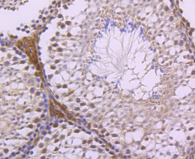 Paraformaldehyde-fixed, paraffin embedded (mouse testis tissue); Antigen retrieval by boiling in sodium citrate buffer (pH6.0) for 15min; Block endogenous peroxidase by 3% hydrogen peroxide for 20 minutes; Blocking buffer (normal goat serum) at 37°C for 30min; Antibody incubation with (LXR alpha) Monoclonal Antibody, Unconjugated (SLM-54023R) at 1:50 overnight at 4°C, followed by operating according to SP Kit(Rabbit) (sp-0023) instructionsand DAB staining.
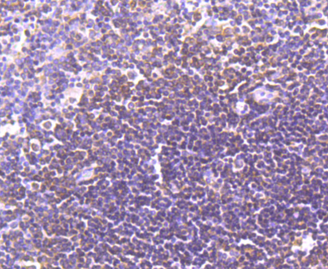 Paraformaldehyde-fixed, paraffin embedded (human spleen tissue); Antigen retrieval by boiling in sodium citrate buffer (pH6.0) for 15min; Block endogenous peroxidase by 3% hydrogen peroxide for 20 minutes; Blocking buffer (normal goat serum) at 37°C for 30min; Antibody incubation with (LXR alpha) Monoclonal Antibody, Unconjugated (SLM-54023R) at 1:50 overnight at 4°C, followed by operating according to SP Kit(Rabbit) (sp-0023) instructionsand DAB staining.
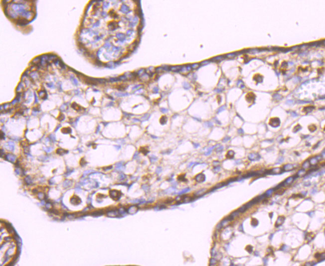 Paraformaldehyde-fixed, paraffin embedded (human placenta tissue); Antigen retrieval by boiling in sodium citrate buffer (pH6.0) for 15min; Block endogenous peroxidase by 3% hydrogen peroxide for 20 minutes; Blocking buffer (normal goat serum) at 37°C for 30min; Antibody incubation with (LXR alpha) Monoclonal Antibody, Unconjugated (SLM-54023R) at 1:50 overnight at 4°C, followed by operating according to SP Kit(Rabbit) (sp-0023) instructionsand DAB staining.
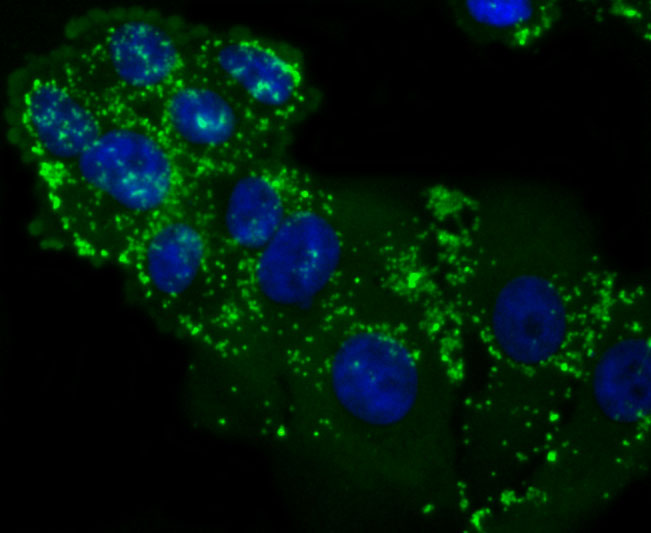 HepG2 cell; 4% Paraformaldehyde-fixed; Triton X-100 at room temperature for 20 min; Blocking buffer (normal goat serum, C-0005) at 37°C for 20 min; Antibody incubation with (LXR alpha) monoclonal Antibody, Unconjugated (SLM-54023R) 1:50, 90 minutes at 37°C; followed by a conjugated Goat Anti-Rabbit IgG antibody at 37°C for 90 minutes, DAPI (blue, C02-04002) was used to stain the cell nuclei.
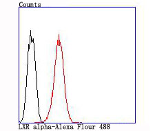 Blank control:HepG2 .
Primary Antibody (green line): Rabbit Anti-LXR alpha antibody (SLM-54023R)
Dilution: 1:50;
Isotype Control Antibody (orange line): Rabbit IgG .
Secondary Antibody : Goat anti-rabbit IgG-AF488
Dilution: 1:1000.
Protocol
The cells were fixed with 4% PFA (10min at room temperature)and then permeabilized with 90% ice-cold methanol for 20 min at -20℃. The cells were then incubated in 5%BSA to block non-specific protein-protein interactions for 30 min at room temperature .Cells stained with Primary Antibody for 30 min at room temperature. The secondary antibody used for 40 min at room temperature. Acquisition of 20,000 events was performed.
Cartpieces
Totalgoods,subtotals:¥Checkout
References (0)
No References
Bought notes(bought amounts latest0)
No one bought this product
User Comment(Total0User Comment Num)
- No comment


 +86 571 56623320
 +86 18668110335

