
Rabbit Anti-LYVE1 antibody
lymphalic vessel endotheilial hyaluronan receptor 1; CRSBP 1; CRSBP1; extracellular link domain containing 1; extracellular link domain-containing 1; HAR; hyaluronic acid receptor; Cell surface retention sequence-binding protein 1; CRSBP-1; extracellular
View History [Clear]
Details
Product Name LYVE1 Chinese Name 淋巴管内皮透明质酸受体Recombinant rabbit monoclonal anti Alias lymphalic vessel endotheilial hyaluronan receptor 1; CRSBP 1; CRSBP1; extracellular link domain containing 1; extracellular link domain-containing 1; HAR; hyaluronic acid receptor; Cell surface retention sequence-binding protein 1; CRSBP-1; extracellular link domain-containing 1; Extracellular link domain-containing protein 1; Lymphatic vessel endothelial hyaluronic acid receptor 1; LYVE1_HUMAN; Lymphatic endothelium specific hyaluronan receptor; LYVE 1; LYVE-1; XLKD1. Research Area Cardiovascular Cell biology immunology Cell type markers Immunogen Species Rabbit Clonality Monoclonal Clone NO. 5C1 React Species Human, Applications IHC-P=1:50-200 IHC-F=1:50-200 Flow-Cyt=1:50 ICC=1:50 (Paraffin sections need antigen repair)
not yet tested in other applications.
optimal dilutions/concentrations should be determined by the end user.Theoretical molecular weight 32kDa Cellular localization cytoplasmic The cell membrane Extracellular matrix Form Liquid Concentration 1mg/ml immunogen Recombinant human LYVE1 protein Lsotype IgG Purification affinity purified by Protein A Buffer Solution 0.01M TBS(pH7.4) with 1% BSA, 0.03% Proclin300 and 50% Glycerol. Storage Shipped at 4℃. Store at -20 °C for one year. Avoid repeated freeze/thaw cycles. Attention This product as supplied is intended for research use only, not for use in human, therapeutic or diagnostic applications. PubMed PubMed Product Detail The lymphatic vasculature forms a second circulatory system that drains extracellular fluid from the tissues and provides an exclusive environment in which immune cells can encounter and respond to foreign antigen. Recently a number of interesting molecules have been identified that may be exploited as markers for lymphatic endothelium, including the hyaluronan receptor LYVE1, PALE, VEGFR3, podoplanin. LYVE1 has been identified as a major receptor for HA (extracellular matrix glycosaminoglycan hyaluronan) on the lymph vessel wall. The deduced amino acid sequence of LYVE1 predicts a 322-residue type I integral membrane polypeptide 41% similar to the CD44 HA receptor with a 212-residue extracellular domain containing a single Link module the prototypic HA binding domain of the Link protein superfamily. Like CD44, the LYVE1 molecule binds both soluble and immobilized HA. However, unlike CD44, the LYVE1 molecule colocalizes with HA on the luminal face of the lymph vessel wall and is completely absent from blood vessels. Hence, LYVE1 is the first lymph-specific HA receptor to be characterized and is a uniquely powerful marker for lymph vessels themselves.
Function:
Ligand-specific transporter trafficking between intracellular organelles (TGN) and the plasma membrane. Plays a role in autocrine regulation of cell growth mediated by growth regulators containing cell surface retention sequence binding (CRS). May act as an hyaluronan (HA) transporter, either mediating its uptake for catabolism within lymphatic endothelial cells themselves, or its transport into the lumen of afferent lymphatic vessels for subsequent re-uptake and degradation in lymph nodes.
Subunit:
Homodimer; disulfide-linked. Interacts with PDGFB and IGFBP3. Forms a transient ternary complex with PDGFB and PDGFRB in TGN.
Subcellular Location:
Membrane; Single-pass type I membrane protein. Note=Localized to the plasma membrane and in vesicles near extranuclear membranes which may represent trans-Golgi network (TGN) and endosomes/prelysosomeal compartments. Undergoes ligand-dependent internalization and recycling at the cell surface.
Post-translational modifications:
O-glycosylated.
Similarity:
Contains 1 Link domain.
SWISS:
Q9Y5Y7
Gene ID:
10894
Database links:Entrez Gene: 10894 Human
Entrez Gene: 114332 Mouse
Omim: 605702 Human
SwissProt: Q9Y5Y7 Human
SwissProt: Q8BHC0 Mouse
Unigee: 246769 Human
Unigene: 655332 Human
Unigene: 396078 Mouse
Product Picture 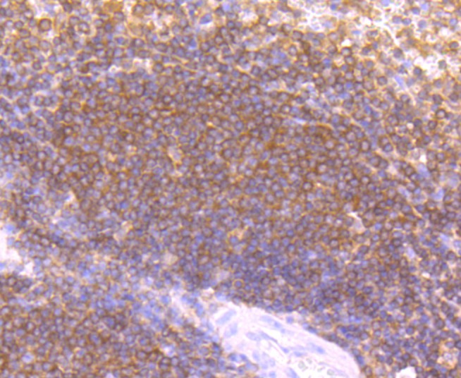 Paraformaldehyde-fixed, paraffin embedded (human spleen tissue); Antigen retrieval by boiling in sodium citrate buffer (pH6.0) for 15min; Block endogenous peroxidase by 3% hydrogen peroxide for 20 minutes; Blocking buffer (normal goat serum) at 37°C for 30min; Antibody incubation with (LYVE1) Monoclonal Antibody, Unconjugated (SLM-52811R) at 1:50 overnight at 4°C, followed by operating according to SP Kit(Rabbit) (sp-0023) instructionsand DAB staining.
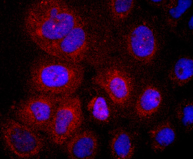 A431 cell; 4% Paraformaldehyde-fixed; Triton X-100 at room temperature for 20 min; Blocking buffer (normal goat serum,C-0005) at 37°C for 20 min; Antibody incubation with (LYVE1) monoclonal Antibody, Unconjugated (SLM-52811R) 1:50, 90 minutes at 37°C; followed by a conjugated Goat Anti-Rabbit IgG antibody at 37°C for 90 minutes, DAPI (blue, C02-04002) was used to stain the cell nuclei.
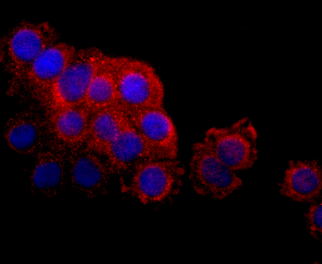 SW480 cell; 4% Paraformaldehyde-fixed; Triton X-100 at room temperature for 20 min; Blocking buffer (normal goat serum,C-0005) at 37°C for 20 min; Antibody incubation with (LYVE1) monoclonal Antibody, Unconjugated (SLM-52811R) 1:50, 90 minutes at 37°C; followed by a conjugated Goat Anti-Rabbit IgG antibody at 37°C for 90 minutes, DAPI (blue, C02-04002) was used to stain the cell nuclei.
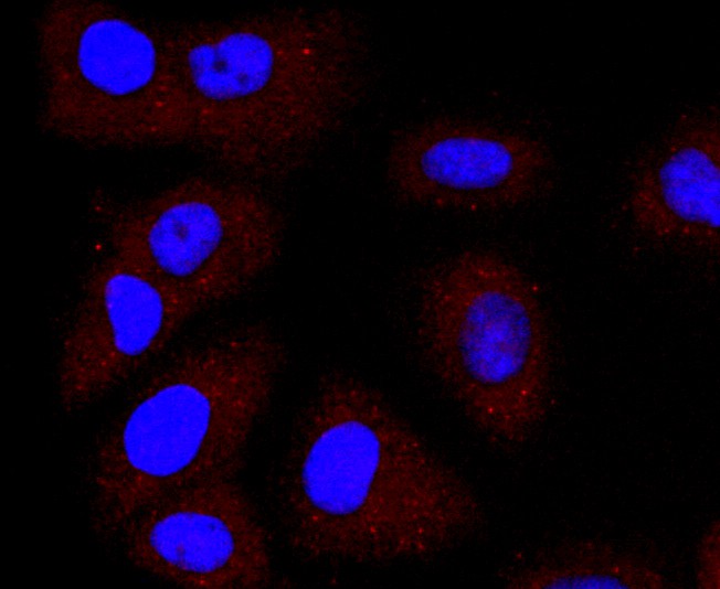 HUVEC cell; 4% Paraformaldehyde-fixed; Triton X-100 at room temperature for 20 min; Blocking buffer (normal goat serum,C-0005) at 37°C for 20 min; Antibody incubation with (LYVE1) monoclonal Antibody, Unconjugated (SLM-52811R) 1:50, 90 minutes at 37°C; followed by a conjugated Goat Anti-Rabbit IgG antibody at 37°C for 90 minutes, DAPI (blue, C02-04002) was used to stain the cell nuclei.
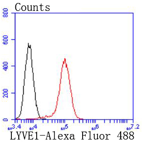 Blank control:Hela.
Primary Antibody (green line): Rabbit Anti-LYVE1 antibody (SLM-52811R)
Dilution: 1:50;
Isotype Control Antibody (orange line): Rabbit IgG .
Secondary Antibody : Goat anti-rabbit IgG-AF488
Dilution: 1:1000.
Protocol
The cells were fixed with 4% PFA (10min at room temperature)and then permeabilized with 0.1% PBST for 20 min at room temperature.The cells were then incubated in 5%BSA to block non-specific protein-protein interactions for 30 min at room temperature .Cells stained with Primary Antibody for 30 min at room temperature. The secondary antibody used for 40 min at room temperature. Acquisition of 20,000 events was performed.
Cartpieces
Totalgoods,subtotals:¥Checkout
References (0)
No References
Bought notes(bought amounts latest0)
No one bought this product
User Comment(Total0User Comment Num)
- No comment


 +86 571 56623320
 +86 18668110335

