
Rabbit Anti-Tissue factor antibody
CD 142; CD-142; CD142; CD142 antigen; Coagulation factor III (thromboplastin tissue factor); Coagulation factor III; F3; TF; TFA; Thromboplastin; Tissue factor; TF_HUMAN.
View History [Clear]
Details
Product Name Tissue factor Chinese Name 组织因子(CD142)兔单克隆抗体 Alias CD 142; CD-142; CD142; CD142 antigen; Coagulation factor III (thromboplastin tissue factor); Coagulation factor III; F3; TF; TFA; Thromboplastin; Tissue factor; TF_HUMAN. literatures Research Area Cardiovascular Cell biology immunology Growth factors and hormones Cell Surface Molecule Immunogen Species Rabbit Clonality Monoclonal Clone NO. 6A4 React Species Human, Mouse, Rat, Applications WB=1:500-2000 IHC-P=1:100-500 ICC=1:50 IF=1:100-500 (Paraffin sections need antigen repair)
not yet tested in other applications.
optimal dilutions/concentrations should be determined by the end user.Theoretical molecular weight 35kDa Cellular localization The cell membrane Form Liquid Concentration 1mg/ml immunogen KLH conjugated synthetic peptide derived from human Tissue factor: 1-100/295 Lsotype IgG Purification affinity purified by Protein A Buffer Solution 0.01M TBS(pH7.4) with 1% BSA, 0.03% Proclin300 and 50% Glycerol. Storage Shipped at 4℃. Store at -20 °C for one year. Avoid repeated freeze/thaw cycles. Attention This product as supplied is intended for research use only, not for use in human, therapeutic or diagnostic applications. PubMed PubMed Product Detail This gene encodes coagulation factor III which is a cell surface glycoprotein. This factor enables cells to initiate the blood coagulation cascades, and it functions as the high-affinity receptor for the coagulation factor VII. The resulting complex provides a catalytic event that is responsible for initiation of the coagulation protease cascades by specific limited proteolysis. Unlike the other cofactors of these protease cascades, which circulate as nonfunctional precursors, this factor is a potent initiator that is fully functional when expressed on cell surfaces. There are 3 distinct domains of this factor: extracellular, transmembrane, and cytoplasmic. This protein is the only one in the coagulation pathway for which a congenital deficiency has not been described. Alternate splicing results in multiple transcript variants.[provided by RefSeq, May 2010]
Function:
Initiates blood coagulation by forming a complex with circulating factor VII or VIIa. The [TF:VIIa] complex activates factors IX or X by specific limited protolysis. TF plays a role in normal hemostasis by initiating the cell-surface assembly and propagation of the coagulation protease cascade.
Subunit:
Interacts with HSPE; the interaction, inhibited by heparin, promotes the generation of activated factor X and activates coagulation in the presence of activated factor VII.
Subcellular Location:
Isoform 1: Membrane; Single-pass type I membrane protein.
Isoform 2: Secreted.
Tissue Specificity:
Lung, placenta and pancreas.
Similarity:
Belongs to the tissue factor family.
SWISS:
P13726
Gene ID:
2152
Database links:Entrez Gene: 2152 Human
Entrez Gene: 14066 Mouse
Omim: 134390 Human
SwissProt: P13726 Human
SwissProt: P20352 Mouse
Unigene: 62192 Human
Unigene: 273188 Mouse
Unigene: 9980 Rat
Product Picture 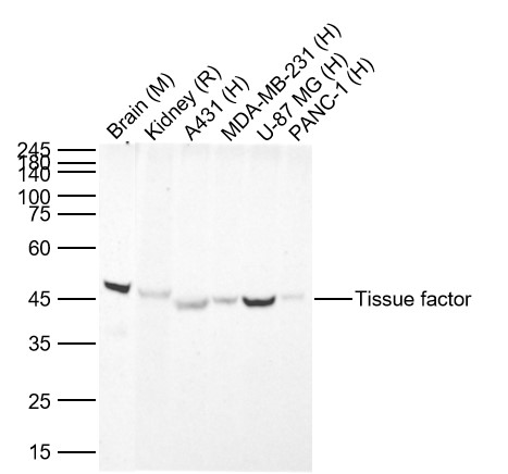 Sample:
Lane 1: Mouse Brain Lysates
Lane 2: Rat Kidney Lysates
Lane 3: Human A431 cell Lysates
Lane 4: Human MDA-MB-231 cell Lysates
Lane 5: Human U-87 MG cell Lysates
Lane 6: Human PANC-1 cell Lysates
Primary: Anti-Tissue factor (SLM-52647R) at 1/1000 dilution
Secondary: IRDye800CW Goat Anti-Rabbit IgG at 1/20000 dilution
Predicted band size: 35kDa
Observed band size: 45kDa
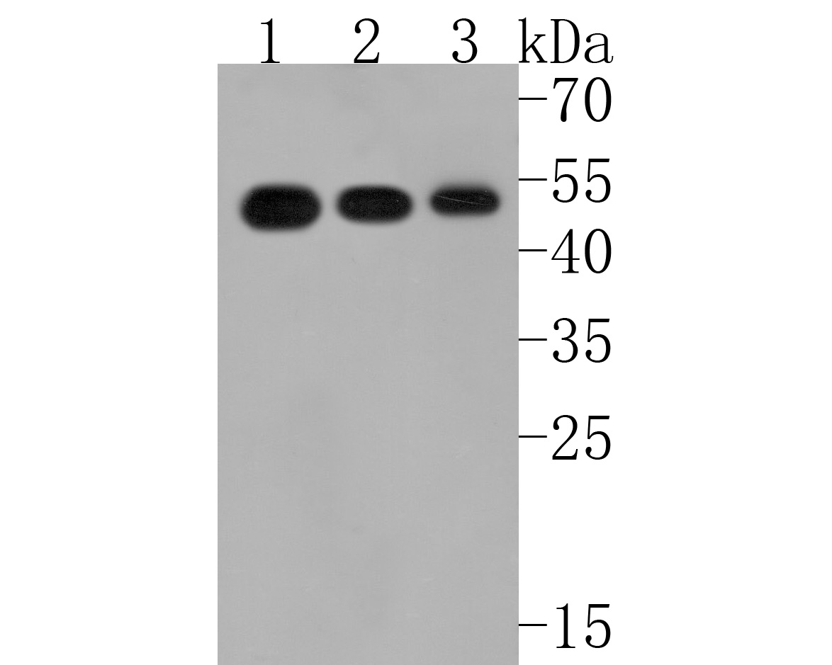 Sample:
Lane 1: mouse brain tissue lysate
Lane 2: U937 cell lysate
Lane 3: SH-SY5Y cell lysate
Primary: Anti-Tissue factor (SLM-52647R) at 1:500 dilution
Secondary: Goat Anti-Rabbit IgG - HRP at 1:5000 dilution
Predicted band size: 35 kD
Observed band size: 50 kD
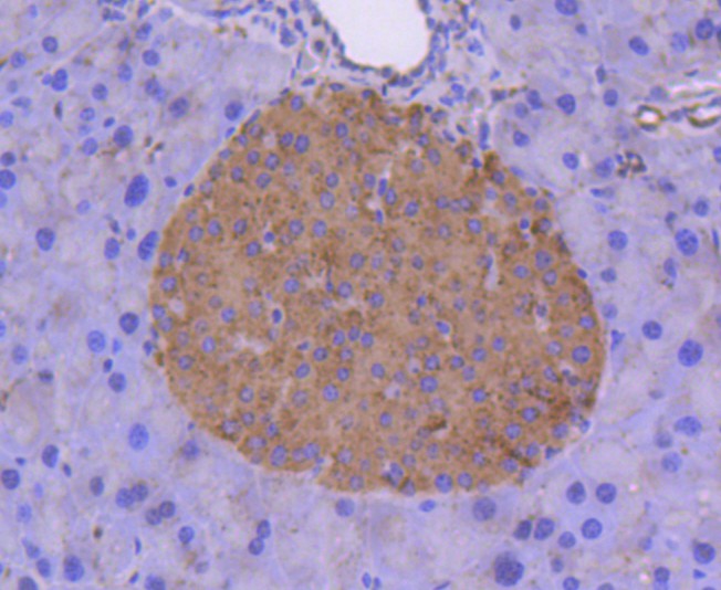 Paraformaldehyde-fixed, paraffin embedded (mouse pancreas); Antigen retrieval by boiling in sodium citrate buffer (pH6.0) for 15min; Block endogenous peroxidase by 3% hydrogen peroxide for 20 minutes; Blocking buffer (normal goat serum) at 37°C for 30min; Antibody incubation with (Tissue factor) Monoclonal Antibody, Unconjugated (SLM-52647R) at 1:50 overnight at 4°C, followed by operating according to SP Kit(Rabbit) (sp-0023) instructionsand DAB staining.
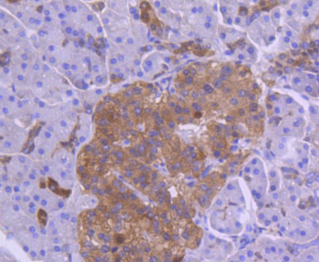 Paraformaldehyde-fixed, paraffin embedded (human pancreas tissue); Antigen retrieval by boiling in sodium citrate buffer (pH6.0) for 15min; Block endogenous peroxidase by 3% hydrogen peroxide for 20 minutes; Blocking buffer (normal goat serum) at 37°C for 30min; Antibody incubation with (Tissue factor) Monoclonal Antibody, Unconjugated (SLM-52647R) at 1:50 overnight at 4°C, followed by operating according to SP Kit(Rabbit) (sp-0023) instructionsand DAB staining.
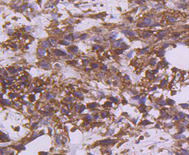 Paraformaldehyde-fixed, paraffin embedded (human breast carcinoma); Antigen retrieval by boiling in sodium citrate buffer (pH6.0) for 15min; Block endogenous peroxidase by 3% hydrogen peroxide for 20 minutes; Blocking buffer (normal goat serum) at 37°C for 30min; Antibody incubation with (Tissue factor) Monoclonal Antibody, Unconjugated (SLM-52647R) at 1:50 overnight at 4°C, followed by operating according to SP Kit(Rabbit) (sp-0023) instructionsand DAB staining.
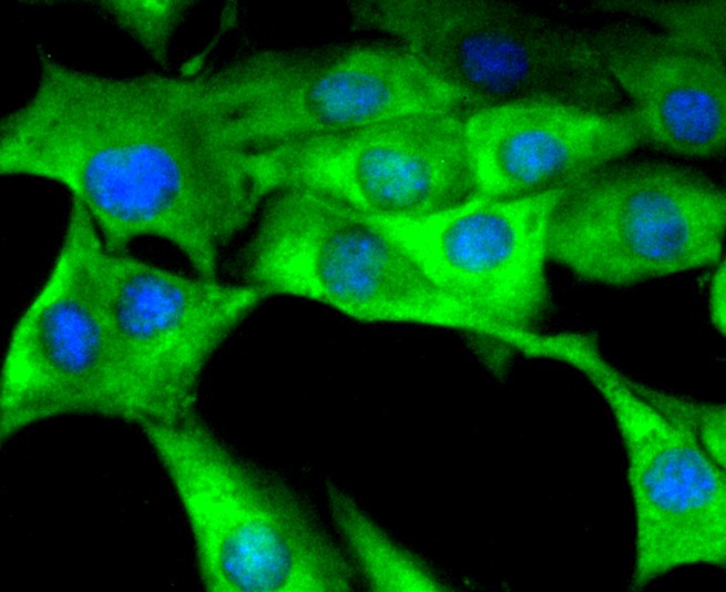 SHG-44 Hela cell; 4% Paraformaldehyde-fixed; Triton X-100 at room temperature for 20 min; Blocking buffer (normal goat serum,C-0005) at 37°C for 20 min; Antibody incubation with (Tissue Factor) monoclonal Antibody, Unconjugated (SLM-52647R) 1:50, 90 minutes at 37°C; followed by a conjugated Goat Anti-Rabbit IgG antibody at 37°C for 90 minutes, DAPI (blue, C02-04002) was used to stain the cell nuclei.
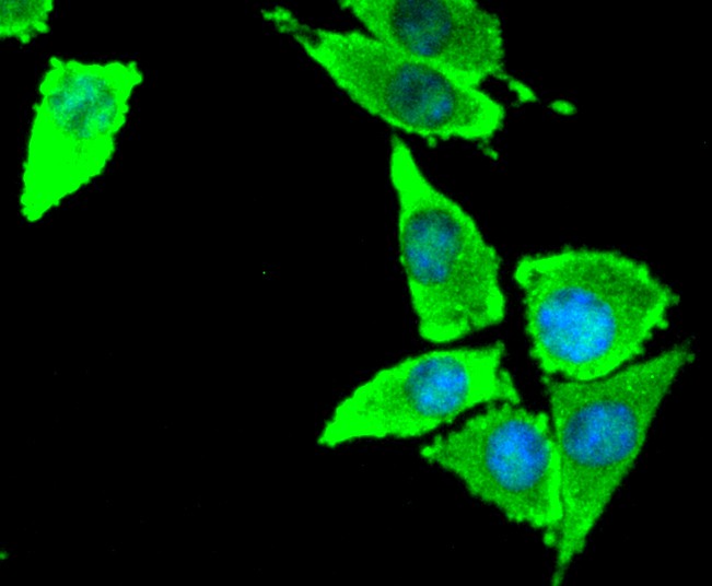 SH-SY5Y cell; 4% Paraformaldehyde-fixed; Triton X-100 at room temperature for 20 min; Blocking buffer (normal goat serum,C-0005) at 37°C for 20 min; Antibody incubation with (Tissue Factor) monoclonal Antibody, Unconjugated (SLM-52647R) 1:50, 90 minutes at 37°C; followed by a conjugated Goat Anti-Rabbit IgG antibody at 37°C for 90 minutes, DAPI (blue, C02-04002) was used to stain the cell nuclei.
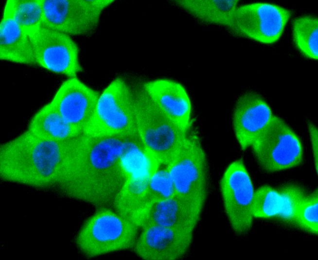 PANC-1 Hela cell; 4% Paraformaldehyde-fixed; Triton X-100 at room temperature for 20 min; Blocking buffer (normal goat serum,C-0005) at 37°C for 20 min; Antibody incubation with (Tissue Factor) monoclonal Antibody, Unconjugated (SLM-52647R) 1:50, 90 minutes at 37°C; followed by a conjugated Goat Anti-Rabbit IgG antibody at 37°C for 90 minutes, DAPI (blue, C02-04002) was used to stain the cell nuclei.
Cartpieces
Totalgoods,subtotals:¥Checkout
References (0)
No References
Bought notes(bought amounts latest0)
No one bought this product
User Comment(Total0User Comment Num)
- No comment


 +86 571 56623320
 +86 18668110335

