
Rabbit Anti-Calnexin antibody
Calnexin-ER membrane marker; CANX; Cell adhesion molecule with homology to l1cam precursor; CNX; D11Ertd153e; 1110069N15Rik; AI988026; FLJ26570; IP90; P90; Similar to calnexin; Major histocompatibility complex class I antigen-binding protein p88; CALX_HUM
View History [Clear]
Details
Product Name Calnexin Chinese Name 钙连蛋白Recombinant rabbit monoclonal anti Alias Calnexin-ER membrane marker; CANX; Cell adhesion molecule with homology to l1cam precursor; CNX; D11Ertd153e; 1110069N15Rik; AI988026; FLJ26570; IP90; P90; Similar to calnexin; Major histocompatibility complex class I antigen-binding protein p88; CALX_HUMAN. literatures Research Area immunology Chromatin and nuclear signals Binding protein Immunogen Species Rabbit Clonality Monoclonal Clone NO. 24G6 React Species Human, Rat, Applications WB=1:500-2000 IHC-P=1:50-200 Flow-Cyt=1:50 ICC=1:50 IF=1:100-500 (Paraffin sections need antigen repair)
not yet tested in other applications.
optimal dilutions/concentrations should be determined by the end user.Theoretical molecular weight 65kDa Cellular localization cytoplasmic The cell membrane Form Liquid Concentration 1mg/ml immunogen KLH conjugated synthetic peptide derived from human Calnexin Lsotype IgG Purification affinity purified by Protein A Buffer Solution 0.01M TBS(pH7.4) with 1% BSA, 0.03% Proclin300 and 50% Glycerol. Storage Shipped at 4℃. Store at -20℃ for one year. Avoid repeated freeze/thaw cycles. Attention This product as supplied is intended for research use only, not for use in human, therapeutic or diagnostic applications. PubMed PubMed Product Detail This gene encodes a member of the calnexin family of molecular chaperones. The encoded protein is a calcium-binding, endoplasmic reticulum (ER)-associated protein that interacts transiently with newly synthesized N-linked glycoproteins, facilitating protein folding and assembly. It may also play a central role in the quality control of protein folding by retaining incorrectly folded protein subunits within the ER for degradation. Alternatively spliced transcript variants encoding the same protein have been described. [provided by RefSeq]
Function:
Calcium-binding protein that interacts with newly synthesized glycoproteins in the endoplasmic reticulum. It may act in assisting protein assembly and/or in the retention within the ER of unassembled protein subunits. It seems to play a major role in the quality control apparatus of the ER by the retention of incorrectly folded proteins.
Subunit:
Interacts with MAPK3/ERK1. Interacts with KCNH2. Associates with ribosomes. Interacts with SGIP1; involved in negative regulation of endocytosis (By similarity). The palmitoylated form interacts with the ribosome-translocon complex component SSR1, promoting efficient folding of glycoproteins.
Subcellular Location:
Endoplasmic reticulum membrane. Melanosome. Identified by mass spectrometry in melanosome fractions from stage I to stage IV.
Post-translational modifications:
Phosphorylated at Ser-564 by MAPK3/ERK1. phosphorylation by MAPK3/ERK1 increases its association with ribosomes (By similarity). br>Palmitoylation by DHHC6 leads to the preferential localization to the perinuclear rough ER. It mediates the association of calnexin with the ribosome-translocon complex (RTC) which is required for efficient folding of glycosylated proteins.
Similarity:
Belongs to the calreticulin family.
SWISS:
P27824
Gene ID:
821
Database links:Entrez Gene: 821 Human
Entrez Gene: 12330 Mouse
Omim: 114217 Human
SwissProt: P27824 Human
SwissProt: P35564 Mouse
Unigene: 567968 Human
Unigene: 248827 Mouse
Unigene: 1762 Rat
Product Picture  Sample:
Lane 1: Hela cell lysate
Primary: Anti-Calnexin (SLM-52639R) at 1:500 dilution
Secondary: Goat Anti-Rabbit IgG - HRP at 1:5000 dilution
Predicted band size: 65 kD
Observed band size: 100 kD
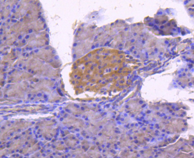 Paraformaldehyde-fixed, paraffin embedded (rat pancreas); Antigen retrieval by boiling in sodium citrate buffer (pH6.0) for 15min; Block endogenous peroxidase by 3% hydrogen peroxide for 20 minutes; Blocking buffer (normal goat serum) at 37°C for 30min; Antibody incubation with (Calnexin) Monoclonal Antibody, Unconjugated (SLM-52639R) at 1:50 overnight at 4°C, followed by operating according to SP Kit(Rabbit) (sp-0023) instructionsand DAB staining.
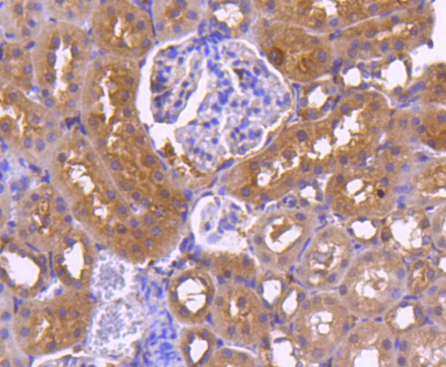 Paraformaldehyde-fixed, paraffin embedded (rat kidney); Antigen retrieval by boiling in sodium citrate buffer (pH6.0) for 15min; Block endogenous peroxidase by 3% hydrogen peroxide for 20 minutes; Blocking buffer (normal goat serum) at 37°C for 30min; Antibody incubation with (Calnexin) Monoclonal Antibody, Unconjugated (SLM-52639R) at 1:50 overnight at 4°C, followed by operating according to SP Kit(Rabbit) (sp-0023) instructionsand DAB staining.
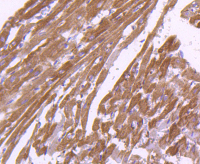 Paraformaldehyde-fixed, paraffin embedded (rat heart tissue); Antigen retrieval by boiling in sodium citrate buffer (pH6.0) for 15min; Block endogenous peroxidase by 3% hydrogen peroxide for 20 minutes; Blocking buffer (normal goat serum) at 37°C for 30min; Antibody incubation with (Calnexin) Monoclonal Antibody, Unconjugated (SLM-52639R) at 1:50 overnight at 4°C, followed by operating according to SP Kit(Rabbit) (sp-0023) instructionsand DAB staining.
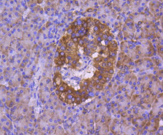 Paraformaldehyde-fixed, paraffin embedded (human pancreas tissue); Antigen retrieval by boiling in sodium citrate buffer (pH6.0) for 15min; Block endogenous peroxidase by 3% hydrogen peroxide for 20 minutes; Blocking buffer (normal goat serum) at 37°C for 30min; Antibody incubation with (Calnexin) Monoclonal Antibody, Unconjugated (SLM-52639R) at 1:50 overnight at 4°C, followed by operating according to SP Kit(Rabbit) (sp-0023) instructionsand DAB staining.
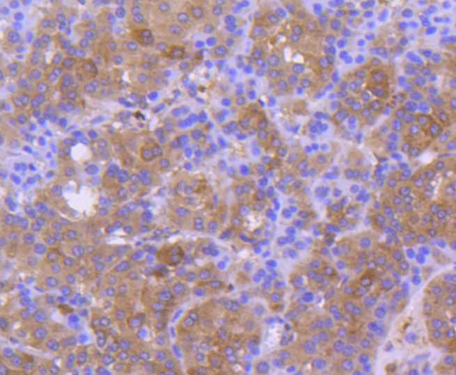 Paraformaldehyde-fixed, paraffin embedded (human liver carcinoma); Antigen retrieval by boiling in sodium citrate buffer (pH6.0) for 15min; Block endogenous peroxidase by 3% hydrogen peroxide for 20 minutes; Blocking buffer (normal goat serum) at 37°C for 30min; Antibody incubation with (Calnexin) Monoclonal Antibody, Unconjugated (SLM-52639R) at 1:50 overnight at 4°C, followed by operating according to SP Kit(Rabbit) (sp-0023) instructionsand DAB staining.
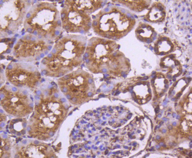 Paraformaldehyde-fixed, paraffin embedded (human kidney tissue); Antigen retrieval by boiling in sodium citrate buffer (pH6.0) for 15min; Block endogenous peroxidase by 3% hydrogen peroxide for 20 minutes; Blocking buffer (normal goat serum) at 37°C for 30min; Antibody incubation with (Calnexin) Monoclonal Antibody, Unconjugated (SLM-52639R) at 1:50 overnight at 4°C, followed by operating according to SP Kit(Rabbit) (sp-0023) instructionsand DAB staining.
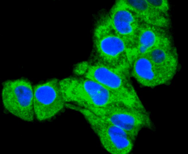 Hela cell; 4% Paraformaldehyde-fixed; Triton X-100 at room temperature for 20 min; Blocking buffer (normal goat serum,C-0005) at 37°C for 20 min; Antibody incubation with (Calnexin) monoclonal Antibody, Unconjugated (SLM-52639R) 1:50, 90 minutes at 37°C; followed by a conjugated Goat Anti-Rabbit IgG antibody at 37°C for 90 minutes, DAPI (blue, C02-04002) was used to stain the cell nuclei.
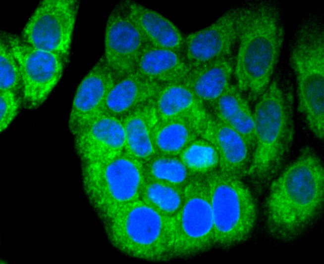 HepG2 cell; 4% Paraformaldehyde-fixed; Triton X-100 at room temperature for 20 min; Blocking buffer (normal goat serum,C-0005) at 37°C for 20 min; Antibody incubation with (Calnexin) monoclonal Antibody, Unconjugated (SLM-52639R) 1:50, 90 minutes at 37°C; followed by a conjugated Goat Anti-Rabbit IgG antibody at 37°C for 90 minutes, DAPI (blue, C02-04002) was used to stain the cell nuclei.
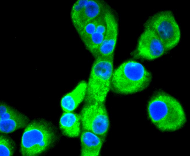 PANC-1 cell; 4% Paraformaldehyde-fixed; Triton X-100 at room temperature for 20 min; Blocking buffer (normal goat serum,C-0005) at 37°C for 20 min; Antibody incubation with (Calnexin) monoclonal Antibody, Unconjugated (SLM-52639R) 1:50, 90 minutes at 37°C; followed by a conjugated Goat Anti-Rabbit IgG antibody at 37°C for 90 minutes, DAPI (blue, C02-04002) was used to stain the cell nuclei.
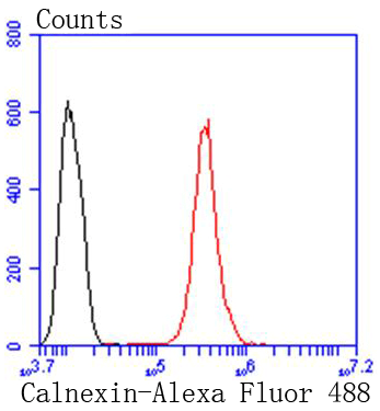 Blank control:Hela.
Primary Antibody (green line): Rabbit Anti-Calnexin antibody (SLM-52639R)
Dilution: 1:50;
Isotype Control Antibody (orange line): Rabbit IgG .
Secondary Antibody : Goat anti-rabbit IgG-AF488
Dilution: 1:1000.
Protocol
The cells were fixed with 4% PFA (10min at room temperature)and then permeabilized with 0.1% PBST for 20 min at room temperature.The cells were then incubated in 5%BSA to block non-specific protein-protein interactions for 30 min at room temperature .Cells stained with Primary Antibody for 30 min at room temperature. The secondary antibody used for 40 min at room temperature. Acquisition of 20,000 events was performed.
Cartpieces
Totalgoods,subtotals:¥Checkout
References (0)
No References
Bought notes(bought amounts latest0)
No one bought this product
User Comment(Total0User Comment Num)
- No comment


 +86 571 56623320
 +86 18668110335

