
Rabbit Anti-SGK1 antibody
Serum/glucocorticoid regulated kinase 1; SGK 1; SGK-1; Serine/threonine protein kinase SGK; Serine/threonine protein kinase Sgk1; Serum and glucocorticoid regulated kinase; Serum/glucocorticoid regulated kinase; SGK 1; SGK; SGK1_HUMAN.
View History [Clear]
Details
Product Name SGK1 Chinese Name 糖皮质激素调节激酶1Recombinant rabbit monoclonal anti Alias Serum/glucocorticoid regulated kinase 1; SGK 1; SGK-1; Serine/threonine protein kinase SGK; Serine/threonine protein kinase Sgk1; Serum and glucocorticoid regulated kinase; Serum/glucocorticoid regulated kinase; SGK 1; SGK; SGK1_HUMAN. Research Area Tumour Cell biology immunology Neurobiology Signal transduction Growth factors and hormones transcriptional regulatory factor Kinases and Phosphatases Immunogen Species Rabbit Clonality Monoclonal Clone NO. 6D1 React Species (predicted: Human, Mouse, Rat, ) Applications WB=1:500-1000 IHC-P=1:100-500 (Paraffin sections need antigen repair)
not yet tested in other applications.
optimal dilutions/concentrations should be determined by the end user.Theoretical molecular weight 55kDa Cellular localization The nucleus cytoplasmic The cell membrane Form Liquid Concentration 1mg/ml immunogen KLH conjugated synthetic peptide derived from human SGK1 Lsotype IgG Purification affinity purified by Protein A Buffer Solution 0.01M TBS(pH7.4) with 1% BSA, 0.03% Proclin300 and 50% Glycerol. Storage Shipped at 4℃. Store at -20 °C for one year. Avoid repeated freeze/thaw cycles. Attention This product as supplied is intended for research use only, not for use in human, therapeutic or diagnostic applications. PubMed PubMed Product Detail SGK1 is a protein kinase that plays an important role in cellular stress response. SGK1 activates certain potassium, sodium, and chloride channels, suggesting an involvement in the regulation of processes such as cell survival, neuronal excitability, and renal sodium excretion. Sustained high levels of SGK1 and activity may contribute to conditions such as hypertension and diabetic nephropathy. This protein also mediates cell survival signals, as it has been shown to phosphorylate and negatively regulate the pro apoptotic FOXO3A protein. Ser 422 is a critical site on the protein and may be involved in its activation.
Function:
Protein kinase that plays an important role in cellular stress response. Activates certain potassium, sodium, and chloride channels, suggesting an involvement in the regulation of processes such as cell survival, neuronal excitability and renal sodium excretion. Sustained high levels and activity may contribute to conditions such as hypertension and diabetic nephropathy. Mediates cell survival signals, phosphorylates and negatively regulates pro-apoptotic FOXO3A. Phosphorylates NEDD4L, which leads to its inactivation and to the subsequent activation of various channels and transporters such as ENaC, KCNA3/Kv1.3 or EAAT1. Isoform 2 exhibited a greater effect on cell plasma membrane expression of ENaC and Na(+) transport than isoform 1. Subunit : Homodimer; disulfide-linked. Forms a trimeric complex with FBXW7 and NOTCH1. Interacts with MAPK3/ERK1, MAPK1/ERK2, MAP2K1/MEK1, MAP2K2/MEK2, NEDD4, NEDD4L, MAPT/TAU, MAPK7, CREB1, SLC9A3R2/NHERF2 and KCNJ1/ROMK1. Associates with the mammalian target of rapamycin complex 2 (mTORC2) via an interaction with MAPKAP1/SIN1.
Subcellular Location:
Cell membrane and Cytoplasm. Nucleus. Endoplasmic reticulum. Nuclear, upon phosphorylation.
Tissue Specificity:
Expressed in most tissues with highest levels in the pancreas, followed by placenta, kidney and lung. Isoform 2 is strongly expressed in brain and pancreas, weaker in heart, placenta, lung, liver and skeletal muscle.
Post-translational modifications:
Regulated by phosphorylation. Phosphoinositide 3-kinase (PI3-kinase) pathway promotes phosphorylation at Ser-422 which in turn increases the phosphorylation of Thr-256 by PDPK1.
Similarity:
Belongs to the protein kinase superfamily. AGC Ser/Thr protein kinase family.
Contains 1 AGC-kinase C-terminal domain.
Contains 1 protein kinase domain.
SWISS:
O00141
Gene ID:
6446
Database links:Entrez Gene: 6446 Human
Entrez Gene: 20393 Mouse
Omim: 602958 Human
SwissProt: O00141 Human
SwissProt: Q9WVC6 Mouse
Unigene: 510078 Human
Unigene: 28405 Mouse
Unigene: 4636 Rat
Product Picture 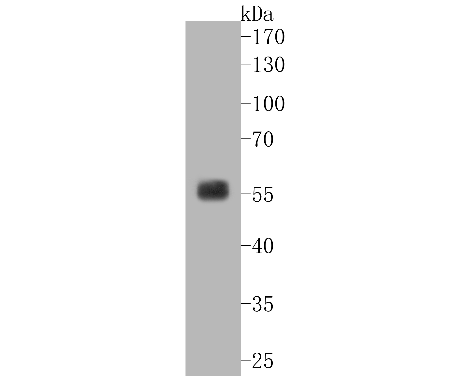 Western blot analysis of SGK1 on human kidney tissue lysates. Proteins were transferred to a PVDF membrane and blocked with 5% BSA in PBS for 1 hour at room temperature. The primary antibody (SLM-52499R, 1/500) was used in 5% BSA at room temperature for 2 hours. Goat Anti-Rabbit IgG - HRP Secondary Antibody (HA1001) at 1:5,000 dilution was used for 1 hour at room temperature.
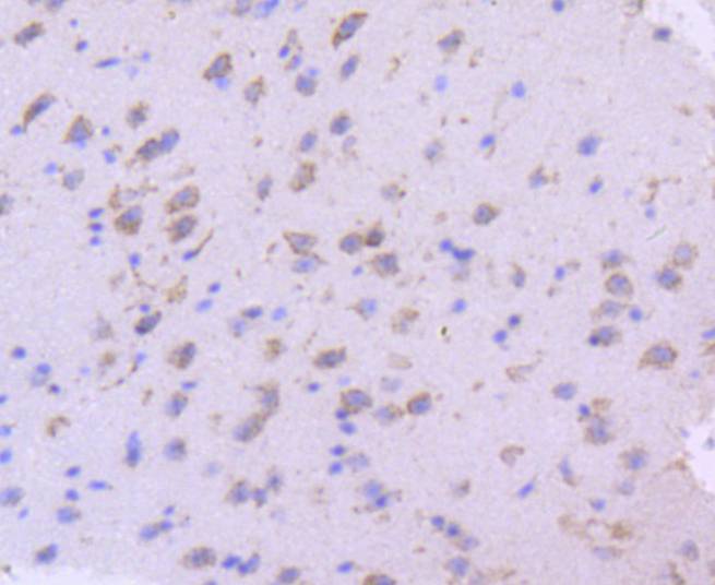 Immunohistochemical analysis of paraffin-embedded mouse brain tissue using anti-SGK1 antibody. The section was pre-treated using heat mediated antigen retrieval with Tris-EDTA buffer (pH 8.0-8.4) for 20 minutes.The tissues were blocked in 5% BSA for 30 minutes at room temperature, washed with ddH2O and PBS, and then probed with the primary antibody (SLM-52499R, 1/50) for 30 minutes at room temperature. The detection was performed using an HRP conjugated compact polymer system. DAB was used as the chromogen. Tissues were counterstained with hematoxylin and mounted with DPX.
 Immunohistochemical analysis of paraffin-embedded mouse liver tissue using anti-SGK1 antibody. The section was pre-treated using heat mediated antigen retrieval with Tris-EDTA buffer (pH 8.0-8.4) for 20 minutes.The tissues were blocked in 5% BSA for 30 minutes at room temperature, washed with ddH2O and PBS, and then probed with the primary antibody (SLM-52499R, 1/50) for 30 minutes at room temperature. The detection was performed using an HRP conjugated compact polymer system. DAB was used as the chromogen. Tissues were counterstained with hematoxylin and mounted with DPX.
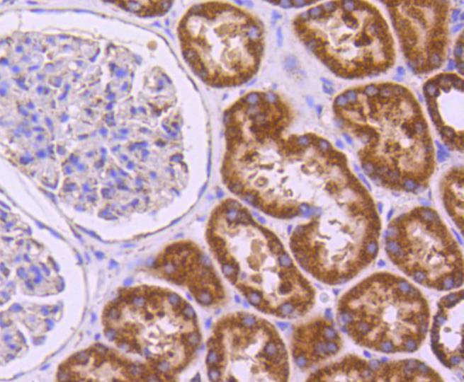 Immunohistochemical analysis of paraffin-embedded human kidney tissue using anti-SGK1 antibody. The section was pre-treated using heat mediated antigen retrieval with Tris-EDTA buffer (pH 8.0-8.4) for 20 minutes.The tissues were blocked in 5% BSA for 30 minutes at room temperature, washed with ddH2O and PBS, and then probed with the primary antibody (SLM-52499R, 1/50) for 30 minutes at room temperature. The detection was performed using an HRP conjugated compact polymer system. DAB was used as the chromogen. Tissues were counterstained with hematoxylin and mounted with DPX.
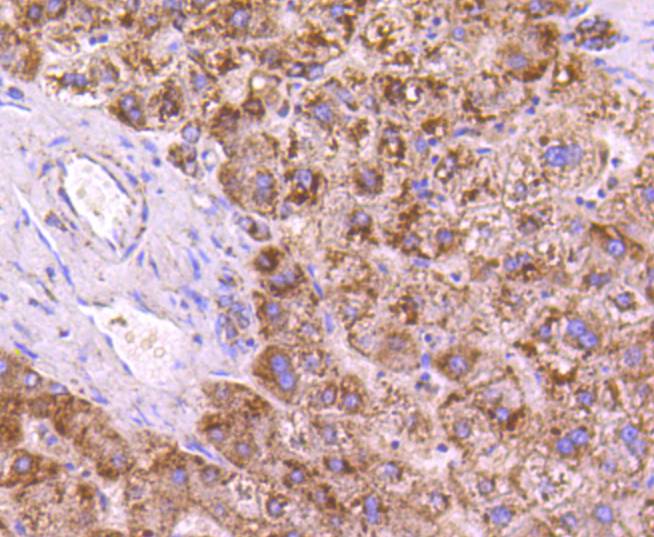 Immunohistochemical analysis of paraffin-embedded human liver tissue using anti-SGK1 antibody. The section was pre-treated using heat mediated antigen retrieval with Tris-EDTA buffer (pH 8.0-8.4) for 20 minutes.The tissues were blocked in 5% BSA for 30 minutes at room temperature, washed with ddH2O and PBS, and then probed with the primary antibody (SLM-52499R, 1/50) for 30 minutes at room temperature. The detection was performed using an HRP conjugated compact polymer system. DAB was used as the chromogen. Tissues were counterstained with hematoxylin and mounted with DPX.
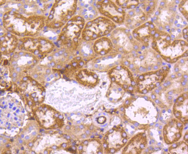 Immunohistochemical analysis of paraffin-embedded mouse kidney tissue using anti-SGK1 antibody. The section was pre-treated using heat mediated antigen retrieval with Tris-EDTA buffer (pH 8.0-8.4) for 20 minutes.The tissues were blocked in 5% BSA for 30 minutes at room temperature, washed with ddH2O and PBS, and then probed with the primary antibody (SLM-52499R, 1/50) for 30 minutes at room temperature. The detection was performed using an HRP conjugated compact polymer system. DAB was used as the chromogen. Tissues were counterstained with hematoxylin and mounted with DPX.
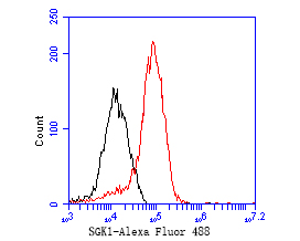 Flow cytometric analysis of SGK1 was done on 293 cells. The cells were fixed, permeabilized and stained with the primary antibody (SLM-52499R, 1/50) (red). After incubation of the primary antibody at room temperature for an hour, the cells were stained with a Alexa Fluor 488-conjugated Goat anti-Rabbit IgG Secondary antibody at 1/1000 dilution for 30 minutes.Unlabelled sample was used as a control (cells without incubation with primary antibody; black).
Cartpieces
Totalgoods,subtotals:¥Checkout
References (0)
No References
Bought notes(bought amounts latest0)
No one bought this product
User Comment(Total0User Comment Num)
- No comment


 +86 571 56623320
 +86 18668110335

