
Rabbit Anti-CD4 antibody
CD4 (L3T4); CD4 antigen (p55); CD4 Antigen ; CD4 molecule; CD4 Receptor; CD4+ Lymphocyte deficiency, included; CD4mut; L3T4; Leu3; Ly-4; Lymphocyte antigen CD4; MGC165891; p55; T Cell Antigen T4 ; T cell antigen T4/LEU3; T cell differentiation antigen L3T
View History [Clear]
Details
Product Name CD4 Chinese Name CD4Recombinant rabbit monoclonal anti Alias CD4 (L3T4); CD4 antigen (p55); CD4 Antigen ; CD4 molecule; CD4 Receptor; CD4+ Lymphocyte deficiency, included; CD4mut; L3T4; Leu3; Ly-4; Lymphocyte antigen CD4; MGC165891; p55; T Cell Antigen T4 ; T cell antigen T4/LEU3; T cell differentiation antigen L3T4; T cell OKT4 deficiency, included; T cell surface antigen T4/Leu 3 ; T cell surface antigen T4/Leu3; T Cell Surface Glycoprotein CD4; W3/25; W3/25 antigen; T-cell surface glycoprotein CD4 isoform 1 precursor; CD4_Human. literatures Research Area Cell biology immunology Stem cells Cell Surface Molecule lymphocyte t-lymphocyte Immunogen Species Rabbit Clonality Monoclonal Clone NO. 65H6 React Species Human, Applications WB=1:500-2000 IHC-P=1:100-500 IHC-F=1:50-200 Flow-Cyt=1ug/Test ICC=1:50-200 IF=1:50-200 (Paraffin sections need antigen repair)
not yet tested in other applications.
optimal dilutions/concentrations should be determined by the end user.Theoretical molecular weight 48kDa Cellular localization The cell membrane Form Liquid Concentration 1mg/ml immunogen KLH conjugated synthetic peptide derived from human CD4 Lsotype IgG Purification affinity purified by Protein A Buffer Solution 0.01M TBS(pH7.4) with 1% BSA, 0.03% Proclin300 and 50% Glycerol. Storage Shipped at 4℃. Store at -20 °C for one year. Avoid repeated freeze/thaw cycles. Attention This product as supplied is intended for research use only, not for use in human, therapeutic or diagnostic applications. PubMed PubMed Product Detail This gene encodes a membrane glycoprotein of T lymphocytes that interacts with major histocompatibility complex class II antigenes and is also a receptor for the human immunodeficiency virus. This gene is expressed not only in T lymphocytes, but also in B cells, macrophages, and granulocytes. It is also expressed in specific regions of the brain. The protein functions to initiate or augment the early phase of T-cell activation, and may function as an important mediator of indirect neuronal damage in infectious and immune-mediated diseases of the central nervous system. Multiple alternatively spliced transcript variants encoding different isoforms have been identified in this gene. [provided by RefSeq, Aug 2010].
Function:
Accessory protein for MHC class-II antigen/T-cell receptor interaction. May regulate T-cell activation. Induces the aggregation of lipid rafts.
Subunit:
Associates with LCK. Binds to HIV-1 gp120 and to P4HB/PDI and upon HIV-1 binding to the cell membrane, is part of P4HB/PDI-CD4-CXCR4-gp120 complex. Interacts with HIV-1 Envelope polyprotein gp160 and protein Vpu. Interacts with Human Herpes virus 7 capsid proteins. Interacts with PTK2/FAK1; this interaction requires the presence of HIV-1 gp120.
Subcellular Location:
Cell membrane; Single-pass type I membrane protein. Note=Localizes to lipid rafts. Removed from plasma membrane by HIV-1 Nef protein that increases clathrin-dependent endocytosis of this antigen to target it to lysosomal degradation. Cell surface expression is also down-modulated by HIV-1 Envelope polyprotein gp160 that interacts with, and sequesters CD4 in the endoplasmic reticulum.
Post-translational modifications:
Palmitoylation and association with LCK contribute to the enrichment of CD4 in lipid rafts.
Similarity:
Contains 3 Ig-like C2-type (immunoglobulin-like) domains.
Contains 1 Ig-like V-type (immunoglobulin-like) domain.
SWISS:
P01730
Gene ID:
920
Database links:Entrez Gene: 920 Human
Entrez Gene: 12504 Mouse
Omim: 186940 Human
SwissProt: P06332 Mouse
SwissProt: P01730 Human
Unigene: 631659 Human
Unigene: 2209 Mouse
Product Picture 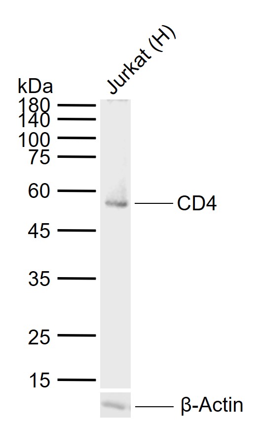 Sample:
Lane 1: Human Jurkat cell lysates
Primary: Anti-CD4 (SLM-52469R) at 1/1000 dilution
Secondary: IRDye800CW Goat Anti-Rabbit IgG at 1/20000 dilution
Predicted band size: 48 kDa
Observed band size: 56 kDa
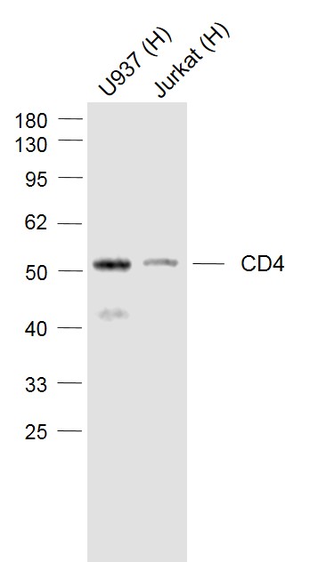 Sample:
Lane 1: U937 (Human) Cell Lysate at 30 ug
Lane 2: Jurkat (Human) Cell Lysate at 30 ug
Primary: Anti-CD4 (SLM-52469R) at 1/1000 dilution
Secondary: IRDye800CW Goat Anti-Rabbit IgG at 1/20000 dilution
Predicted band size: 55 kD
Observed band size: 52 kD
 Paraformaldehyde-fixed, paraffin embedded (human gastric carcinoma); Antigen retrieval by boiling in sodium citrate buffer (pH6.0) for 15min; Block endogenous peroxidase by 3% hydrogen peroxide for 20 minutes; Blocking buffer (normal goat serum) at 37°C for 30min; Antibody incubation with (CD4) Monoclonal Antibody, Unconjugated (SLM-52469R) at 1:200 overnight at 4°C, followed by operating according to SP Kit(Rabbit) (sp-0023) instructionsand DAB staining.
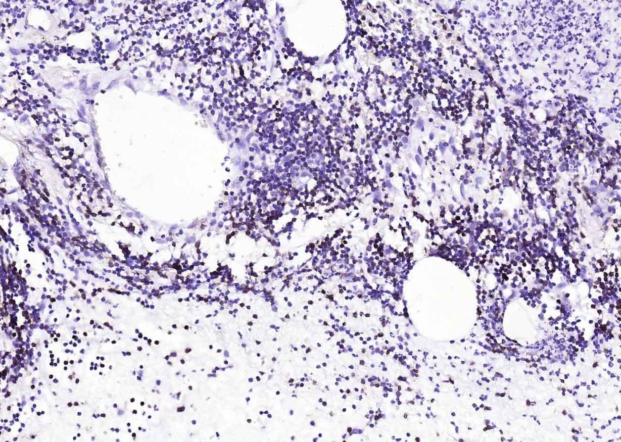 Paraformaldehyde-fixed, paraffin embedded (human appendix); Antigen retrieval by boiling in sodium citrate buffer (pH6.0) for 15min; Block endogenous peroxidase by 3% hydrogen peroxide for 20 minutes; Blocking buffer (normal goat serum) at 37°C for 30min; Antibody incubation with (CD4) Monoclonal Antibody, Unconjugated (SLM-52469R) at 1:200 overnight at 4°C, followed by operating according to SP Kit(Rabbit) (sp-0023) instructionsand DAB staining.
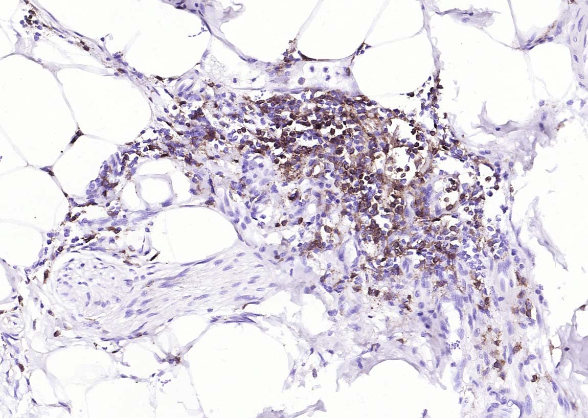 Paraformaldehyde-fixed, paraffin embedded (human colon carcinoma); Antigen retrieval by boiling in sodium citrate buffer (pH6.0) for 15min; Block endogenous peroxidase by 3% hydrogen peroxide for 20 minutes; Blocking buffer (normal goat serum) at 37°C for 30min; Antibody incubation with (CD4) Monoclonal Antibody, Unconjugated (SLM-52469R) at 1:200 overnight at 4°C, followed by operating according to SP Kit(Rabbit) (sp-0023) instructionsand DAB staining.
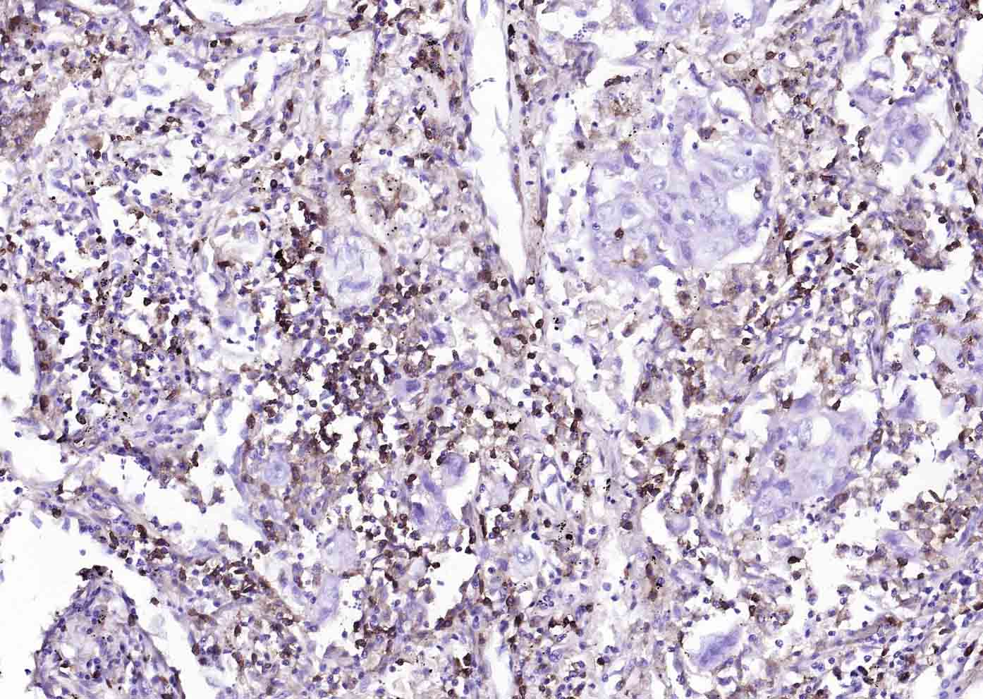 Paraformaldehyde-fixed, paraffin embedded (human lung carcinoma); Antigen retrieval by boiling in sodium citrate buffer (pH6.0) for 15min; Block endogenous peroxidase by 3% hydrogen peroxide for 20 minutes; Blocking buffer (normal goat serum) at 37°C for 30min; Antibody incubation with (CD4) Monoclonal Antibody, Unconjugated (SLM-52469R) at 1:200 overnight at 4°C, followed by operating according to SP Kit(Rabbit) (sp-0023) instructionsand DAB staining.
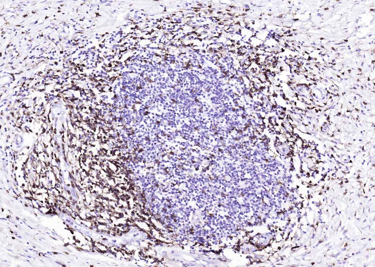
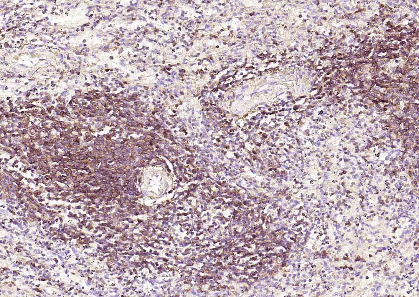 Paraformaldehyde-fixed, paraffin embedded (human spleen); Antigen retrieval by boiling in sodium citrate buffer (pH6.0) for 15min; Block endogenous peroxidase by 3% hydrogen peroxide for 20 minutes; Blocking buffer (normal goat serum) at 37°C for 30min; Antibody incubation with (CD4) Monoclonal Antibody, Unconjugated (SLM-52469R) at 1:200 overnight at 4°C, followed by operating according to SP Kit(Rabbit) (sp-0023)instructionsand DAB staining.
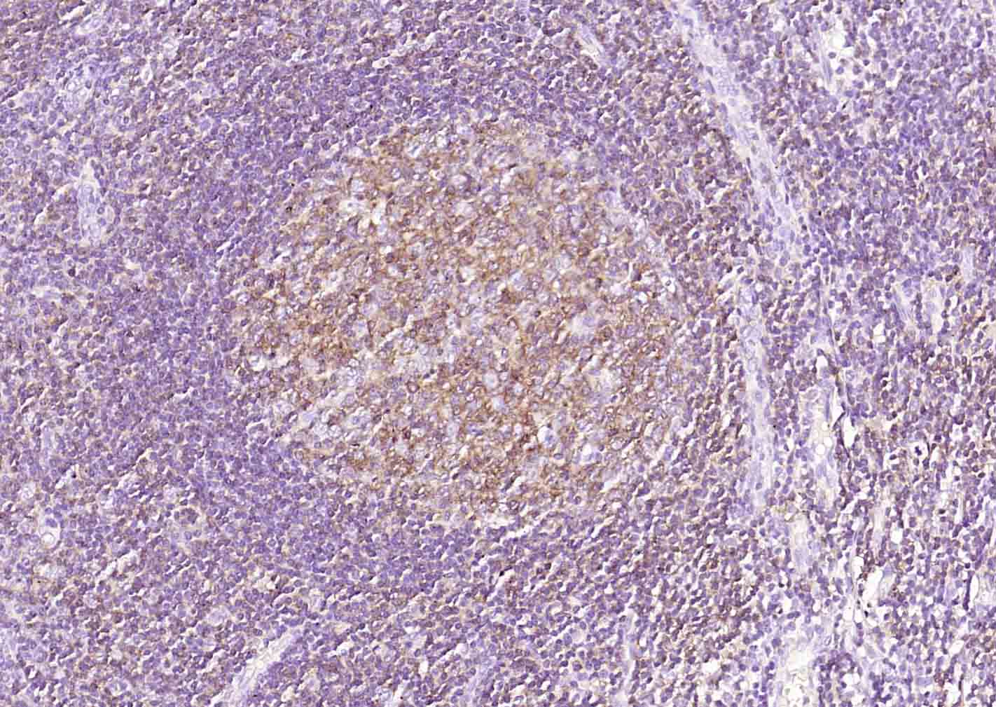 Paraformaldehyde-fixed, paraffin embedded (human tonsil); Antigen retrieval by boiling in sodium citrate buffer (pH6.0) for 15min; Block endogenous peroxidase by 3% hydrogen peroxide for 20 minutes; Blocking buffer (normal goat serum) at 37°C for 30min; Antibody incubation with (CD4) Monoclonal Antibody, Unconjugated (SLM-52469R) at 1:200 overnight at 4°C, followed by operating according to SP Kit(Rabbit) (sp-0023)instructionsand DAB staining.
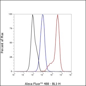 Cell line: Jurkat
Fixative: 4% Paraformaldehyde
Permeabilization: 90% methanol
Primary ab dilution: 1:100
Secondary ab: Goat anti Rabbit IgG
Unlabelled control: The cell without incubation
with primary antibody and secondary antibody(Black line).
Isotype control: Rabbit monoclonal IgG (Blue line).
Comment: Line red is the positive signal for SLM-52469R
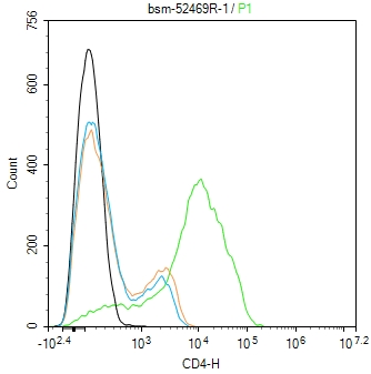 Blank control:Jurkat.
Primary Antibody (green line): Rabbit Anti-CD4 antibody (SLM-52469R)
Dilution: 1ug/Test;
Secondary Antibody (white blue line) : Goat anti-rabbit IgG-AF488
Dilution: 0.5ug/Test.
Isotype control(orange line):Normal Rabbit IgG
Protocol
The cells were incubated in 5%BSA to block non-specific protein-protein interactions for 30 min at room temperature .Cells stained with Primary Antibody for 30 min at room temperature. The secondary antibody used for 40 min at room temperature. Acquisition of 20,000 events was performed.
Cartpieces
Totalgoods,subtotals:¥Checkout
References (0)
No References
Bought notes(bought amounts latest0)
No one bought this product
User Comment(Total0User Comment Num)
- No comment


 +86 571 56623320
 +86 18668110335

