
Rabbit Anti-HDAC8 antibody
HD 8; HD8; HDAC 8; HDACL 1; HDACL1; Histone deacetylase 8; Histone deacetylase like 1; RPD 3; RPD3; CDA07; Hdac8; HDAC8_HUMAN.
View History [Clear]
Details
Product Name HDAC8 Chinese Name 组蛋白去乙酰化酶8Recombinant rabbit monoclonal anti Alias HD 8; HD8; HDAC 8; HDACL 1; HDACL1; Histone deacetylase 8; Histone deacetylase like 1; RPD 3; RPD3; CDA07; Hdac8; HDAC8_HUMAN. Research Area Tumour Developmental biology Signal transduction Apoptosis transcriptional regulatory factor Epigenetics Immunogen Species Rabbit Clonality Monoclonal Clone NO. 4C3 React Species Human, Mouse, Rat, Applications WB=1:500-2000 IHC-P=1:100-500 IHC-F=1:400-800 ICC=1:100-500 IF=1:100-500 (Paraffin sections need antigen repair)
not yet tested in other applications.
optimal dilutions/concentrations should be determined by the end user.Theoretical molecular weight 42kDa Cellular localization The nucleus cytoplasmic Form Liquid Concentration 1mg/ml immunogen Recombinant human HDAC8 protein (1-150aa) Lsotype IgG Purification affinity purified by Protein A Buffer Solution 0.01M TBS(pH7.4) with 1% BSA, 0.03% Proclin300 and 50% Glycerol. Storage Shipped at 4℃. Store at -20 °C for one year. Avoid repeated freeze/thaw cycles. Attention This product as supplied is intended for research use only, not for use in human, therapeutic or diagnostic applications. PubMed PubMed Product Detail Histones play a critical role in transcriptional regulation, cell cycle progression, and developmental events. Histone acetylation/deacetylation alters chromosome structure and affects transcription factor access to DNA. The protein encoded by this gene belongs to class I of the histone deacetylase family. It catalyzes the deacetylation of lysine residues in the histone N-terminal tails and represses transcription in large multiprotein complexes with transcriptional co-repressors. Multiple transcript variants encoding different isoforms have been found for this gene. [provided by RefSeq, Oct 2009].
Function:
Responsible for the deacetylation of lysine residues on the N-terminal part of the core histones (H2A, H2B, H3 and H4). Histone deacetylation gives a tag for epigenetic repression and plays an important role in transcriptional regulation, cell cycle progression and developmental events. Histone deacetylases act via the formation of large multiprotein complexes. May play a role in smooth muscle cell contractility.
Subunit:
Interacts with PEPB2-MYH11, a fusion protein consisting of the 165 N-terminal residues of CBF-beta (PEPB2) with the tail region of MYH11 produced by the inversion Inv(16)(p13q22), a translocation associated with acute myeloid leukemia of M4EO subtype. The PEPB2-MYH1 fusion protein also interacts with RUNX1, a well known transcriptional regulator, suggesting that the interaction with HDAC8 may participate in the conversion of RUNX1 into a constitutive transcriptional repressor. Interacts with CBFA2T3. Interacts with phosphorylated SMG5/EST1B; this interaction protects SMG5 from ubiquitin-mediated degradation. Associates with alpha-SMA (smooth muscle alpha-actin).
Subcellular Location:
Nucleus. Cytoplasm. Excluded from the nucleoli. Found in the cytoplasm of cells showing smooth muscle differentiation.
Tissue Specificity:
Weakly expressed in most tissues. Expressed at higher level in heart, brain, kidney and pancreas and also in liver, lung, placenta, prostate and kidney.
Post-translational modifications:
Phosphorylated by PKA on serine 39. Phosphorylation reduces deacetylase activity observed preferentially on histones H3 and H4.
Similarity:
Belongs to the histone deacetylase family. HD type 1 subfamily.
SWISS:
Q9BY41
Gene ID:
55869
Database links:Entrez Gene: 55869 Human
Entrez Gene: 70315 Mouse
Omim: 300269 Human
SwissProt: Q9BY41 Human
SwissProt: Q8VH37 Mouse
Unigene: 310536 Human
Unigene: 328128 Mouse
Unigene: 208476 Rat
Product Picture 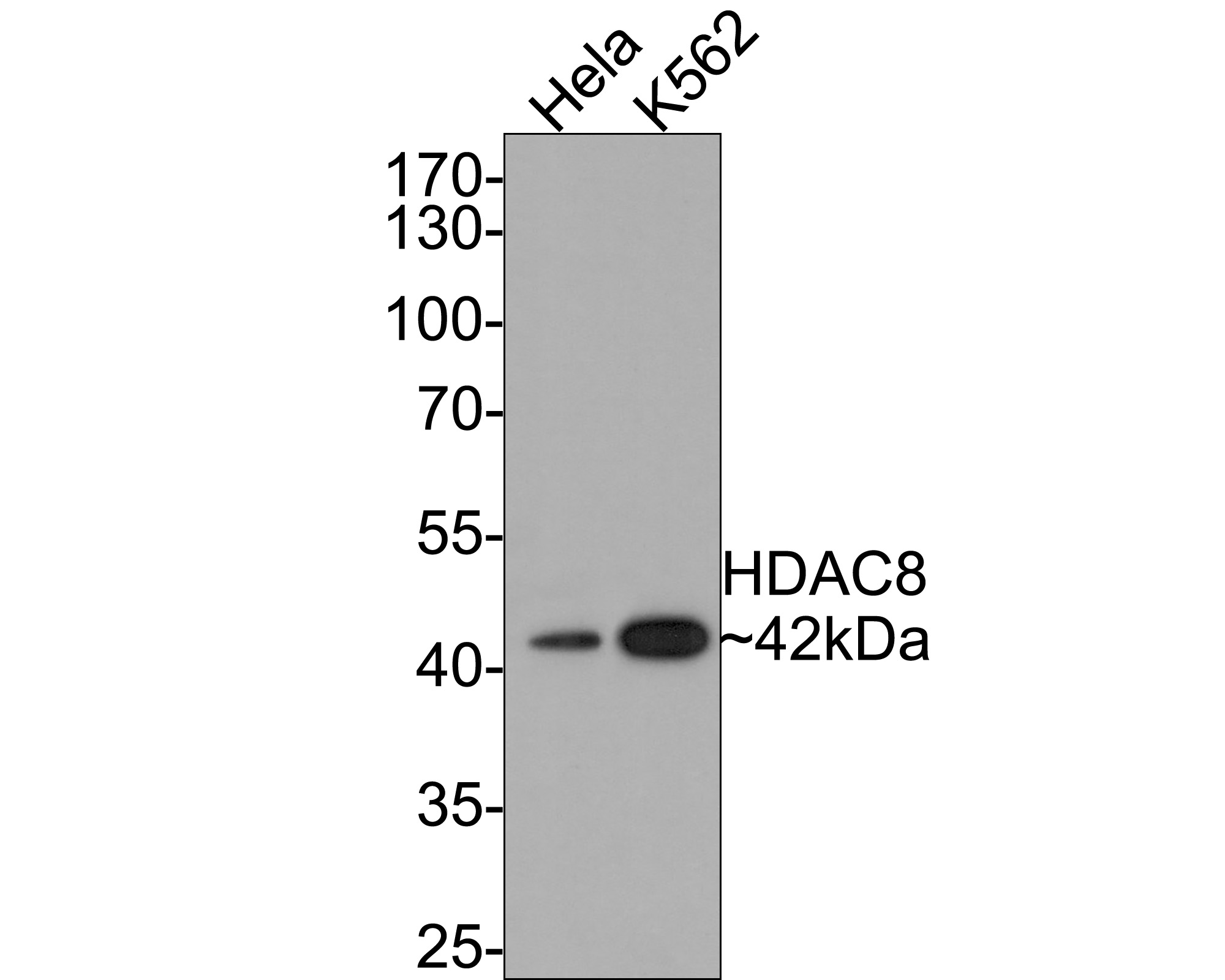 Western blot analysis of HDAC8 on different lysates with Rabbit anti-HDAC8 antibody (SLM-52088R) at 1/500 dilution.
Lane 1: Hela cell lysate
Lane 2: K562 cell lysate
Lysates/proteins at 10 µg/Lane.
Predicted band size: 42 kDa
Observed band size: 42 kDa
Exposure time: 1 minute;
10% SDS-PAGE gel.
Proteins were transferred to a PVDF membrane and blocked with 5% NFDM/TBST for 1 hour at room temperature. The primary antibody (SLM-52088R) at 1/500 dilution was used in 5% NFDM/TBST at room temperature for 2 hours. Goat Anti-Rabbit IgG - HRP Secondary Antibody at 1:300,000 dilution was used for 1 hour at room temperature.
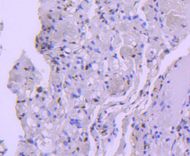 Immunohistochemical analysis of paraffin-embedded human lung tissue using anti-HDAC8 antibody. The section was pre-treated using heat mediated antigen retrieval with Tris-EDTA buffer (pH 8.0-8.4) for 20 minutes.The tissues were blocked in 5% BSA for 30 minutes at room temperature, washed with ddH2O and PBS, and then probed with the primary antibody (SLM-52088R, 1/50) for 30 minutes at room temperature. The detection was performed using an HRP conjugated compact polymer system. DAB was used as the chromogen. Tissues were counterstained with hematoxylin and mounted with DPX.
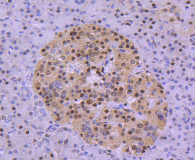 Immunohistochemical analysis of paraffin-embedded human pancreas tissue using anti-HDAC8 antibody. The section was pre-treated using heat mediated antigen retrieval with Tris-EDTA buffer (pH 8.0-8.4) for 20 minutes.The tissues were blocked in 5% BSA for 30 minutes at room temperature, washed with ddH2O and PBS, and then probed with the primary antibody (SLM-52088R, 1/50) for 30 minutes at room temperature. The detection was performed using an HRP conjugated compact polymer system. DAB was used as the chromogen. Tissues were counterstained with hematoxylin and mounted with DPX.
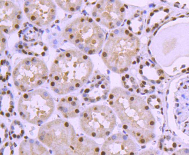 Immunohistochemical analysis of paraffin-embedded human kidney tissue using anti-HDAC8 antibody. The section was pre-treated using heat mediated antigen retrieval with Tris-EDTA buffer (pH 8.0-8.4) for 20 minutes.The tissues were blocked in 5% BSA for 30 minutes at room temperature, washed with ddH2O and PBS, and then probed with the primary antibody (SLM-52088R, 1/50) for 30 minutes at room temperature. The detection was performed using an HRP conjugated compact polymer system. DAB was used as the chromogen. Tissues were counterstained with hematoxylin and mounted with DPX.
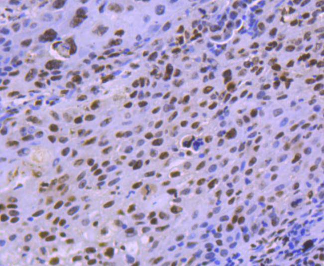 Immunohistochemical analysis of paraffin-embedded human lung carcinoma tissue using anti-HDAC8 antibody. The section was pre-treated using heat mediated antigen retrieval with Tris-EDTA buffer (pH 8.0-8.4) for 20 minutes.The tissues were blocked in 5% BSA for 30 minutes at room temperature, washed with ddH2O and PBS, and then probed with the primary antibody (SLM-52088R, 1/50) for 30 minutes at room temperature. The detection was performed using an HRP conjugated compact polymer system. DAB was used as the chromogen. Tissues were counterstained with hematoxylin and mounted with DPX.
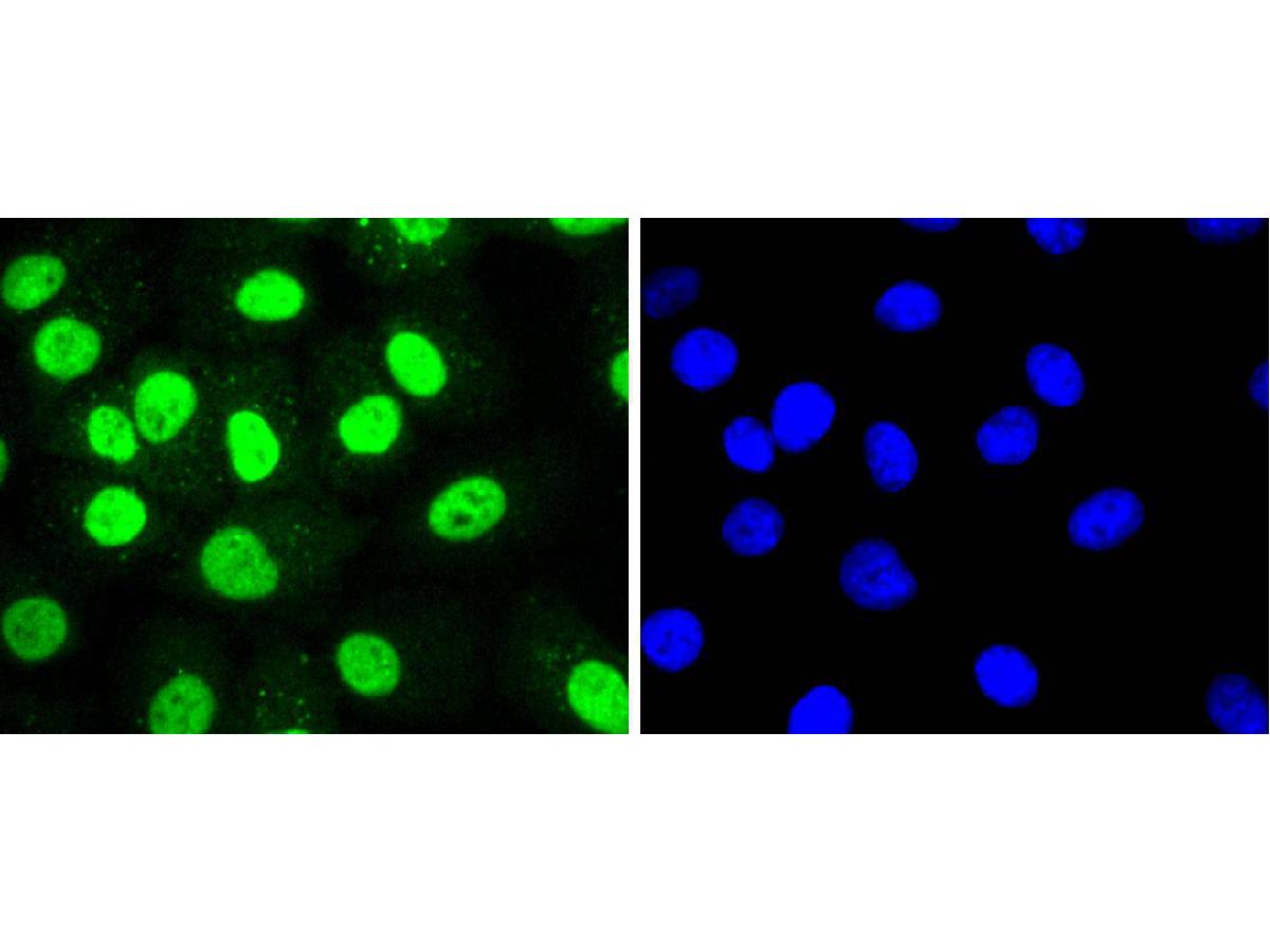 ICC staining of HDAC8 in A549 cells (green). Formalin fixed cells were permeabilized with 0.1% Triton X-100 in TBS for 10 minutes at room temperature and blocked with 1% Blocker BSA for 15 minutes at room temperature. Cells were probed with the primary antibody (SLM-52088R, 1/50) for 1 hour at room temperature, washed with PBS. Alexa Fluor®488 Goat anti-Rabbit IgG was used as the secondary antibody at 1/1,000 dilution. The nuclear counter stain is DAPI (blue).
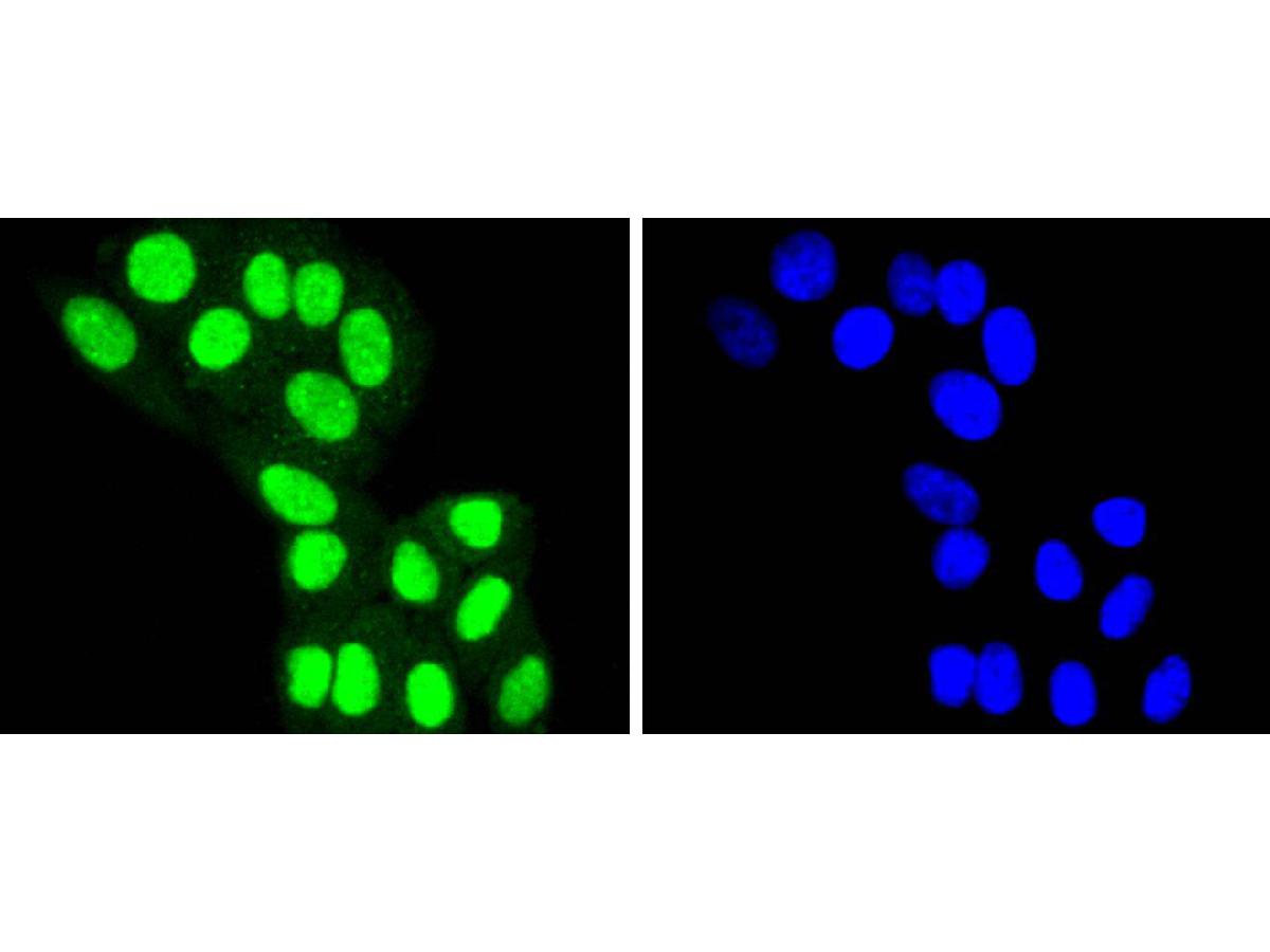 ICC staining of HDAC8 in HepG2 cells (green). Formalin fixed cells were permeabilized with 0.1% Triton X-100 in TBS for 10 minutes at room temperature and blocked with 1% Blocker BSA for 15 minutes at room temperature. Cells were probed with the primary antibody (SLM-52088R, 1/50) for 1 hour at room temperature, washed with PBS. Alexa Fluor®488 Goat anti-Rabbit IgG was used as the secondary antibody at 1/1,000 dilution. The nuclear counter stain is DAPI (blue).
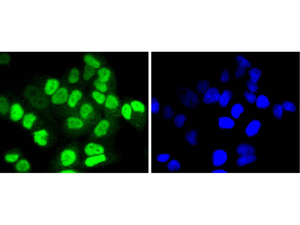 ICC staining of HDAC8 in Hela cells (green). Formalin fixed cells were permeabilized with 0.1% Triton X-100 in TBS for 10 minutes at room temperature and blocked with 1% Blocker BSA for 15 minutes at room temperature. Cells were probed with the primary antibody (SLM-52088R, 1/50) for 1 hour at room temperature, washed with PBS. Alexa Fluor®488 Goat anti-Rabbit IgG was used as the secondary antibody at 1/1,000 dilution. The nuclear counter stain is DAPI (blue).
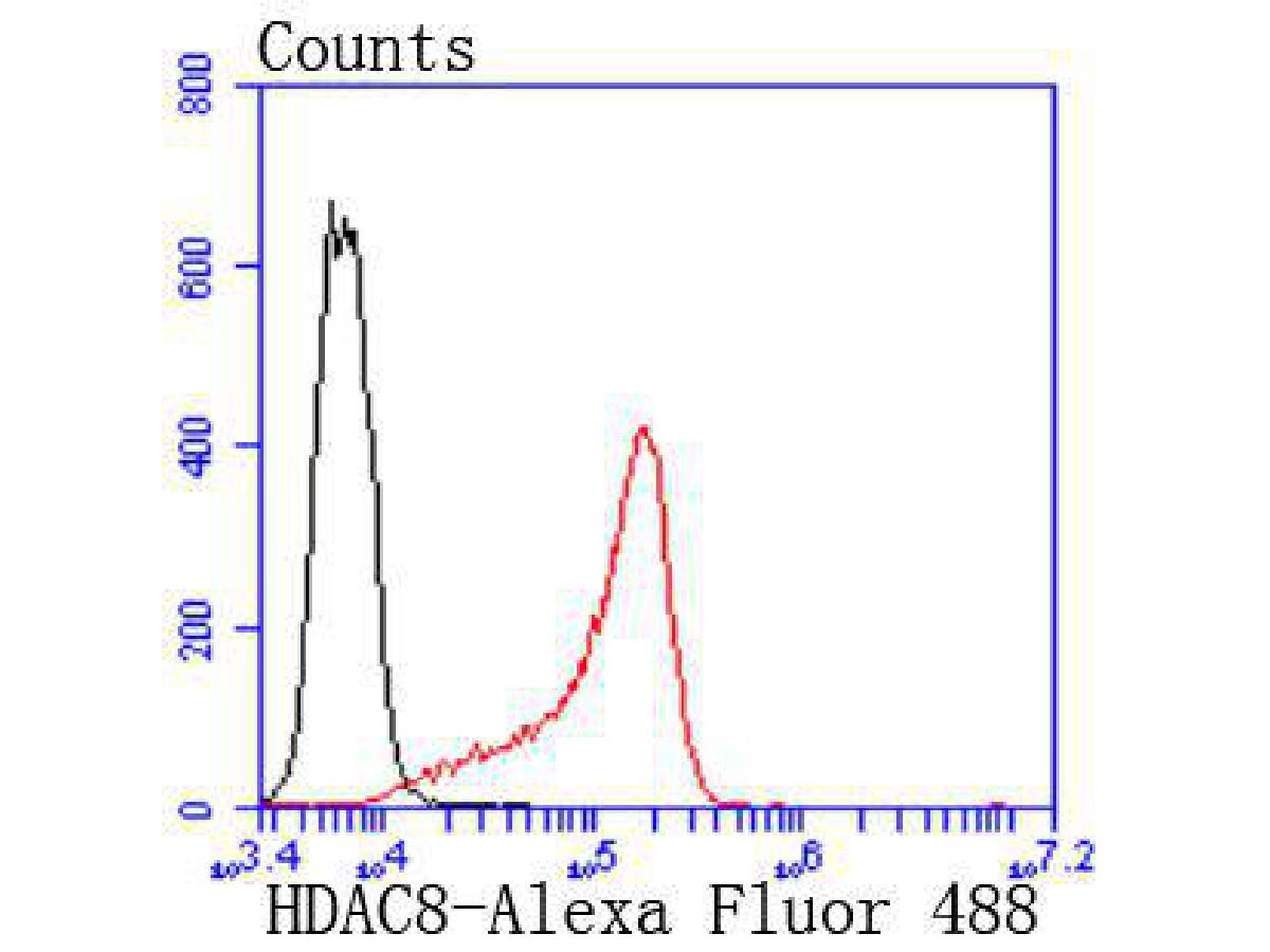 Flow cytometric analysis of HDAC8 was done on K562 cells. The cells were fixed, permeabilized and stained with the primary antibody (SLM-52088R, 1/50) (red). After incubation of the primary antibody at room temperature for an hour, the cells were stained with a Alexa Fluor 488-conjugated Goat anti-Rabbit IgG Secondary antibody at 1/1,000 dilution for 30 minutes.Unlabelled sample was used as a control (cells without incubation with primary antibody; black).
Cartpieces
Totalgoods,subtotals:¥Checkout
References (0)
No References
Bought notes(bought amounts latest0)
No one bought this product
User Comment(Total0User Comment Num)
- No comment


 +86 571 56623320
 +86 18668110335

