
Rabbit Anti-HDAC2 antibody
HDAC2_HUMAN; Histone deacetylase 2; EC:3.5.1.98; HD2; Protein deacylase HDAC2;
View History [Clear]
Details
Product Name [KO validated anti] HDAC2 Chinese Name 组蛋白去乙酰化酶2Recombinant rabbit monoclonal anti Alias HDAC2_HUMAN; Histone deacetylase 2; EC:3.5.1.98; HD2; Protein deacylase HDAC2; Research Area immunology Developmental biology transcriptional regulatory factor Epigenetics Immunogen Species Rabbit Clonality Monoclonal Clone NO. 3B7 React Species Human, Mouse, Rat, Applications WB=1:500-2000 IHC-P=1:50-200 IHC-F=1:50-200 Flow-Cyt=1ug/Test ICC=1:100 IF=1:50-200 (Paraffin sections need antigen repair)
not yet tested in other applications.
optimal dilutions/concentrations should be determined by the end user.Theoretical molecular weight 53kDa Cellular localization The nucleus Form Liquid Concentration 1mg/ml immunogen Recombinant human HDAC2 protein, around C-terminal 150aa Lsotype IgG Purification affinity purified by Protein A Buffer Solution 0.01M TBS(pH7.4) with 1% BSA, 0.03% Proclin300 and 50% Glycerol. Storage Shipped at 4℃. Store at -20 °C for one year. Avoid repeated freeze/thaw cycles. Attention This product as supplied is intended for research use only, not for use in human, therapeutic or diagnostic applications. PubMed PubMed Product Detail This gene product belongs to the histone deacetylase family. Histone deacetylases act via the formation of large multiprotein complexes, and are responsible for the deacetylation of lysine residues at the N-terminal regions of core histones (H2A, H2B, H3 and H4). This protein forms transcriptional repressor complexes by associating with many different proteins, including YY1, a mammalian zinc-finger transcription factor. Thus, it plays an important role in transcriptional regulation, cell cycle progression and developmental events. Alternative splicing results in multiple transcript variants. [provided by RefSeq].
Function:
Responsible for the deacetylation of lysine residues on the N-terminal part of the core histones (H2A, H2B, H3 and H4). Histone deacetylation gives a tag for epigenetic repression and plays an important role in transcriptional regulation, cell cycle progression and developmental events. Histone deacetylases act via the formation of large multiprotein complexes. Forms transcriptional repressor complexes by associating with MAD, SIN3, YY1 and N-COR. Interacts in the late S-phase of DNA-replication with DNMT1 in the other transcriptional repressor complex composed of DNMT1, DMAP1, PCNA, CAF1. Deacetylates TSHZ3 and regulates its transcriptional repressor activity. Component of a RCOR/GFI/KDM1A/HDAC complex that suppresses, via histone deacetylase (HDAC) recruitment, a number of genes implicated in multilineage blood cell development.
Subunit:
Component of a RCOR/GFI/KDM1A/HDAC complex. Interacts directly with GFI1 and GFI1B. Interacts with SNW1, HDAC7, PRDM6, SAP30, SETDB1 and SUV39H1. Interacts with the H2AFY (via the non-histone region) (By similarity). Part of the core histone deacetylase (HDAC) complex composed of HDAC1, HDAC2, RBBP4 and RBBP7. The core complex associates with MTA2, MBD3, MTA1L1, CHD3 and CHD4 to form the nucleosome remodeling and histone deacetylation (NuRD) complex, or with SIN3, SAP18 and SAP30 to form the SIN3 HDAC complex. Component of a BHC histone deacetylase complex that contains HDAC1, HDAC2, HMG20B, KDM1A, RCOR1 and PHF21A. The BHC complex may also contain ZMYM2, ZNF217, ZMYM3, GSE1 and GTF2I. Part of a complex containing the core histones H2A, H2B, H3 and H4, DEK and unphosphorylated DAXX. Part of a complex containing ATR and CHD4. Forms a heterologous complex at least with YY1. Interacts with ATR, CBFA2T3, DNMT1, MINT, HDAC10, HCFC1, NRIP1, KDM4A and PELP1. Component of a mSin3A corepressor complex that contains SIN3A, SAP130, SUDS3, ARID4B, HDAC1 and HDAC2. Interacts with CHFR and SAP30L. Interacts (CK2 phosphorylated form) with SP3. Interacts with TSHZ3 (via its N-terminus). Interacts with APEX1; the interaction is not dependent on the acetylated status of APEX1. Part of a complex composed of TRIM28, HDAC1, HDAC2 and EHMT2.
Subcellular Location:
Nucleus.
Tissue Specificity:
Widely expressed; lower levels in brain and lung.
Post-translational modifications:
S-nitrosylated by GAPDH. In neurons, S-Nitrosylation at Cys-262 and Cys-274 does not affect the enzyme activity but abolishes chromatin-binding, leading to increases acetylation of histones and activate genes that are associated with neuronal development. In embryonic cortical neurons, S-Nitrosylation regulates dendritic growth and branching.
Similarity:
Belongs to the histone deacetylase family. HD type 1 subfamily.
SWISS:
Q92769
Gene ID:
3066
Database links:Entrez Gene: 3066 Human
Entrez Gene: 15182 Mouse
SwissProt: Q92769 Human
SwissProt: P70288 Mouse
Product Picture 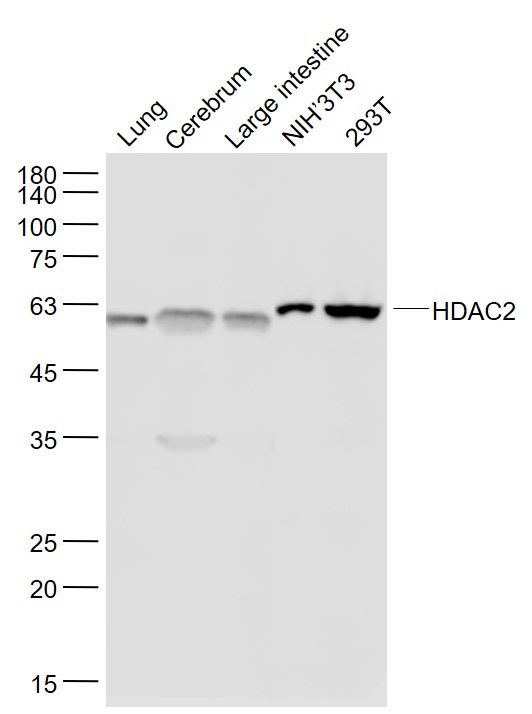 Sample:
Lung (Mouse) Lysate at 40 ug
Cerebrum (Mouse) Lysate at 40 ug
Large intestine (Mouse) Lysate at 40 ug
NIH/3T3(Mouse) Cell Lysate at 30 ug
293T(Human) Cell Lysate at 30 ug
Primary: Anti- HDAC2 (SLM-52082R) at 1/1000 dilution
Secondary: IRDye800CW Goat Anti-Rabbit IgG at 1/20000 dilution
Predicted band size: 53 kD
Observed band size: 56 kD
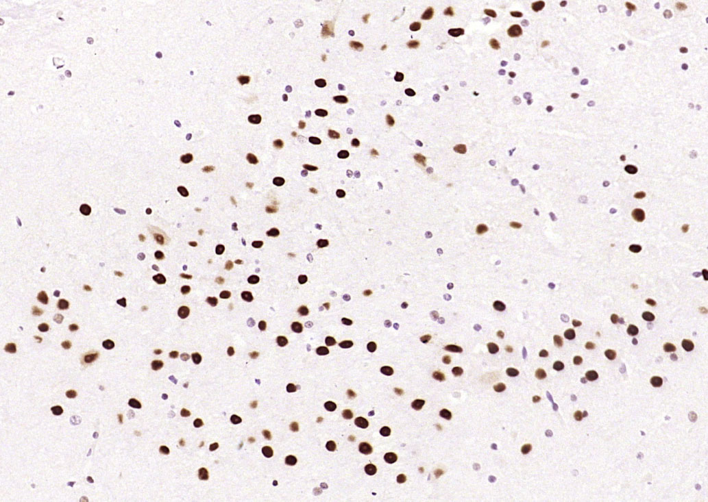 Paraformaldehyde-fixed, paraffin embedded (rat brain); Antigen retrieval by boiling in sodium citrate buffer (pH6.0) for 15min; Block endogenous peroxidase by 3% hydrogen peroxide for 20 minutes; Blocking buffer (normal goat serum) at 37°C for 30min; Antibody incubation with (HDAC2) Monoclonal Antibody, Unconjugated (SLM-52082R) at 1:200 overnight at 4°C, followed by operating according to SP Kit(Rabbit) (sp-0023) instructionsand DAB staining.
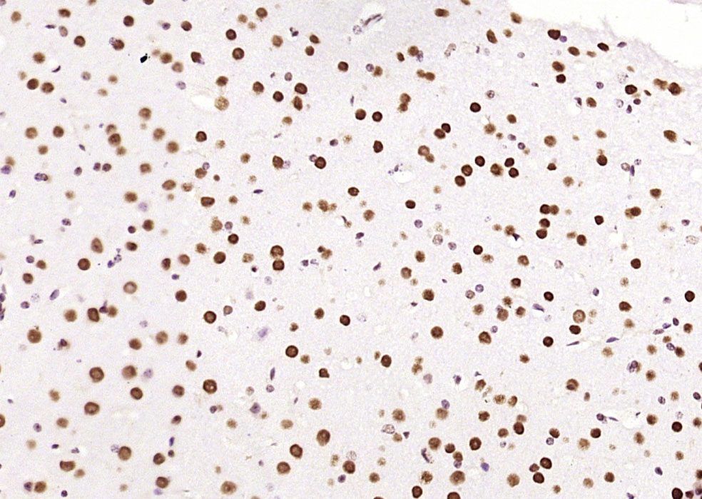 Paraformaldehyde-fixed, paraffin embedded (mouse brain); Antigen retrieval by boiling in sodium citrate buffer (pH6.0) for 15min; Block endogenous peroxidase by 3% hydrogen peroxide for 20 minutes; Blocking buffer (normal goat serum) at 37°C for 30min; Antibody incubation with (HDAC2) Monoclonal Antibody, Unconjugated (SLM-52082R) at 1:200 overnight at 4°C, followed by operating according to SP Kit(Rabbit) (sp-0023) instructionsand DAB staining.
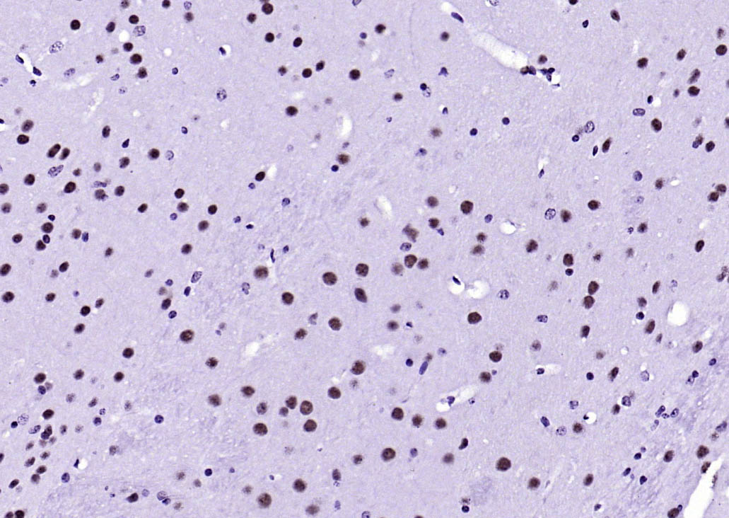 Paraformaldehyde-fixed, paraffin embedded (mouse brain); Antigen retrieval by boiling in sodium citrate buffer (pH6.0) for 15min; Block endogenous peroxidase by 3% hydrogen peroxide for 20 minutes; Blocking buffer (normal goat serum) at 37°C for 30min; Antibody incubation with (HDAC2) Monoclonal Antibody, Unconjugated (SLM-52082R) at 1:200 overnight at 4°C, followed by operating according to SP Kit(Rabbit) (sp-0023) instructionsand DAB staining.
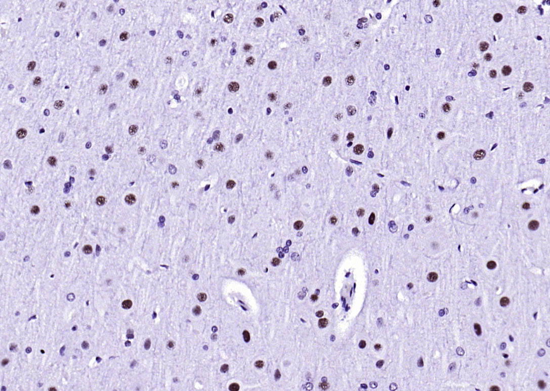 Paraformaldehyde-fixed, paraffin embedded (rat brain); Antigen retrieval by boiling in sodium citrate buffer (pH6.0) for 15min; Block endogenous peroxidase by 3% hydrogen peroxide for 20 minutes; Blocking buffer (normal goat serum) at 37°C for 30min; Antibody incubation with (HDAC2) Monoclonal Antibody, Unconjugated (SLM-52082R) at 1:200 overnight at 4°C, followed by operating according to SP Kit(Rabbit) (sp-0023) instructionsand DAB staining.
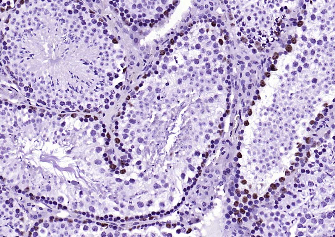 Paraformaldehyde-fixed, paraffin embedded (mouse testis); Antigen retrieval by boiling in sodium citrate buffer (pH6.0) for 15min; Block endogenous peroxidase by 3% hydrogen peroxide for 20 minutes; Blocking buffer (normal goat serum) at 37°C for 30min; Antibody incubation with (HDAC2) Monoclonal Antibody, Unconjugated (SLM-52082R) at 1:200 overnight at 4°C, followed by operating according to SP Kit(Rabbit) (sp-0023) instructionsand DAB staining.
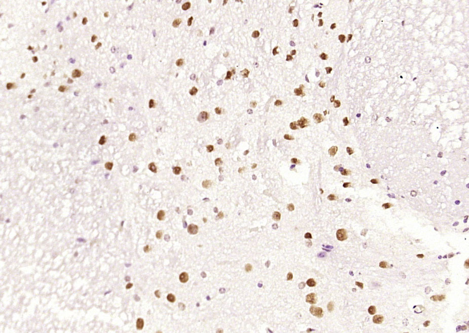 Paraformaldehyde-fixed, paraffin embedded (mouse Spinal Cord); Antigen retrieval by boiling in sodium citrate buffer (pH6.0) for 15min; Block endogenous peroxidase by 3% hydrogen peroxide for 20 minutes; Blocking buffer (normal goat serum) at 37°C for 30min; Antibody incubation with (HDAC2) Monoclonal Antibody, Unconjugated (SLM-52082R) at 1:200 overnight at 4°C, followed by operating according to SP Kit(Rabbit) (sp-0023) instructionsand DAB staining.
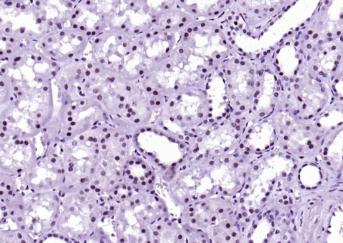 Paraformaldehyde-fixed, paraffin embedded (Human kidney); Antigen retrieval by boiling in sodium citrate buffer (pH6.0) for 15min; Block endogenous peroxidase by 3% hydrogen peroxide for 20 minutes; Blocking buffer (normal goat serum) at 37°C for 30min; Antibody incubation with (HDAC2) Monoclonal Antibody, Unconjugated (SLM-52082R) at 1:200 overnight at 4°C, followed by operating according to SP Kit(Rabbit) (sp-0023) instructionsand DAB staining.
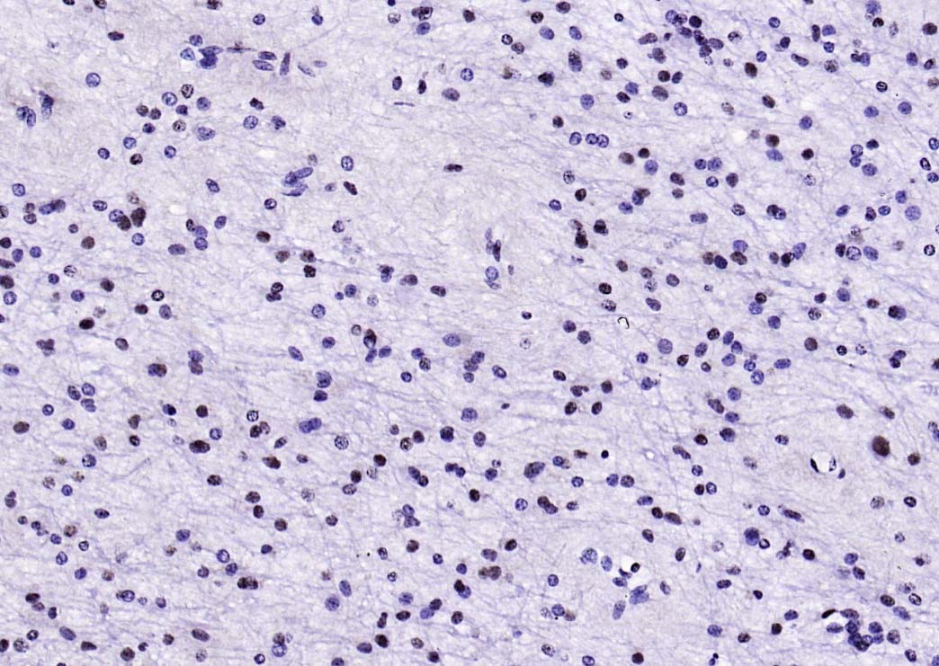 Paraformaldehyde-fixed, paraffin embedded (human brain); Antigen retrieval by boiling in sodium citrate buffer (pH6.0) for 15min; Block endogenous peroxidase by 3% hydrogen peroxide for 20 minutes; Blocking buffer (normal goat serum) at 37°C for 30min; Antibody incubation with (HDAC2) Monoclonal Antibody, Unconjugated (SLM-52082R) at 1:200 overnight at 4°C, followed by operating according to SP Kit(Rabbit) (sp-0023) instructionsand DAB staining.
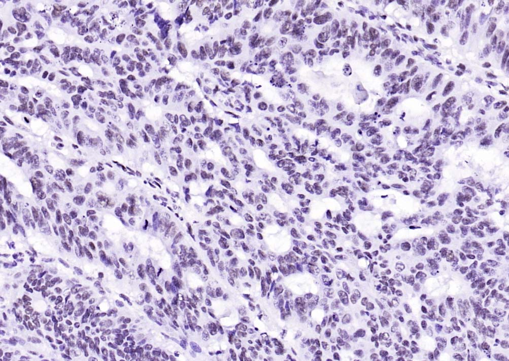 Paraformaldehyde-fixed, paraffin embedded (human colon carcinoma); Antigen retrieval by boiling in sodium citrate buffer (pH6.0) for 15min; Block endogenous peroxidase by 3% hydrogen peroxide for 20 minutes; Blocking buffer (normal goat serum) at 37°C for 30min; Antibody incubation with (HDAC2) Monoclonal Antibody, Unconjugated (SLM-52082R) at 1:200 overnight at 4°C, followed by operating according to SP Kit(Rabbit) (sp-0023) instructionsand DAB staining.
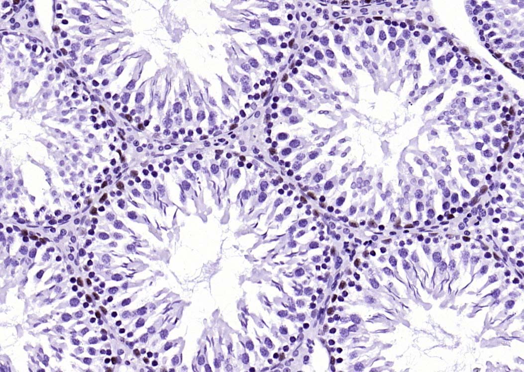 Paraformaldehyde-fixed, paraffin embedded (rat testis); Antigen retrieval by boiling in sodium citrate buffer (pH6.0) for 15min; Block endogenous peroxidase by 3% hydrogen peroxide for 20 minutes; Blocking buffer (normal goat serum) at 37°C for 30min; Antibody incubation with (HDAC2) Monoclonal Antibody, Unconjugated (SLM-52082R) at 1:200 overnight at 4°C, followed by operating according to SP Kit(Rabbit) (sp-0023) instructionsand DAB staining.
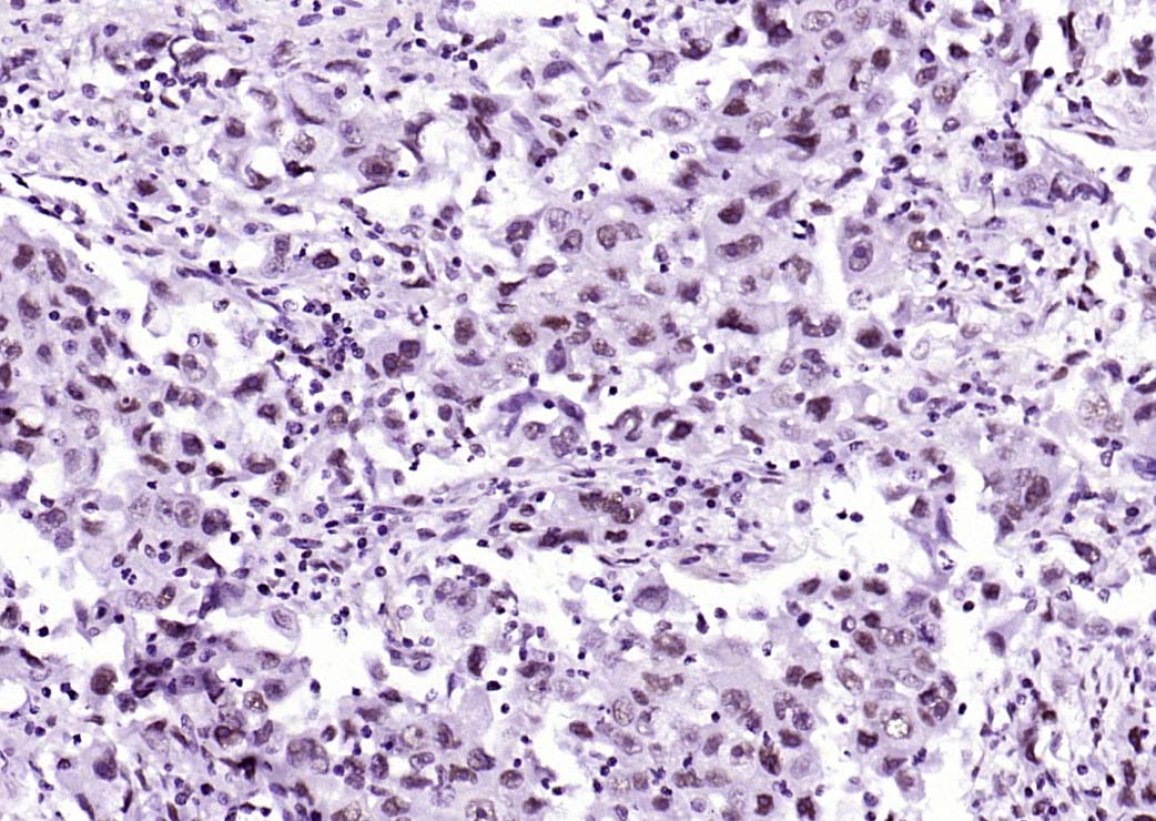 Paraformaldehyde-fixed, paraffin embedded (human lung carcinoma); Antigen retrieval by boiling in sodium citrate buffer (pH6.0) for 15min; Block endogenous peroxidase by 3% hydrogen peroxide for 20 minutes; Blocking buffer (normal goat serum) at 37°C for 30min; Antibody incubation with (HDAC2) Monoclonal Antibody, Unconjugated (SLM-52082R) at 1:200 overnight at 4°C, followed by operating according to SP Kit(Rabbit) (sp-0023) instructionsand DAB staining.
 Paraformaldehyde-fixed, paraffin embedded (mouse colon); Antigen retrieval by boiling in sodium citrate buffer (pH6.0) for 15min; Block endogenous peroxidase by 3% hydrogen peroxide for 20 minutes; Blocking buffer (normal goat serum) at 37°C for 30min; Antibody incubation with (HDAC2) Monoclonal Antibody, Unconjugated (SLM-52082R) at 1:200 overnight at 4°C, followed by operating according to SP Kit(Rabbit) (sp-0023) instructionsand DAB staining.
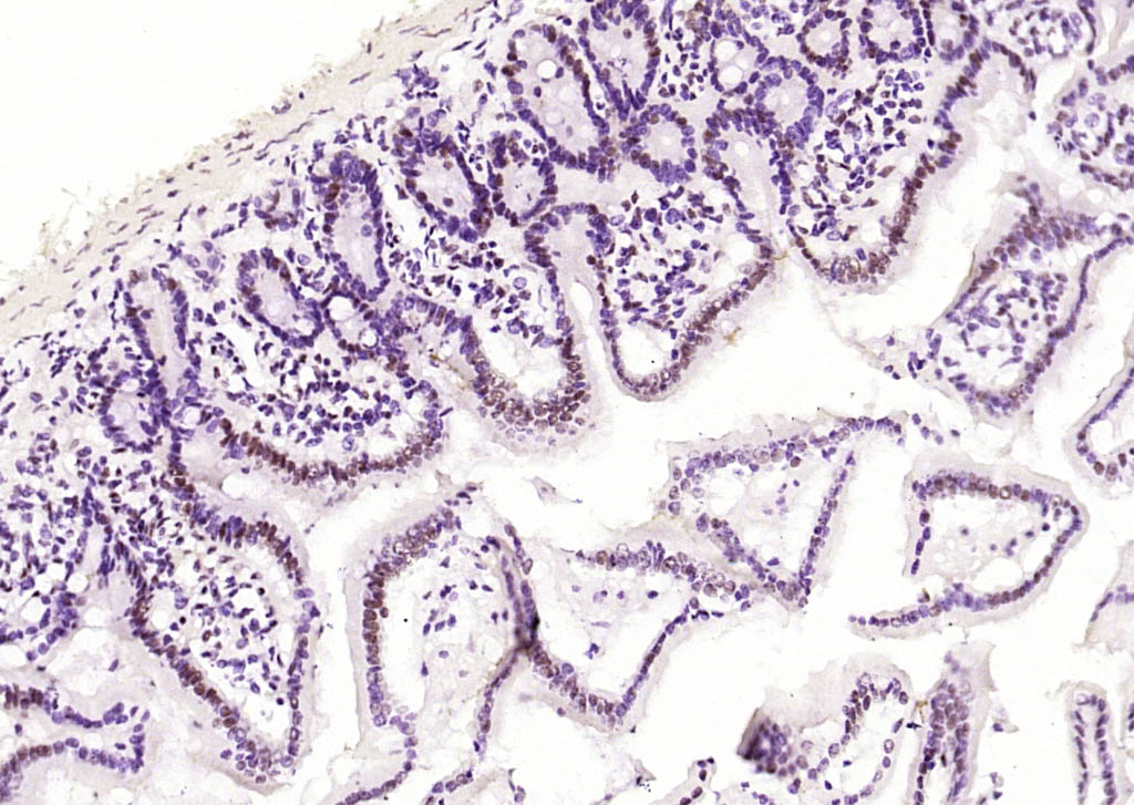 Paraformaldehyde-fixed, paraffin embedded (mouse intestine); Antigen retrieval by boiling in sodium citrate buffer (pH6.0) for 15min; Block endogenous peroxidase by 3% hydrogen peroxide for 20 minutes; Blocking buffer (normal goat serum) at 37°C for 30min; Antibody incubation with (HDAC2) Monoclonal Antibody, Unconjugated (SLM-52082R) at 1:200 overnight at 4°C, followed by operating according to SP Kit(Rabbit) (sp-0023) instructionsand DAB staining.
 Paraformaldehyde-fixed, paraffin embedded (human tonsil); Antigen retrieval by boiling in sodium citrate buffer (pH6.0) for 15min; Block endogenous peroxidase by 3% hydrogen peroxide for 20 minutes; Blocking buffer (normal goat serum) at 37°C for 30min; Antibody incubation with (HDAC2) Monoclonal Antibody, Unconjugated (SLM-52082R) at 1:200 overnight at 4°C, followed by operating according to SP Kit(Rabbit) (sp-0023) instructionsand DAB staining.
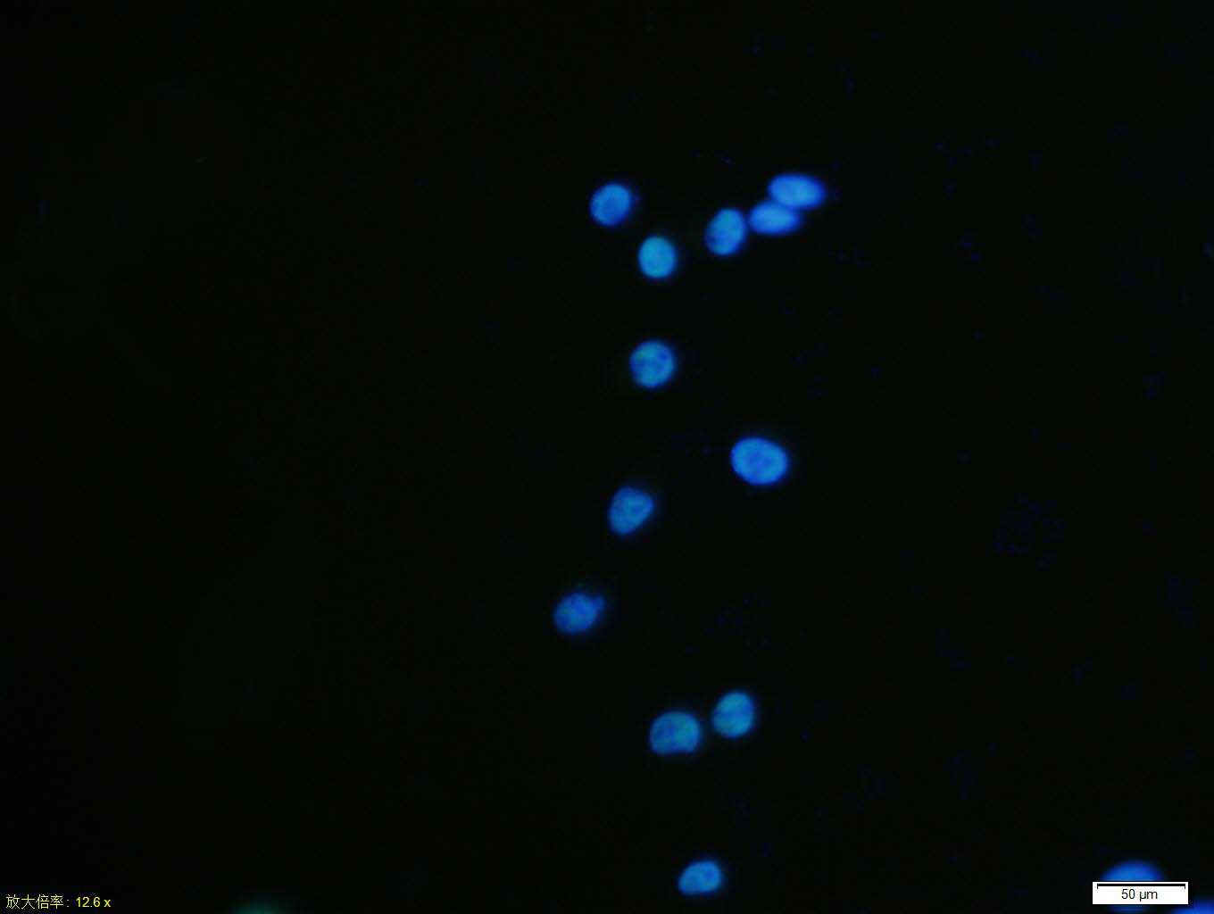 SH-SY5Y cell; 4% Paraformaldehyde-fixed; Triton X-100 at room temperature for 20 min; Blocking buffer (normal goat serum, C-0005) at 37°C for 20 min; Antibody incubation with (HDAC2) monoclonal Antibody, Unconjugated (SLM-52082R) 1:100, 90 minutes at 37°C; followed by a conjugated Goat Anti-Rabbit IgG antibody at 37°C for 90 minutes, DAPI (blue, C02-04002) was used to stain the cell nuclei.
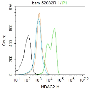 Blank control:K562.
Primary Antibody (green line): Rabbit Anti-HDAC2 antibody (SL52082R)
Dilution: 1μg /10^6 cells;
Isotype Control Antibody (orange line): Rabbit IgG .
Secondary Antibody : Goat anti-rabbit IgG-FITC
Dilution: 0.5μg /test.
Protocol
The cells were fixed with 4% PFA (10min at room temperature)and then permeabilized with 90% ice-cold methanol for 20 min at-20℃. The cells were then incubated in 5%BSA to block non-specific protein-protein interactions for 30 min at room temperature .Cells stained with Primary Antibody for 30 min at room temperature. The secondary antibody used for 40 min at room temperature. Acquisition of 20,000 events was performed.
Cartpieces
Totalgoods,subtotals:¥Checkout
References (0)
No References
Bought notes(bought amounts latest0)
No one bought this product
User Comment(Total0User Comment Num)
- No comment


 +86 571 56623320
 +86 18668110335

