
Rabbit Anti-Oct4 antibody
MGC22487; Oct 3; Oct 4; Oct-3; Oct-4; OCT3; Oct4; Octamer binding protein 3; Octamer-binding protein 3; Octamer binding protein 4; Octamer binding transcription factor 3; Octamer-binding transcription factor 3; Octamer-4; POU domain transcription factor
View History [Clear]
Details
Product Name Oct4 Chinese Name 胚胎Stem cells关键蛋白Recombinant rabbit monoclonal anti Alias MGC22487; Oct 3; Oct 4; Oct-3; Oct-4; OCT3; Oct4; Octamer binding protein 3; Octamer-binding protein 3; Octamer binding protein 4; Octamer binding transcription factor 3; Octamer-binding transcription factor 3; Octamer-4; POU domain transcription factor OCT4; OTF3; OTF4; OTF 3; OTF 4; OTF-3; POU class 5 homeobox 1; POU domain, class 5, transcription factor 1; POU domain class 5 transcription factor 1; PO5F1_HUMAN; POU type homeodomain containing DNA binding protein ; POU5F1. literatures Research Area Cell biology Developmental biology Signal transduction Stem cells Cyclin transcriptional regulatory factor Cell type markers Epigenetics Immunogen Species Rabbit Clonality Monoclonal Clone NO. 2D5 React Species (predicted: Human, Mouse, Rat, ) Applications WB=1:500-2000 IP=1:20-100 IHC-P=1:100-500 IHC-F=1:50-200 ICC=1:50-200 (Paraffin sections need antigen repair)
not yet tested in other applications.
optimal dilutions/concentrations should be determined by the end user.Theoretical molecular weight 39kDa Cellular localization The nucleus Form Liquid Concentration 1mg/ml immunogen Recombinant human Oct 4 protein, around 1-100aa Lsotype IgG Purification affinity purified by Protein A Buffer Solution 0.01M TBS(pH7.4) with 1% BSA, 0.03% Proclin300 and 50% Glycerol. Storage Shipped at 4℃. Store at -20 °C for one year. Avoid repeated freeze/thaw cycles. Attention This product as supplied is intended for research use only, not for use in human, therapeutic or diagnostic applications. PubMed PubMed Product Detail Expression of the POU-domain transcription factor Octamer-4 (Oct-4) is widely regarded as a hallmark of pluripotent stem cells. The relationship of Oct-4 to pluripotent stem cells is indicated by its tightly restricted expression to undifferentiated pluripotent stem cells. Upon differentiation to somatic lineages, the expression of Oct-4 disappears rapidly. Unlike the majority of pluripotent stem cell markers, the biological role of Oct-4 has been well characterized. Studies performed in mice point to the critical role of Oct-4 in the establishment and/or maintenance of pluripotent stem cells in an uncommitted state.
Function:
Transcription factor that binds to the octamer motif (5'-ATTTGCAT-3'). Forms a trimeric complex with SOX2 on DNA and controls the expression of a number of genes involved in embryonic development such as YES1, FGF4, UTF1 and ZFP206.
Subunit:
Interacts with UBE2I and ZSCAN10. Interacts with PKM2. Interacts with WWP2.
Subcellular Location:
Nucleus.Note=Expressed in a diffuse and slightly punctuate pattern.
Tissue Specificity:
Expressed in developing brain. Highest levels found in specific cell layers of the cortex, the olfactory bulb, the hippocampus and the cerebellum. Low levels of expression in adult tissues.
Post-translational modifications:
Belongs to the POU transcription factor family. Class-5 subfamily.
Contains 1 homeobox DNA-binding domain.
Contains 1 POU-specific domain.
Similarity:
Belongs to the POU transcription factor family. Class-5 subfamily.
Contains 1 homeobox DNA-binding domain.
Contains 1 POU-specific domain.
SWISS:
Q01860
Gene ID:
5460
Database links:
Entrez Gene: 5460 Human
Entrez Gene: 18999 Mouse
Omim: 164177 Human
SwissProt: Q01860 Human
SwissProt: P20263 Mouse
Unigene: 249184 Human
Unigene: 632482 Human
Unigene: 646545 Human
Unigene: 17031 Mouse
Unigene: 161748 Rat
Product Picture 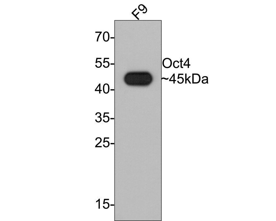 Western blot analysis of Oct4 on F9 cell lysates with Rabbit anti-Oct4 antibody (SLM-52002R) at 1/1,000 dilution.
Lysates/proteins at 10 碌g/Lane.
Predicted band size: 39 kDa
Observed band size: 45 kDa
Exposure time: 2 minutes;
12% SDS-PAGE gel.
Proteins were transferred to a PVDF membrane and blocked with 5% NFDM/TBST for 1 hour at room temperature. The primary antibody (SLM-52002R) at 1/1,000 dilution was used in 5% NFDM/TBST at room temperature for 2 hours. Goat Anti-Rabbit IgG - HRP Secondary Antibody at 1:300,000 dilution was used for 1 hour at room temperature.
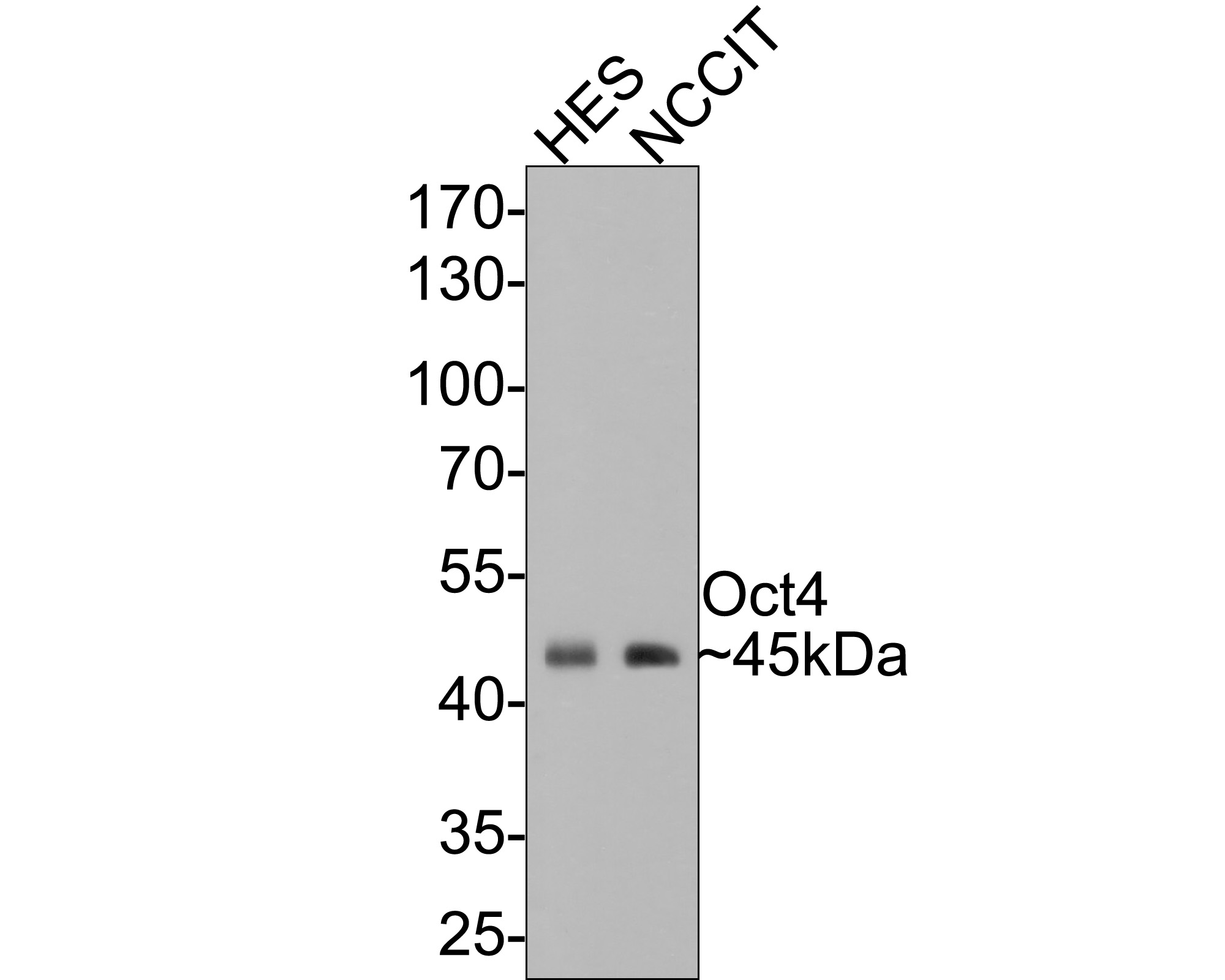 Western blot analysis of Oct4 on different lysates with Rabbit anti-Oct4 antibody (SLM-52002R) at 1/500 dilution.
Lane 1: HES cell lysate
Lane 2: NCCIT cell lysate
Lysates/proteins at 10 碌g/Lane.
Predicted band size: 39 kDa
Observed band size: 45 kDa
Exposure time: 1 minute;
10% SDS-PAGE gel.
Proteins were transferred to a PVDF membrane and blocked with 5% NFDM/TBST for 1 hour at room temperature. The primary antibody (SLM-52002R) at 1/500 dilution was used in 5% NFDM/TBST at room temperature for 2 hours. Goat Anti-Rabbit IgG - HRP Secondary Antibody at 1:300,000 dilution was used for 1 hour at room temperature.
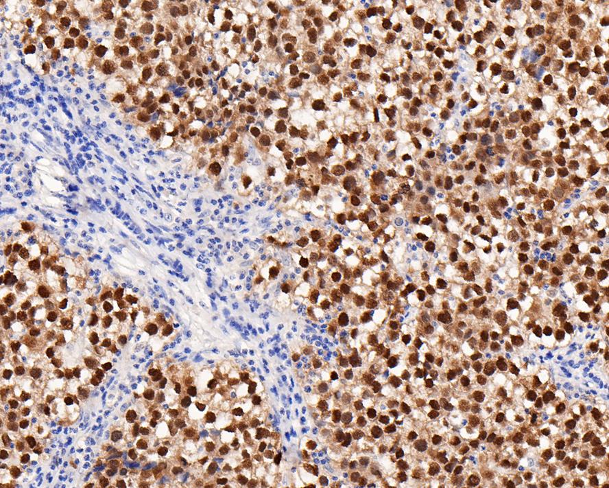 Immunohistochemical analysis of paraffin-embedded human seminoma tissue tissue with Rabbit anti-Oct4 antibody (ET1612-20) at 1/4,000 dilution. The section was pre-treated using heat mediated antigen retrieval with sodium citrate buffer (pH 6.0) for 2 minutes. The tissues were blocked in 1% BSA for 20 minutes at room temperature, washed with ddH2O and PBS, and then probed with the primary antibody (SLM-52002R) at 1/4,000 dilution for 1 hour at room temperature. The detection was performed using an HRP conjugated compact polymer system. DAB was used as the chromogen. Tissues were counterstained with hematoxylin and mounted with DPX.
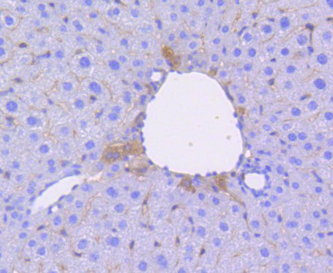 Immunohistochemical analysis of paraffin-embedded mouse liver tissue using anti-Oct4 antibody. The section was pre-treated using heat mediated antigen retrieval with Tris-EDTA buffer (pH 8.0-8.4) for 20 minutes.The tissues were blocked in 5% BSA for 30 minutes at room temperature, washed with ddH2O and PBS, and then probed with the primary antibody (SLM-52002R, 1/50) for 30 minutes at room temperature. The detection was performed using an HRP conjugated compact polymer system. DAB was used as the chromogen. Tissues were counterstained with hematoxylin and mounted with DPX.
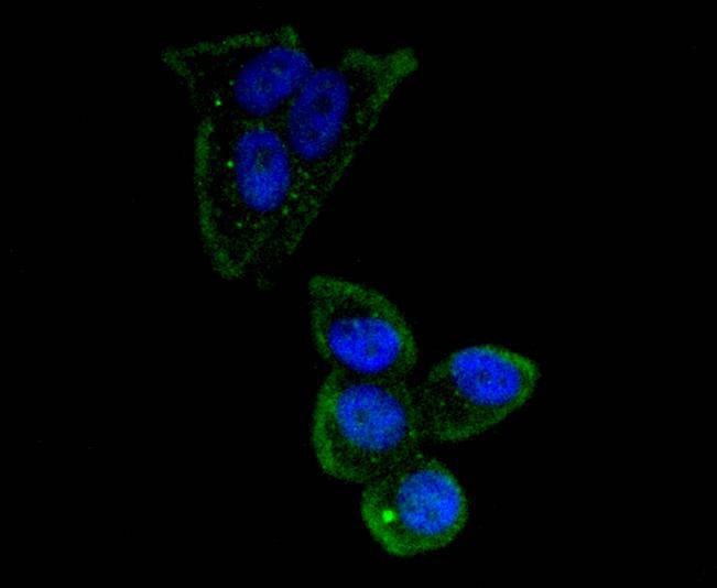 ICC staining of Oct4 in MCF-7 cells (green). Formalin fixed cells were permeabilized with 0.1% Triton X-100 in TBS for 10 minutes at room temperature and blocked with 1% Blocker BSA for 15 minutes at room temperature. Cells were probed with the primary antibody (SLM-52002R, 1/50) for 1 hour at room temperature, washed with PBS. Alexa Fluor®488 Goat anti-Rabbit IgG was used as the secondary antibody at 1/1,000 dilution. The nuclear counter stain is DAPI (blue).
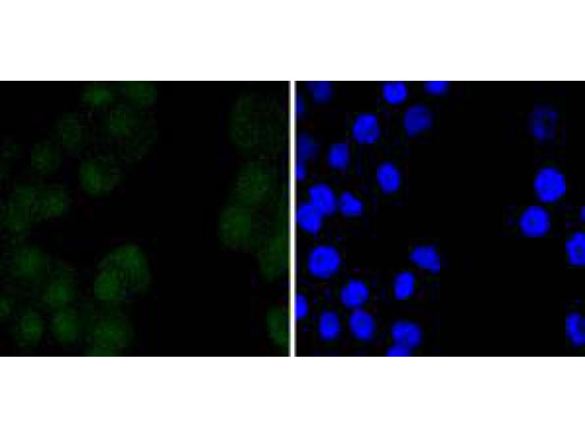 ICC staining of Oct4 in N2A cells (green). Formalin fixed cells were permeabilized with 0.1% Triton X-100 in TBS for 10 minutes at room temperature and blocked with 1% Blocker BSA for 15 minutes at room temperature. Cells were probed with the primary antibody (SLM-52002R, 1/50) for 1 hour at room temperature, washed with PBS. Alexa Fluor®488 Goat anti-Rabbit IgG was used as the secondary antibody at 1/1,000 dilution. The nuclear counter stain is DAPI (blue).
Cartpieces
Totalgoods,subtotals:¥Checkout
Bought notes(bought amounts latest0)
No one bought this product
User Comment(Total0User Comment Num)
- No comment


 +86 571 56623320
 +86 18668110335

