
Mouse Anti-SP1 antibody
Sp1 transcription factor isoform a; TSFP1; TSFP 1; Specificity protein 1; Transcription factor Sp1; SP1_HUMAN.
View History [Clear]
Details
Product Name SP1 Chinese Name 转录生长因子SP1单克隆抗体 Alias Sp1 transcription factor isoform a; TSFP1; TSFP 1; Specificity protein 1; Transcription factor Sp1; SP1_HUMAN. Research Area Chromatin and nuclear signals Signal transduction Stem cells transcriptional regulatory factor The new supersedes the old Zinc finger protein Epigenetics Immunogen Species Mouse Clonality Monoclonal Clone NO. Y5R2 React Species Human, Applications WB=1:500-1000 IHC-P=1:50-100 Flow-Cyt=1:25 ICC=1:25 IF=1:100-500 (Paraffin sections need antigen repair)
not yet tested in other applications.
optimal dilutions/concentrations should be determined by the end user.Theoretical molecular weight 81kDa Cellular localization The nucleus cytoplasmic Form Liquid Concentration 1mg/ml immunogen Recombinant human SP1 Lsotype IgG1,κ Purification affinity purified by Protein G Buffer Solution 0.01M TBS(pH7.4) with 1% BSA, 0.03% Proclin300 and 50% Glycerol. Storage Shipped at 4℃. Store at -20 °C for one year. Avoid repeated freeze/thaw cycles. Attention This product as supplied is intended for research use only, not for use in human, therapeutic or diagnostic applications. PubMed PubMed Product Detail Profound changes in patterns of gene expression can result from relatively small changes in the concentrations of sequence specific transcription factors. Synergistic interaction between factors bound to different sites within a transcriptional control region is supported by the work of Courey et al. (1989). Sp1 is a sequence specific transcription factor that recognizes GGGGCGGGGC and closely related sequences, which are often referred to as GC boxes. Sp1 binds to GC box promoters elements and selectively activates mRNA synthesis from genes that contain functional recognition sites. SP1 can interact with G/C rich motifs from serotonin receptor promoter. Kadonaga et al. (1987) cloned the human Sp1 cDNA and showed that it has contiguous zinc finger motifs and requires zinc for sequence specific binding to DNA. Altername:Sp1 transcription factor isoform a; TSFP1; TSFP 1; Specificity protein 1; Transcription factor Sp1.
Function:
Transcription factor that can activate or repress transcription in response to physiological and pathological stimuli. Binds with high affinity to GC-rich motifs and regulates the expression of a large number of genes involved in a variety of processes such as cell growth, apoptosis, differentiation and immune responses. Highly regulated by post-translational modifications (phosphorylations, sumoylation, proteolytic cleavage, glycosylation and acetylation). Binds also the PDGFR-alpha G-box promoter. May have a role in modulating the cellular response to DNA damage. Implicated in chromatin remodeling. Plays a role in the recruitment of SMARCA4/BRG1 on the c-FOS promoter. Plays an essential role in the regulation of FE65 gene expression. In complex with ATF7IP, maintains telomerase activity in cancer cells by inducing TERT and TERC gene expression.
Subunit:
Interacts with ATF7IP, ATF7IP2, BAHD1, POGZ, HCFC1, AATF and PHC2. Interacts with varicella-zoster virus IE62 protein. Interacts with HIV-1 Vpr; the interaction is inhibited by SP1 O-glycosylation. Interacts with SV40 VP2/3 proteins. Interacts with SV40 major capsid protein VP1; this interaction leads to a cooperativity between the 2 proteins in DNA binding. Interacts with HLTF; the interaction may be required for basal transcriptional activity of HLTF. Interacts (deacetylated form) with EP300; the interaction enhances gene expression. Interacts with HDAC1 and JUN. Interacts with ELF1; the interaction is inhibited by glycosylation of SP1. Interaction with NFYA; the interaction is inhibited by glycosylation of SP1. Interacts with SMARCA4/BRG1. Interacts with ATF7IP and TBP. Interacts with MEIS2 isoform 4 and PBX1 isoform PBX1a.
Subcellular Location:
Nucleus. Cytoplasm. Nuclear location is governed by glycosylated/phosphorylated states. Insulin promotes nuclear location, while glucagon favors cytoplasmic location.
Tissue Specificity:
Up-regulated in adenocarcinomas of the stomach (at protein level).
Post-translational modifications:
Phosphorylated on multiple serine and threonine residues. Phosphorylation is coupled to ubiquitination, sumoylation and proteolytic processing. Phosphorylation on Ser-59 enhances proteolytic cleavage. Phosphorylation on Ser-7 enhances ubiquitination and protein degradation. Hyperphosphorylation on Ser-101 in response to DNA damage has no effect on transcriptional activity. MAPK1/MAPK3-mediated phosphorylation on Thr-453 and Thr-739 enhances VEGF transcription but, represses FGF2-triggered PDGFR-alpha transcription. Also implicated in the repression of RECK by ERBB2. Hyperphosphorylated on Thr-278 and Thr-739 during mitosis by MAPK8 shielding SP1 from degradation by the ubiquitin-dependent pathway. Phosphorylated in the zinc-finger domain by calmodulin-activated PKCzeta. Phosphorylation on Ser-641 by PKCzeta is critical for TSA-activated LHR gene expression through release of its repressor, p107. Phosphorylation on Thr-668, Ser-670 and Thr-681 is stimulated by angiotensin II via the AT1 receptor inducing increased binding to the PDGF-D promoter. This phosphorylation is increased in injured artey wall. Ser-59 and Thr-681 can both be dephosphorylated by PP2A during cell-cycle interphase. Dephosphorylation on Ser-59 leads to increased chromatin association during interphase and increases the transcriptional activity. On insulin stimulation, sequentially glycosylated and phosphorylated on several C-terminal serine and threonine residues.
Acetylated. Acetylation/deacetylation events affect transcriptional activity. Deacetylation leads to an increase in the expression the 12(s)-lipooxygenase gene though recruitment of p300 to the promoter.
Ubiquitinated. Ubiquitination occurs on the C-terminal proteolytically-cleaved peptide and is triggered by phosphorylation.
Sumoylated with SUMO1. Sumoylation modulates proteolytic cleavage of the N-terminal repressor domain. Sumoylation levels are attenuated during tumorigenesis. Phosphorylation mediates SP1 desumoylation.
Proteolytic cleavage in the N-terminal repressor domain is prevented by sumoylation. The C-terminal cleaved product is susceptible to degradation.
O-glycosylated; Contains 8 N-acetylglucosamine side chains. Levels are controlled by insulin and the SP1 phosphorylation states. Insulin-mediated O-glycosylation locates SP1 to the nucleus, where it is sequentially deglycosylated and phosphorylated. O-glycosylation affects transcriptional activity through disrupting the interaction with a number of transcription factors including ELF1 and NFYA. Also inhibits interaction with the HIV1 promoter. Inhibited by peroxisomome proliferator receptor gamma (PPARgamma).
Similarity:
Belongs to the Sp1 C2H2-type zinc-finger protein family.
Contains 3 C2H2-type zinc fingers.
SWISS:
P08047
Gene ID:
6667
Database links:Entrez Gene: 6667 Human
SwissProt: P08047 Human
Product Picture 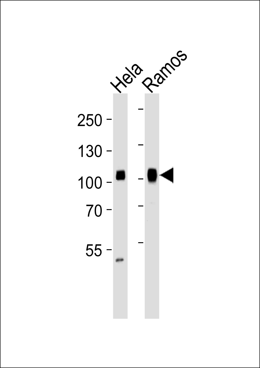 Sample:
Lane 1: Hela cell lysates
Lane 2: Ramos cell lysates
Primary: Anti-SP1 (SLM-51587M) at 1/1000 dilution
Secondary: IRDye800CW Goat Anti-Mouse IgG at 1/20000 dilution
Predicted band size: 81 kD
Observed band size: 105 kD
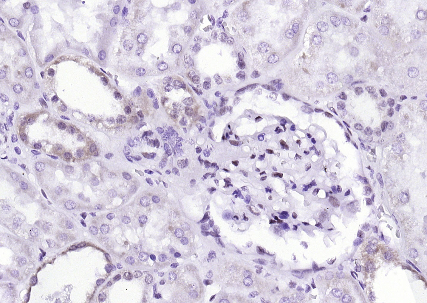 Paraformaldehyde-fixed, paraffin embedded (Human kidney); Antigen retrieval by boiling in sodium citrate buffer (pH6.0) for 15min; Block endogenous peroxidase by 3% hydrogen peroxide for 20 minutes; Blocking buffer (normal goat serum) at 37°C for 30min; Antibody incubation with (SP1 ) Monoclonal Antibody, Unconjugated (SLM-51587M) at 1:200 overnight at 4°C, followed by operating according to SP Kit(Mouse)(sp-0024) instructionsand DAB staining.
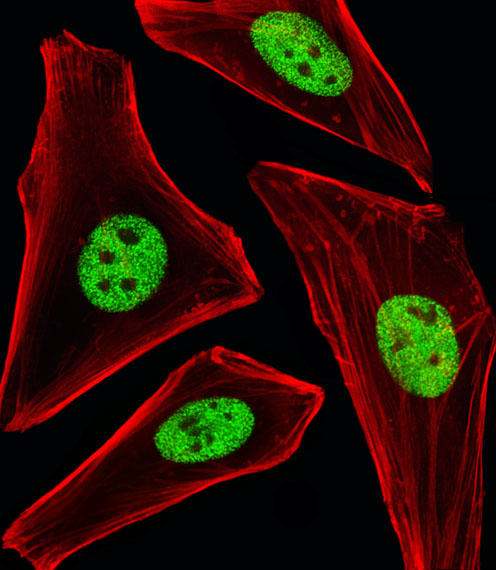 Hela cell; 4% Paraformaldehyde-fixed; Triton X-100 at room temperature for 20 min; Blocking buffer (normal goat serum) at 37°C for 20 min; Antibody incubation with (SP1) monoclonal Antibody, Unconjugated (SLM-51587M) 1:25, 90 minutes at 37°C; followed by a conjugated Goat Anti-Mouse IgG antibody at 37°C for 90 minutes,Alexa Fluor® 555 conjugated with Phalloidin(red) was used to stain the cell Cytoplasmic actin.
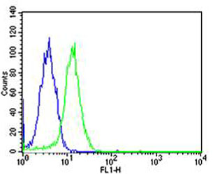 Blank control: Hela.
Primary Antibody (green line): Mouse Anti-SP1 antibody (SLM-51587M)
Dilution: 1:25;
Isotype Control Antibody (blue line): Mouse IgG Secondary Antibody : Goat anti-mouse IgG-AF488
Dilution: 1:400 Protocol
The cells were fixed with 4% PFA (10min at room temperature)and then permeabilized with 90% ice-cold methanol for 20 min at-20℃.The cells were then incubated in 5%BSA to block non-specific protein-protein interactions for 30 min at room temperature .Cells stained with Primary Antibody for 30 min at room temperature. The secondary antibody used for 40 min at room temperature. Acquisition of 20,000 events was performed.
Cartpieces
Totalgoods,subtotals:¥Checkout
References (0)
No References
Bought notes(bought amounts latest0)
No one bought this product
User Comment(Total0User Comment Num)
- No comment


 +86 571 56623320
 +86 18668110335

