
Mouse Anti-NFKB p65 antibody
NF kB P65; NF-kB p65; NFKBp65; NF-κBp65; NF-kBp65; Avian reticuloendotheliosis viral (v rel) oncogene homolog A; MGC131774; NFKB 3; NFKB3; Nuclear Factor NF Kappa B p65 Subunit; Nuclear factor of kappa light polypeptide gene enhancer in B cells 3; Nuclear
View History [Clear]
Details
Product Name [KO validated anti] NFKB p65 Chinese Name The nucleus因子/k基因结合核因子单克隆抗体 Alias NF kB P65; NF-kB p65; NFKBp65; NF-κBp65; NF-kBp65; Avian reticuloendotheliosis viral (v rel) oncogene homolog A; MGC131774; NFKB 3; NFKB3; Nuclear Factor NF Kappa B p65 Subunit; Nuclear factor of kappa light polypeptide gene enhancer in B cells 3; Nuclear Factor Of Kappa Light Polypeptide Gene Enhancer In B Cells; p65; p65 NF kappaB; p65 NFkB; RELA; Transcription Factor p65; v rel avian reticuloendotheliosis viral oncogene homolog A (nuclear factor of kappa light polypeptide gene enhancer in B cells 3 (p65)); V Rel Avian Reticuloendotheliosis Viral Oncogene Homolog A; v rel reticuloendotheliosis viral oncogene homolog A (avian); v-rel reticuloendotheliosis viral oncogene homolog A; p65NFKB; TF65_HUMAN. NFκB-p65; NFκB p65; NF κB-p65; NFκBp65; literatures Research Area Cell biology immunology Neurobiology Signal transduction Apoptosis transcriptional regulatory factor Immunogen Species Mouse Clonality Monoclonal Clone NO. 7G6 React Species Human, Mouse, Rat, Applications WB=1:500-10000 ELISA=1:5000-10000 IHC-P=1:100-500 IHC-F=1:100-500 IF=1:100-500 (Paraffin sections need antigen repair)
not yet tested in other applications.
optimal dilutions/concentrations should be determined by the end user.Theoretical molecular weight 61kDa Cellular localization The nucleus cytoplasmic Form Liquid Concentration 1mg/ml immunogen Recombinant human NFKB p65 Protein Lsotype IgG1 Purification affinity purified by Protein G Buffer Solution 0.01M TBS(pH7.4) with 1% BSA, 0.03% Proclin300 and 50% Glycerol. Storage Shipped at 4℃. Store at -20 °C for one year. Avoid repeated freeze/thaw cycles. Attention This product as supplied is intended for research use only, not for use in human, therapeutic or diagnostic applications. PubMed PubMed Product Detail NF-kappa-B is a ubiquitous transcription factor involved in several biological processes. It is held in the cytoplasm in an inactive state by specific inhibitors. Upon degradation of the inhibitor, NF-kappa-B moves to the nucleus and activates transcription of specific genes. NF-kappa-B is composed of NFKB1 or NFKB2 bound to either REL, RELA, or RELB. The most abundant form of NF-kappa-B is NFKB1 complexed with the product of this gene, RELA. Four transcript variants encoding different isoforms have been found for this gene. [provided by RefSeq, Sep 2011].
Function:
NF-kappa-B is a pleiotropic transcription factor present in almost all cell types and is the endpoint of a series of signal transduction events that are initiated by a vast array of stimuli related to many biological processes such as inflammation, immunity, differentiation, cell growth, tumorigenesis and apoptosis. NF-kappa-B is a homo- or heterodimeric complex formed by the Rel-like domain-containing proteins RELA/p65, RELB, NFKB1/p105, NFKB1/p50, REL and NFKB2/p52 and the heterodimeric p65-p50 complex appears to be most abundant one. The dimers bind at kappa-B sites in the DNA of their target genes and the individual dimers have distinct preferences for different kappa-B sites that they can bind with distinguishable affinity and specificity. Different dimer combinations act as transcriptional activators or repressors, respectively. NF-kappa-B is controlled by various mechanisms of post-translational modification and subcellular compartmentalization as well as by interactions with other cofactors or corepressors. NF-kappa-B complexes are held in the cytoplasm in an inactive state complexed with members of the NF-kappa-B inhibitor (I-kappa-B) family. In a conventional activation pathway, I-kappa-B is phosphorylated by I-kappa-B kinases (IKKs) in response to different activators, subsequently degraded thus liberating the active NF-kappa-B complex which translocates to the nucleus. NF-kappa-B heterodimeric p65-p50 and p65-c-Rel complexes are transcriptional activators. The NF-kappa-B p65-p65 complex appears to be involved in invasin-mediated activation of IL-8 expression. The inhibitory effect of I-kappa-B upon NF-kappa-B the cytoplasm is exerted primarily through the interaction with p65. p65 shows a weak DNA-binding site which could contribute directly to DNA binding in the NF-kappa-B complex. Associates with chromatin at the NF-kappa-B promoter region via association with DDX1.
Subunit:
Component of the NF-kappa-B p65-p50 complex. Component of the NF-kappa-B p65-c-Rel complex. Homodimer; component of the NF-kappa-B p65-p65 complex. Component of the NF-kappa-B p65-p52 complex. May interact with ETHE1. Binds AES and TLE1. Interacts with TP53BP2. Binds to and is phosphorylated by the activated form of either RPS6KA4 or RPS6KA5. Interacts with ING4 and this interaction may be indirect. Interacts with CARM1, USP48 and UNC5CL. Interacts with IRAK1BP1. Interacts with NFKBID. Interacts with NFKBIA. Interacts with GSK3B. Interacts with NFKBIB. Interacts with NFKBIE. Interacts with NFKBIZ. Interacts with EHMT1 (via ANK repeats). Part of a 70-90 kDa complex at least consisting of CHUK, IKBKB, NFKBIA, RELA, IKBKAP and MAP3K14. Interacts with HDAC3; HDAC3 mediates the deacetylation of RELA. Interacts with HDAC1; the interaction requires non-phosphorylated RELA. Interacts with CBP; the interaction requires phosphorylated RELA. Interacts (phosphorylated at 'Thr-254') with PIN1; the interaction inhibits p65 binding to NFKBIA. Interacts with SOCS1. Interacts with UXT. Interacts with MTDH and PHF11. Interacts with ARRB2. Interacts with human respiratory syncytial virus (HRSV) protein M2-1. Interacts with NFKBIA (when phosphorylated), the interaction is direct; phosphorylated NFKBIA is part of a SCF(BTRC)-like complex lacking CUL1. Interacts with RNF25. Interacts (via C-terminus) with DDX1. Interacts with UFL1 and COMMD1. Interacts with BRMS1; this promotes deacetylation of 'Lys-310'. Interacts with NOTCH2. Directly interacts with MEN1; this interaction represses NFKB-mediated transactivation. Interacts with AKIP1, which promotes the phosphorylation and nuclear retention of RELA. Interacts (via the RHD) with GFI1; the interaction, after bacterial lipopolysaccharide (LPS) stimulation, inhibits the transcriptional activity by interfering with the DNA-binding activity to target gene promoter DNA.
Subcellular Location:
Nucleus. Cytoplasm. Note=Colocalized with DDX1 in the nucleus upon TNF-alpha induction. Nuclear, but also found in the cytoplasm in an inactive form complexed to an inhibitor (I-kappa-B). Colocalizes with GFI1 in the nucleus after LPS stimulation.
Post-translational modifications:
Ubiquitinated, leading to its proteasomal degradation. Degradation is required for termination of NF-kappa-B response.
Monomethylated at Lys-310 by SETD6. Monomethylation at Lys-310 is recognized by the ANK repeats of EHMT1 and promotes the formation of repressed chromatin at target genes, leading to down-regulation of NF-kappa-B transcription factor activity. Phosphorylation at Ser-311 disrupts the interaction with EHMT1 without preventing monomethylation at Lys-310 and relieves the repression of target genes.
Phosphorylation at Ser-311 disrupts the interaction with EHMT1 and promotes transcription factor activity. Phosphorylation on Ser-536 stimulates acetylation on Lys-310 and interaction with CBP; the phosphorylated and acetylated forms show enhanced transcriptional activity. Phosphorylation at Ser-276 by RPS6KA4 and RPS6KA5 promotes its transactivation and transcriptional activities.
Reversibly acetylated; the acetylation seems to be mediated by CBP, the deacetylation by HDAC3 and SIRT2. Acetylation at Lys-122 enhances DNA binding and impairs association with NFKBIA. Acetylation at Lys-310 is required for full transcriptional activity in the absence of effects on DNA binding and NFKBIA association. Acetylation can also lower DNA-binding and results in nuclear export. Interaction with BRMS1 promotes deacetylation of Lys-310. Lys-310 is deacetylated by SIRT2.
S-nitrosylation of Cys-38 inactivates the enzyme activity.
Sulfhydration at Cys-38 mediates the anti-apoptotic activity by promoting the interaction with RPS3 and activating the transcription factor activity.
Sumoylation by PIAS3 negatively regulates DNA-bound activated NF-kappa-B.
Similarity:
Contains 1 RHD (Rel-like) domain.
SWISS:
Q04206
Gene ID:
5970
Database links:Entrez Gene: 5970 Human
Entrez Gene: 19697 Mouse
Omim: 164014 Human
SwissProt: Q04206 Human
SwissProt: Q04207 Mouse
Unigene: 502875 Human
Unigene: 249966 Mouse
Unigene: 19480 Rat
Product Picture 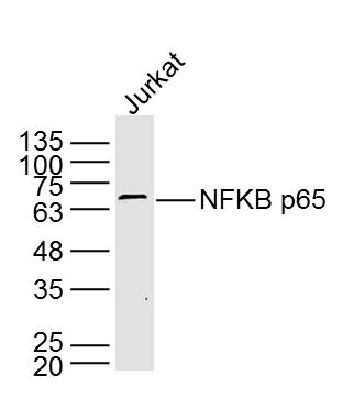 Sample: Jurkat Cell (Human) Lysate at 40 ug
Primary: Anti- NFKB p65 (SLM-33117M) at 1/2 000 dilution
Secondary: IRDye800CW Goat Anti-Mouse IgG at 1/20000 dilution
Predicted band size: 61 kD
Observed band size: 65 kD
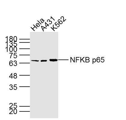 Sample:
Hela Cell (Human) Lysate at 40 ug
A431 Cell (Human) Lysate at 40 ug
K562 Cell (Human) Lysate at 40 ug
Primary: Anti- NFKB p65 (SLM-33117M) at 1/1000 dilution
Secondary: IRDye800CW Goat Anti-Mouse IgG at 1/20000 dilution
Predicted band size: 61 kD
Observed band size: 65 kD
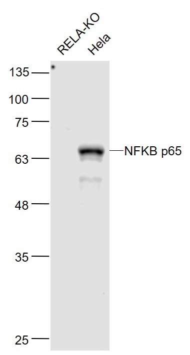 Sample:
RELA-KO(Human) Cell Lysate at 30 ug
Hela(Human) Cell Lysate at 30 ug
Primary: Anti-NFKB p65 (SLM-33117M) at 1/1000 dilution
Secondary: IRDye800CW Goat Anti-Mouse IgG at 1/20000 dilution
Predicted band size: 65 kD
Observed band size: 65 kD
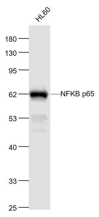 Sample:
HL60A549(Human) Cell Lysate at 30 ug
Primary: Anti-NFKB p65 (SLM-33117M) at 1/1000 dilution
Secondary: IRDye800CW Goat Anti-MouseIgG at 1/20000 dilution
Predicted band size: 65 kD
Observed band size: 62 kD
 Sample:
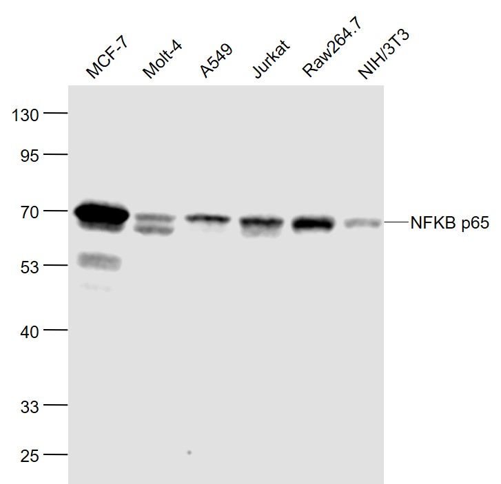
MCF-7(Human) Cell Lysate at 30 ug
Molt-4(Human) Cell Lysate at 30 ug
A549(Human) Cell Lysate at 30 ug
Jurkat(Human) Cell Lysate at 30 ug
Raw264.7(Mouse) Cell Lysate at 30 ug
NIH/3T3(Mouse) Cell Lysate at 30 ug
Primary: Anti-NFKB p65 (SLM-33117M) at 1/1000 dilution
Secondary: IRDye800CW Goat Anti-Mouse IgG at 1/20000 dilution
Predicted band size: 65 kD
Observed band size: 62 kD
 Paraformaldehyde-fixed, paraffin embedded (Rat colon); Antigen retrieval by boiling in sodium citrate buffer (pH6.0) for 15min; Block endogenous peroxidase by 3% hydrogen peroxide for 20 minutes; Blocking buffer (normal goat serum) at 37°C for 30min; Antibody incubation with (NFKB p65) Monoclonal Antibody, Unconjugated (SLM-33117M) at 1:500 overnight at 4°C, followed by a conjugated secondary (sp-0023) for 20 minutes and DAB staining.
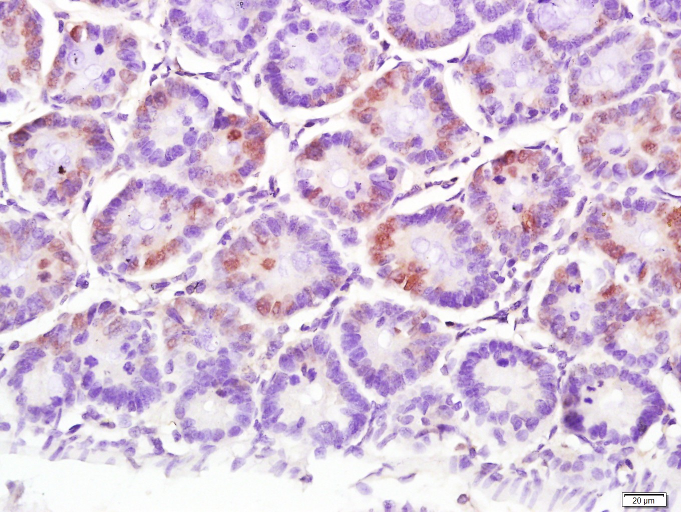
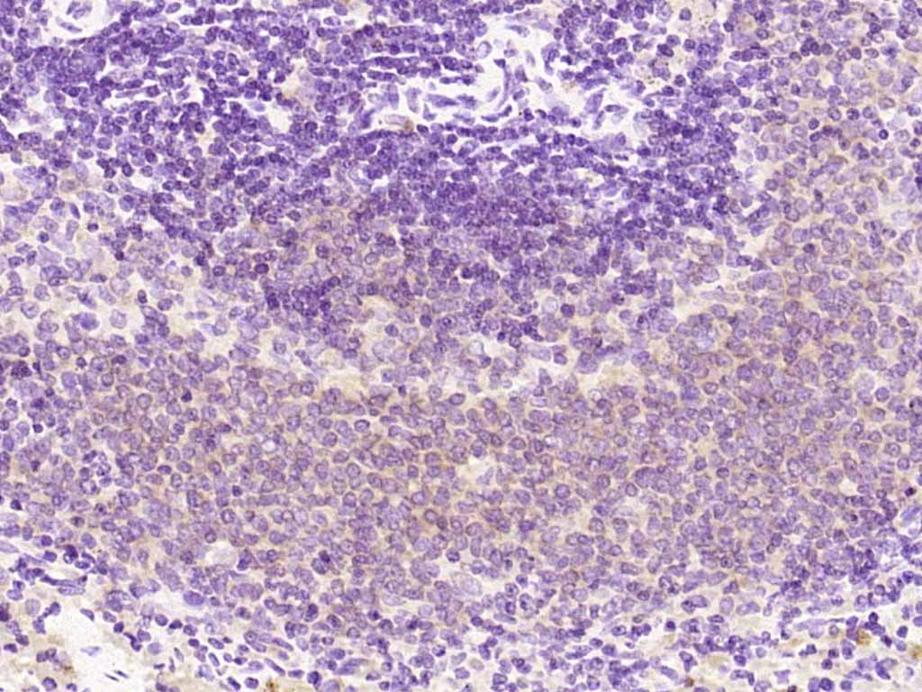 Paraformaldehyde-fixed, paraffin embedded (rat spleen); Antigen retrieval by boiling in sodium citrate buffer (pH6.0) for 15min; Block endogenous peroxidase by 3% hydrogen peroxide for 20 minutes; Blocking buffer (normal goat serum) at 37°C for 30min; Antibody incubation with (NFKB p65) Monoclonal Antibody, Unconjugated (SLM-33117M) at 1:200 overnight at 4°C, followed by operating according to SP Kit(Mouse)(sp-0024) instructionsand DAB staining.
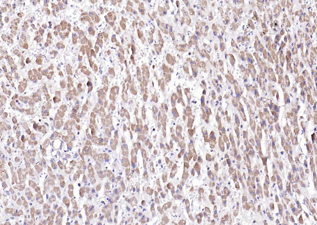 Paraformaldehyde-fixed, paraffin embedded (human liver); Antigen retrieval by boiling in sodium citrate buffer (pH6.0) for 15min; Block endogenous peroxidase by 3% hydrogen peroxide for 20 minutes; Blocking buffer (normal goat serum) at 37°C for 30min; Antibody incubation with (NFKB p65) Monoclonal Antibody, Unconjugated (ascites of SLM-33117M 7G6) at 1:2000 overnight at 4°C, followed by operating according to SP Kit(Mouse) (sp-0024) instructions and DAB staining.
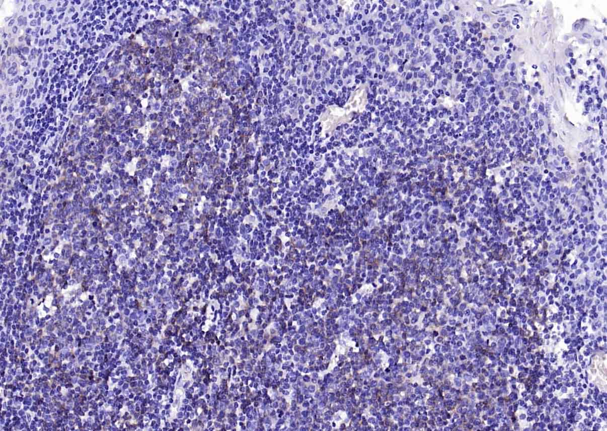 Paraformaldehyde-fixed, paraffin embedded (human tonsil); Antigen retrieval by boiling in sodium citrate buffer (pH6.0) for 15min; Block endogenous peroxidase by 3% hydrogen peroxide for 20 minutes; Blocking buffer (normal goat serum) at 37°C for 30min; Antibody incubation with (NFKB p65) Monoclonal Antibody, Unconjugated (ascites of SLM-33117M 7G6) at 1:2000 overnight at 4°C, followed by operating according to SP Kit(Mouse) (sp-0024) instructions and DAB staining.
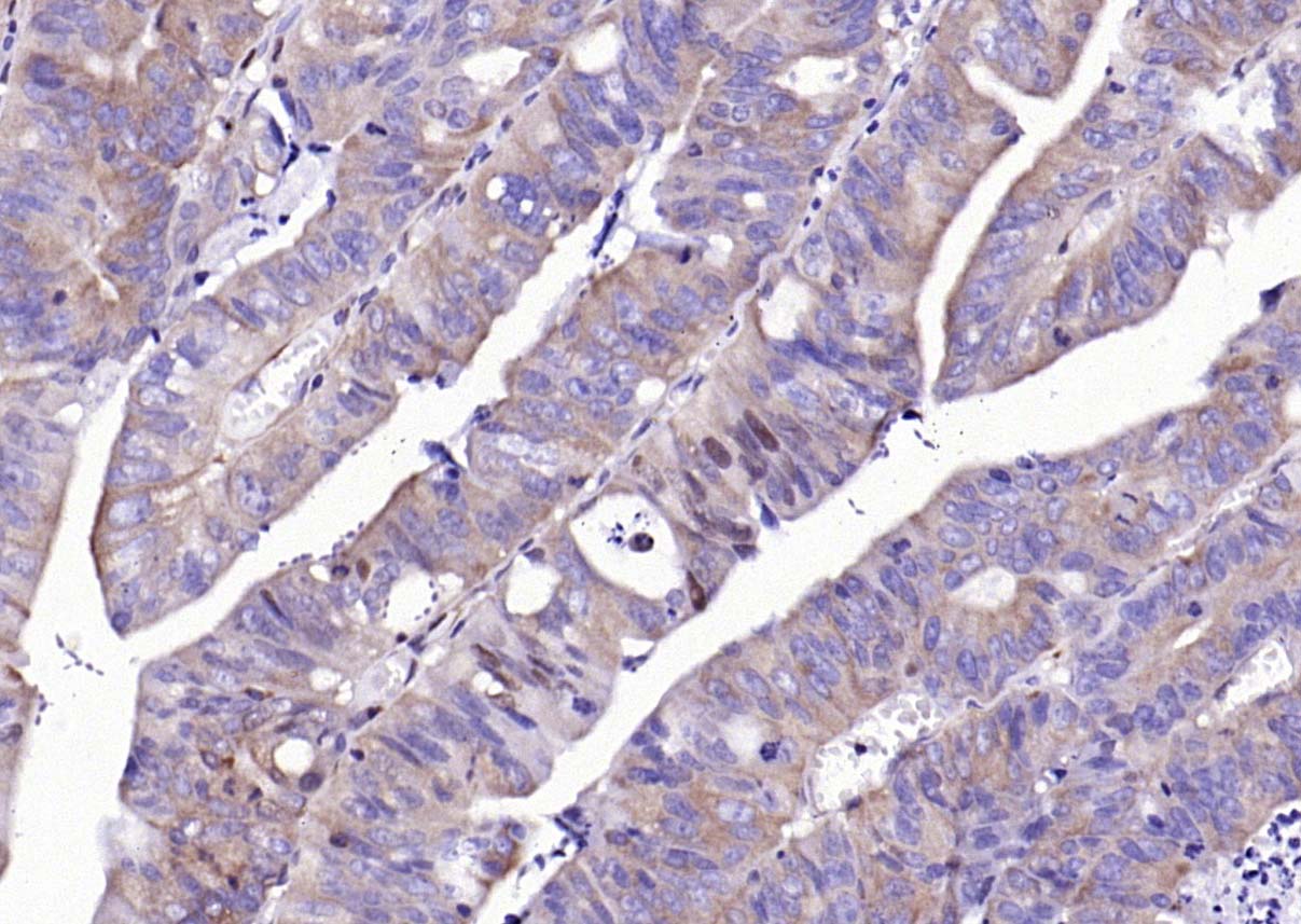 Paraformaldehyde-fixed, paraffin embedded (human colon carcinoma); Antigen retrieval by boiling in sodium citrate buffer (pH6.0) for 15min; Block endogenous peroxidase by 3% hydrogen peroxide for 20 minutes; Blocking buffer (normal goat serum) at 37°C for 30min; Antibody incubation with (NFKB p65) Monoclonal Antibody, Unconjugated (ascites of SLM-33117M 7G6) at 1:2000 overnight at 4°C, followed by operating according to SP Kit(Mouse) (sp-0024) instructions and DAB staining.
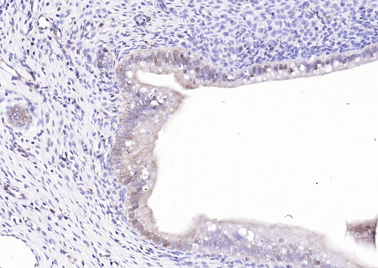 Paraformaldehyde-fixed, paraffin embedded (rat uterus); Antigen retrieval by boiling in sodium citrate buffer (pH6.0) for 15min; Block endogenous peroxidase by 3% hydrogen peroxide for 20 minutes; Blocking buffer (normal goat serum) at 37°C for 30min; Antibody incubation with (NFKB p65) Monoclonal Antibody, Unconjugated (ascites of SLM-33117M 7G6) at 1:2000 overnight at 4°C, followed by operating according to SP Kit(Mouse) (sp-0024) instructions and DAB staining.
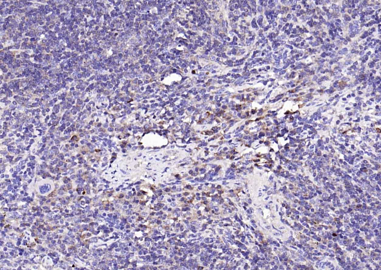 Paraformaldehyde-fixed, paraffin embedded (mouse spleen); Antigen retrieval by boiling in sodium citrate buffer (pH6.0) for 15min; Block endogenous peroxidase by 3% hydrogen peroxide for 20 minutes; Blocking buffer (normal goat serum) at 37°C for 30min; Antibody incubation with (NFKB p65) Monoclonal Antibody, Unconjugated (ascites of SLM-33117M 7G6) at 1:2000 overnight at 4°C, followed by operating according to SP Kit(Mouse) (sp-0024) instructions and DAB staining.
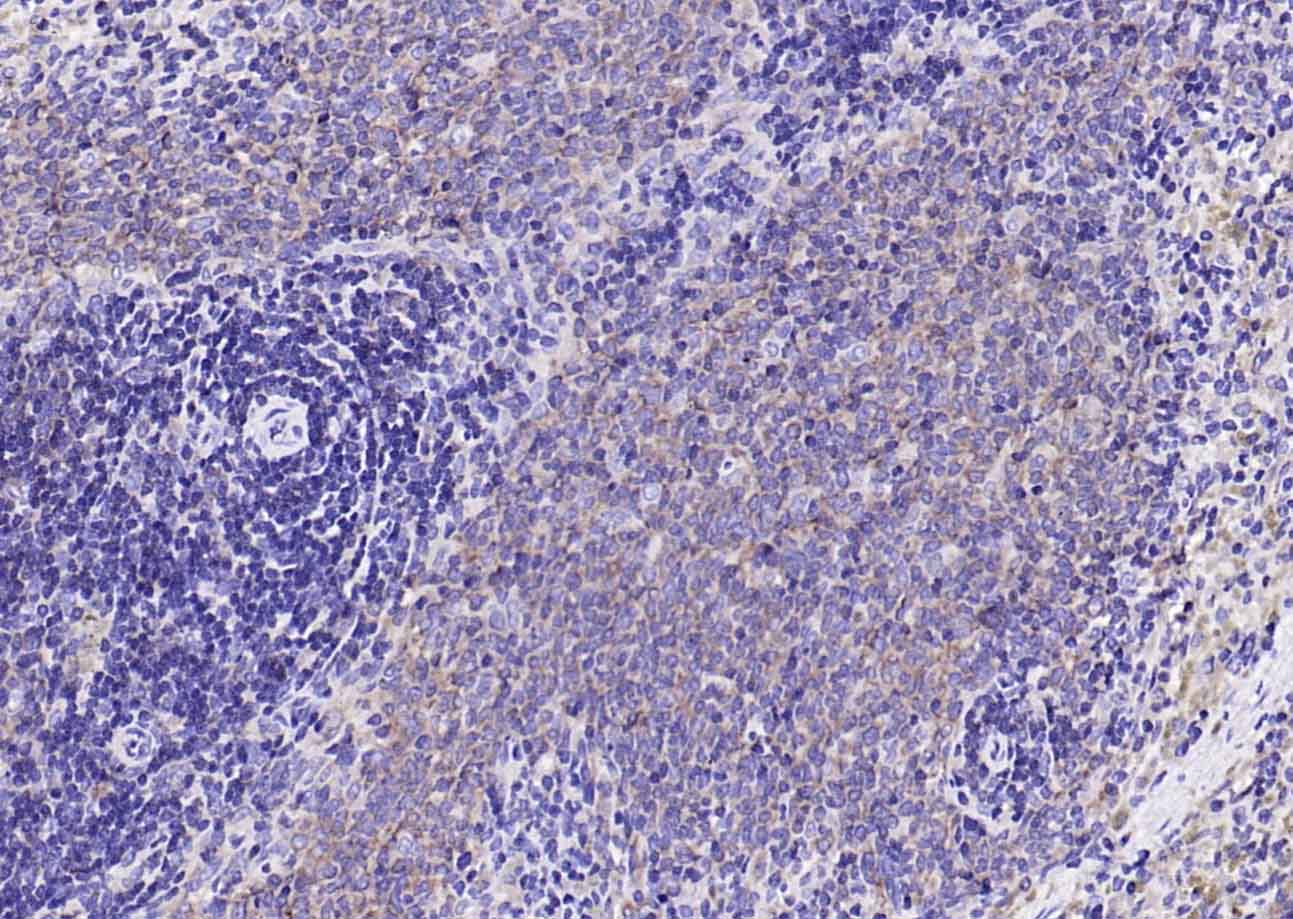 Paraformaldehyde-fixed, paraffin embedded (rat spleen); Antigen retrieval by boiling in sodium citrate buffer (pH6.0) for 15min; Block endogenous peroxidase by 3% hydrogen peroxide for 20 minutes; Blocking buffer (normal goat serum) at 37°C for 30min; Antibody incubation with (NFKB p65) Monoclonal Antibody, Unconjugated (ascites of SLM-33117M 7G6) at 1:2000 overnight at 4°C, followed by operating according to SP Kit(Mouse) (sp-0024) instructions and DAB staining.
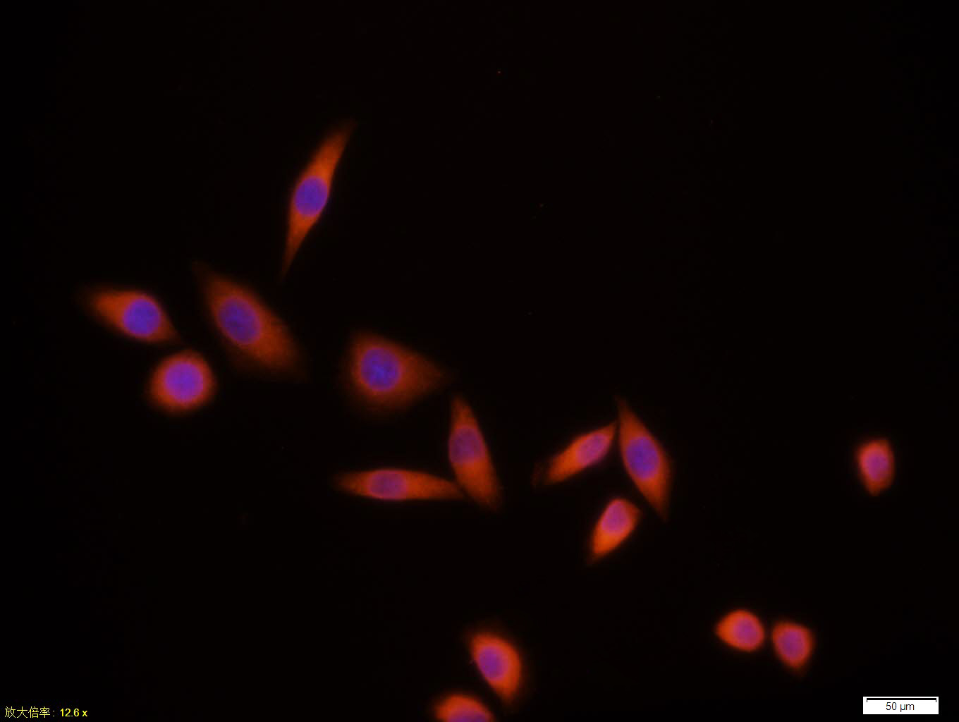 Tissue/cell:MCF7 cell; 4% Paraformaldehyde-fixed; Triton X-100 at room temperature for 20 min; Blocking buffer (normal goat serum,C-0005) at 37°C for 20 min; Antibody incubation with (NFKB p65) monoclonal Antibody, Unconjugated (SLM-33117M)1:100, 90 minutes at 37°C; followed by a CY3 conjugated Goat Anti-Mouse IgG antibody at 37°C for 90 minutes, DAPI (blue, C02-04002) was used to stain the cell nuclei.
Cartpieces
Totalgoods,subtotals:¥Checkout
References (0)
No References
Bought notes(bought amounts latest0)
No one bought this product
User Comment(Total0User Comment Num)
- No comment


 +86 571 56623320
 +86 18668110335

