
Mouse Anti-GFAP antibody
Astrocyte; FLJ45472; GFAP; Glial Fibrillary Acidic Protein; Intermediate filament protein; GFAP_HUMAN.
View History [Clear]
Details
Product Name GFAP Chinese Name 胶质纤维酸性蛋白单克隆抗体 Alias Astrocyte; FLJ45472; GFAP; Glial Fibrillary Acidic Protein; Intermediate filament protein; GFAP_HUMAN. literatures Research Area Tumour Cell biology Neurobiology Immunogen Species Mouse Clonality Monoclonal Clone NO. 7D8 React Species Human, Mouse, Rat, Applications WB=1:500-1000 IHC-P=1:100-500 IHC-F=1:100-500 ICC=1:100-500 (Paraffin sections need antigen repair)
not yet tested in other applications.
optimal dilutions/concentrations should be determined by the end user.Theoretical molecular weight 49kDa Cellular localization cytoplasmic Form Liquid Concentration 1mg/ml immunogen Recombinant mouse GFAP full length Lsotype IgG Purification affinity purified by Protein G Buffer Solution 0.01M TBS(pH7.4) with 1% BSA, 0.03% Proclin300 and 50% Glycerol. Storage Shipped at 4℃. Store at -20 °C for one year. Avoid repeated freeze/thaw cycles. Attention This product as supplied is intended for research use only, not for use in human, therapeutic or diagnostic applications. PubMed PubMed Product Detail This gene encodes one of the major intermediate filament proteins of mature astrocytes. It is used as a marker to distinguish astrocytes from other glial cells during development. Mutations in this gene cause Alexander disease, a rare disorder of astrocytes in the central nervous system. Alternative splicing results in multiple transcript variants encoding distinct isoforms. [provided by RefSeq, Oct 2008]
Function:
GFAP, a class-III intermediate filament, is a cell-specific marker that, during the development of the central nervous system, distinguishes astrocytes from other glial cells.
Subunit:
Interacts with SYNM. Isoform 3 interacts with PSEN1 (via N-terminus).
Subcellular Location:
Cytoplasm. Note=Associated with intermediate filaments.
Tissue Specificity:
Expressed in cells lacking fibronectin.
Post-translational modifications:
Phosphorylated by PKN1.
DISEASE:
Defects in GFAP are a cause of Alexander disease (ALEXD) [MIM:203450]. Alexander disease is a rare disorder of the central nervous system. It is a progressive leukoencephalopathy whose hallmark is the widespread accumulation of Rosenthal fibers which are cytoplasmic inclusions in astrocytes. The most common form affects infants and young children, and is characterized by progressive failure of central myelination, usually leading to death usually within the first decade. Infants with Alexander disease develop a leukoencephalopathy with macrocephaly, seizures, and psychomotor retardation. Patients with juvenile or adult forms typically experience ataxia, bulbar signs and spasticity, and a more slowly progressive course.
Similarity:
Belongs to the intermediate filament family.
SWISS:
P14136
Gene ID:
2670
Database links:
Entrez Gene: 2670 Human
Entrez Gene: 14580 Mouse
Omim: 137780 Human
SwissProt: P14136 Human
SwissProt: P03995 Mouse
星形胶质细胞Maker (Astrocyte Marker)
GFAP是一个56kDa的中间丝蛋白(intermediate filament,IF),在中枢神经系统发育期是一个特异性的Maker,以区别星形细胞和其它胶质细胞。GFAP表达在皮层和海马,急、慢性皮质酮治疗时表达减少。
GFAP可以和人、大鼠、小鼠的GFAP反应,在正常和Tumour性的星形胶质细胞阳性表达,而神经节细胞、神经元、成纤维细胞、少突胶质细胞和这些细胞来源的Tumour细胞阴性表达,主要用于星形胶质瘤等中枢神经系统Tumour的诊断和鉴别诊断,GFAP的缺乏可导致AD病。Product Picture 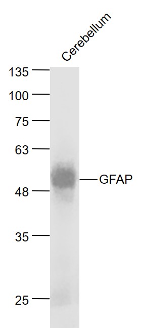 Sample:
Cerebellum (Mouse) Lysate at 40 ug
Primary: Anti- GFAP (SLM-33065M) at 1/1000 dilution
Secondary: IRDye800CW Goat Anti-Rabbit IgG at 1/20000 dilution
Predicted band size: 50 kD
Observed band size: 50 kD
 Sample:
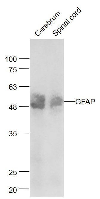
Cerebrum (Mouse) Lysate at 40 ug
Spinal cord (Mouse) Lysate at 40 ug
Primary: Anti- TBX1 (SLM-33065M) at 1/1000 dilution
Secondary: IRDye800CW Goat Anti-Rabbit IgG at 1/20000 dilution
Predicted band size: 50 kD
Observed band size: 50 kD
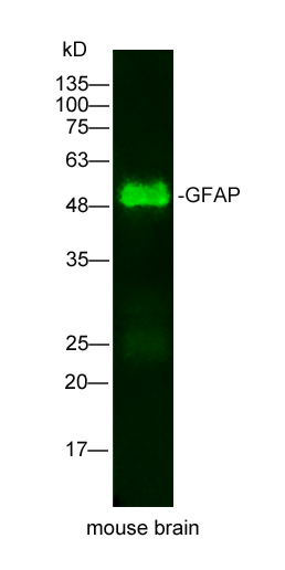 Sample: mouse brain Lysate at 25 ug
Primary: Mouse Anti-GFAP(SLM-33065M) at 1/500 dilution
Secondary: IRDye800CW Goat Anti-Mouse IgG at 1/20000 dilution
Predicted band size: 49kD
Observed band size: 49kD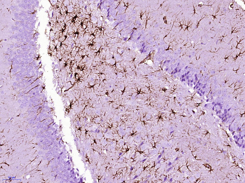 Paraformaldehyde-fixed, paraffin embedded (Rat brain); Antigen retrieval by boiling in sodium citrate buffer (pH6.0) for 15min; Block endogenous peroxidase by 3% hydrogen peroxide for 20 minutes; Blocking buffer (normal goat serum) at 37°C for 30min; Antibody incubation with (GFAP) Monoclonal Antibody, Unconjugated (SLM-33065M) at 1:400 overnight at 4°C, followed by operating according to SP Kit(Mouse) (sp-0024) instructionsand DAB staining.
 Paraformaldehyde-fixed, paraffin embedded (mouse cerebellum); Antigen retrieval by boiling in sodium citrate buffer (pH6.0) for 15min; Block endogenous peroxidase by 3% hydrogen peroxide for 20 minutes; Blocking buffer (normal goat serum) at 37°C for 30min; Antibody incubation with (GFAP) Monoclonal Antibody, Unconjugated (SLM-33065M) at 1:200 overnight at 4°C, followed by operating according to SP Kit(Mouse)(sp-0024) instructionsand DAB staining.
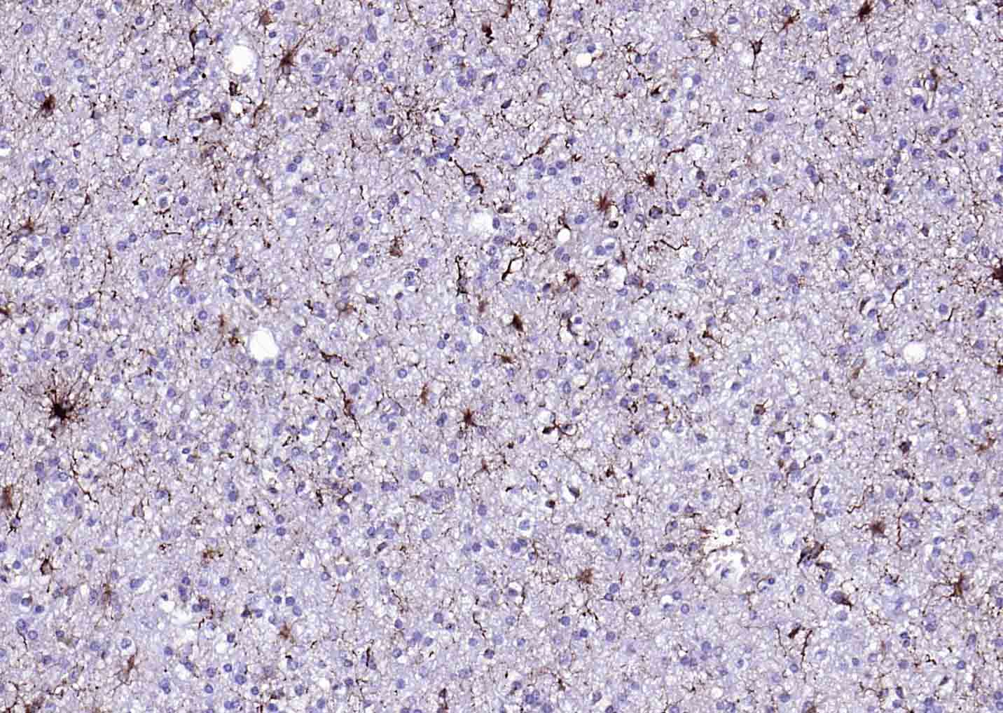 Paraformaldehyde-fixed, paraffin embedded (human brain glioma); Antigen retrieval by boiling in sodium citrate buffer (pH6.0) for 15min; Block endogenous peroxidase by 3% hydrogen peroxide for 20 minutes; Blocking buffer (normal goat serum) at 37°C for 30min; Antibody incubation with (GFAP) Monoclonal Antibody, Unconjugated (SLM-33065M) at 1:200 overnight at 4°C, followed by operating according to SP Kit(Mouse)(sp-0024) instructionsand DAB staining.
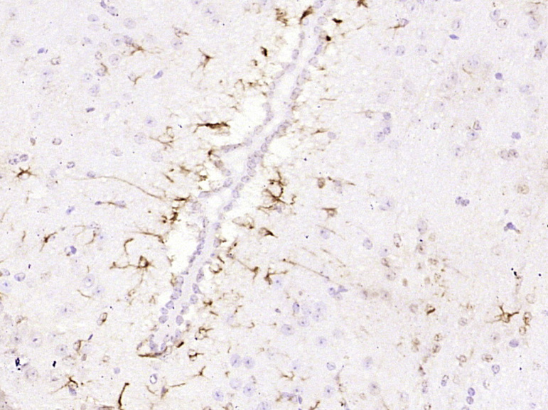 Paraformaldehyde-fixed, paraffin embedded (Mouse brain); Antigen retrieval by boiling in sodium citrate buffer (pH6.0) for 15min; Block endogenous peroxidase by 3% hydrogen peroxide for 20 minutes; Blocking buffer (normal goat serum) at 37°C for 30min; Antibody incubation with (GFAP) Monoclonal Antibody, Unconjugated (SLM-33065M) at 1:800 overnight at 4°C, followed by operating according to SP Kit(Mouse) (sp-0024) instructions and DAB staining.
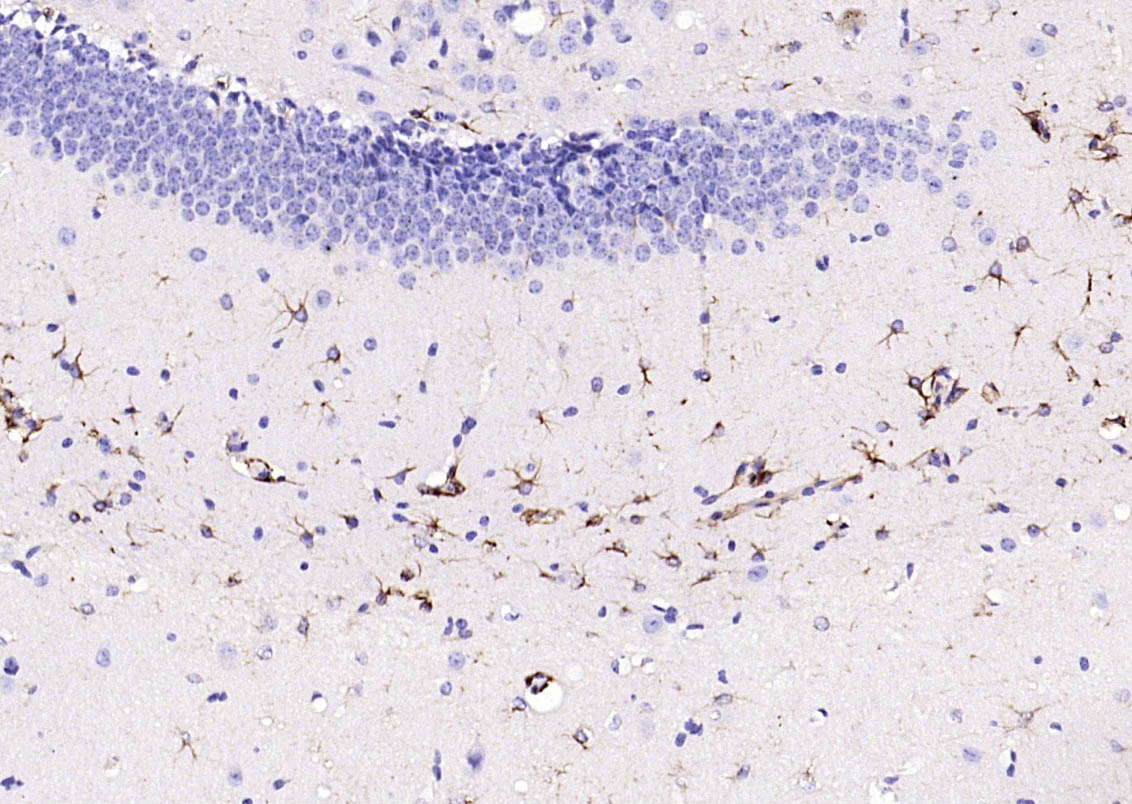 Paraformaldehyde-fixed, paraffin embedded (mouse brain); Antigen retrieval by boiling in sodium citrate buffer (pH6.0) for 15min; Block endogenous peroxidase by 3% hydrogen peroxide for 20 minutes; Blocking buffer (normal goat serum) at 37°C for 30min; Antibody incubation with (GFAP) Monoclonal Antibody, Unconjugated (SLM-33065M) at 1:200 overnight at 4°C, followed by operating according to SP Kit(Mouse)(sp-0024) instructionsand DAB staining.
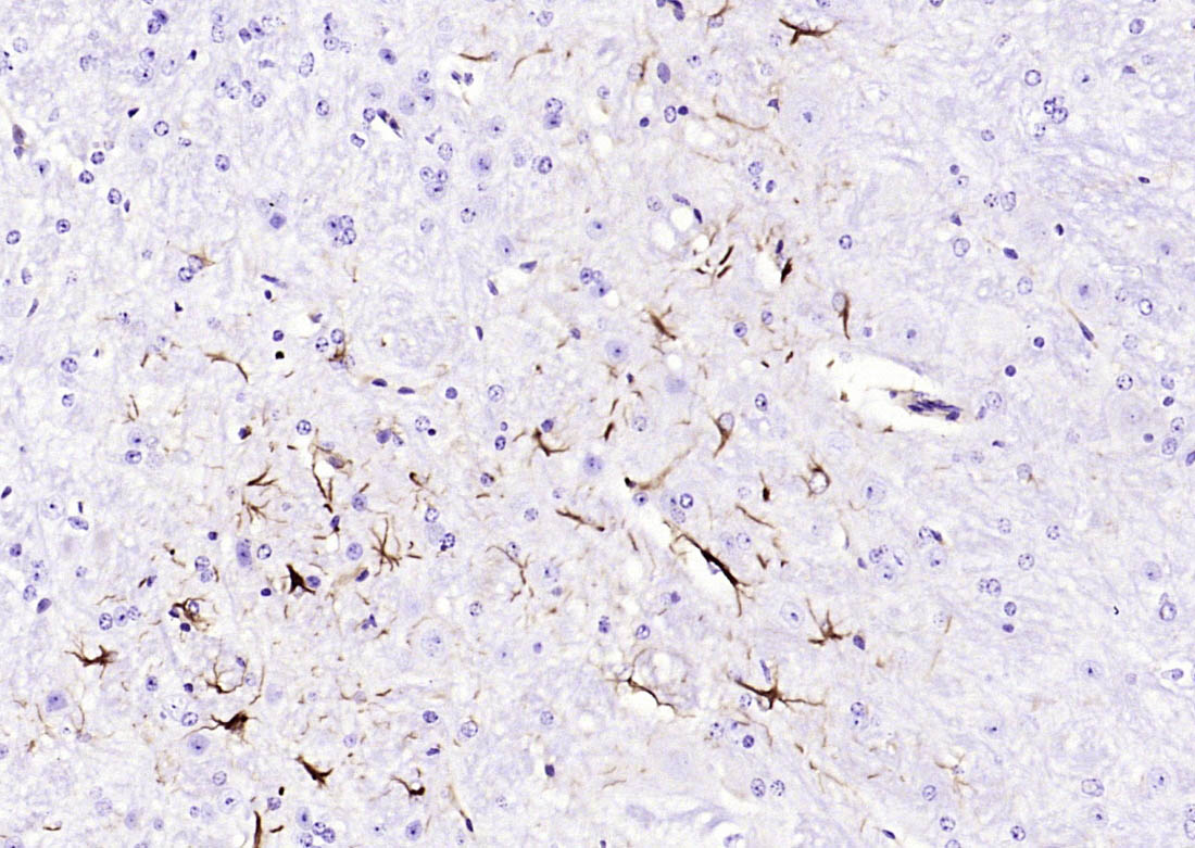 Paraformaldehyde-fixed, paraffin embedded (mouse cerebellum); Antigen retrieval by boiling in sodium citrate buffer (pH6.0) for 15min; Block endogenous peroxidase by 3% hydrogen peroxide for 20 minutes; Blocking buffer (normal goat serum) at 37°C for 30min; Antibody incubation with (GFAP) Monoclonal Antibody, Unconjugated (SLM-33065M) at 1:2000 overnight at 4°C, followed by operating according to SP Kit(Mouse)(sp-0024) instructionsand DAB staining.
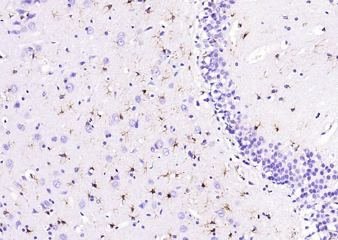 Paraformaldehyde-fixed, paraffin embedded (rat brain); Antigen retrieval by boiling in sodium citrate buffer (pH6.0) for 15min; Block endogenous peroxidase by 3% hydrogen peroxide for 20 minutes; Blocking buffer (normal goat serum) at 37°C for 30min; Antibody incubation with (GFAP) Monoclonal Antibody, Unconjugated (SLM-33065M) at 1:200 overnight at 4°C, followed by operating according to SP Kit(Mouse)(sp-0024) instructionsand DAB staining.
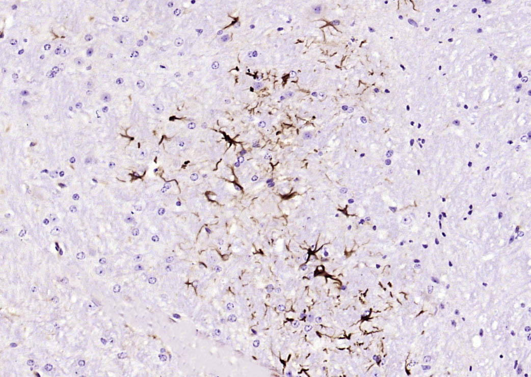 Paraformaldehyde-fixed, paraffin embedded (mouse cerebellum); Antigen retrieval by boiling in sodium citrate buffer (pH6.0) for 15min; Block endogenous peroxidase by 3% hydrogen peroxide for 20 minutes; Blocking buffer (normal goat serum) at 37°C for 30min; Antibody incubation with (GFAP) Monoclonal Antibody, Unconjugated (SLM-33065M) at 1:200 overnight at 4°C, followed by operating according to SP Kit(Mouse)(sp-0024) instructionsand DAB staining.
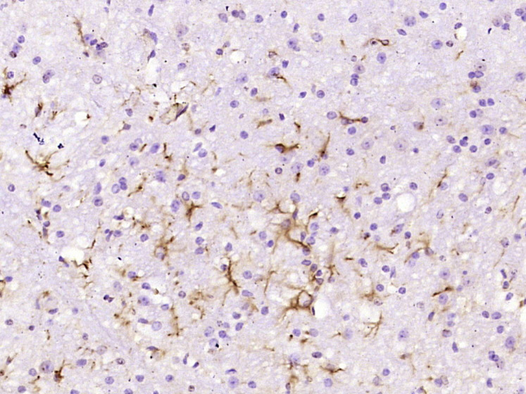 Paraformaldehyde-fixed, paraffin embedded (Mouse cerebellum); Antigen retrieval by boiling in sodium citrate buffer (pH6.0) for 15min; Block endogenous peroxidase by 3% hydrogen peroxide for 20 minutes; Blocking buffer (normal goat serum) at 37°C for 30min; Antibody incubation with (GFAP) Monoclonal Antibody, Unconjugated (SLM-33065M) at 1:800 overnight at 4°C, followed by operating according to SP Kit(Mouse) (sp-0024) instructions and DAB staining.
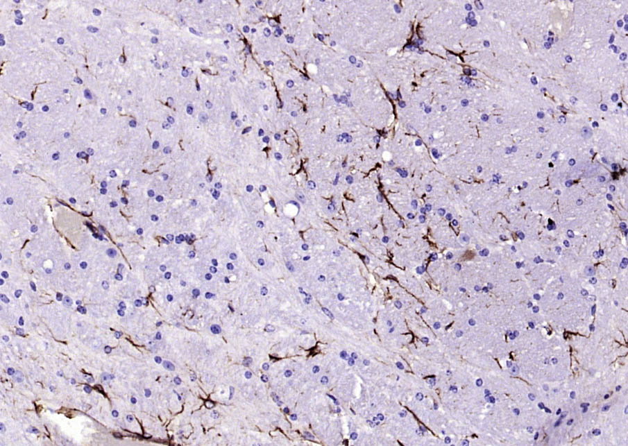 Paraformaldehyde-fixed, paraffin embedded (mouse brain); Antigen retrieval by boiling in sodium citrate buffer (pH6.0) for 15min; Block endogenous peroxidase by 3% hydrogen peroxide for 20 minutes; Blocking buffer (normal goat serum) at 37°C for 30min; Antibody incubation with (GFAP) Monoclonal Antibody, Unconjugated (SLM-33065M) at 1:200 overnight at 4°C, followed by operating according to SP Kit(Mouse)(sp-0024) instructionsand DAB staining.
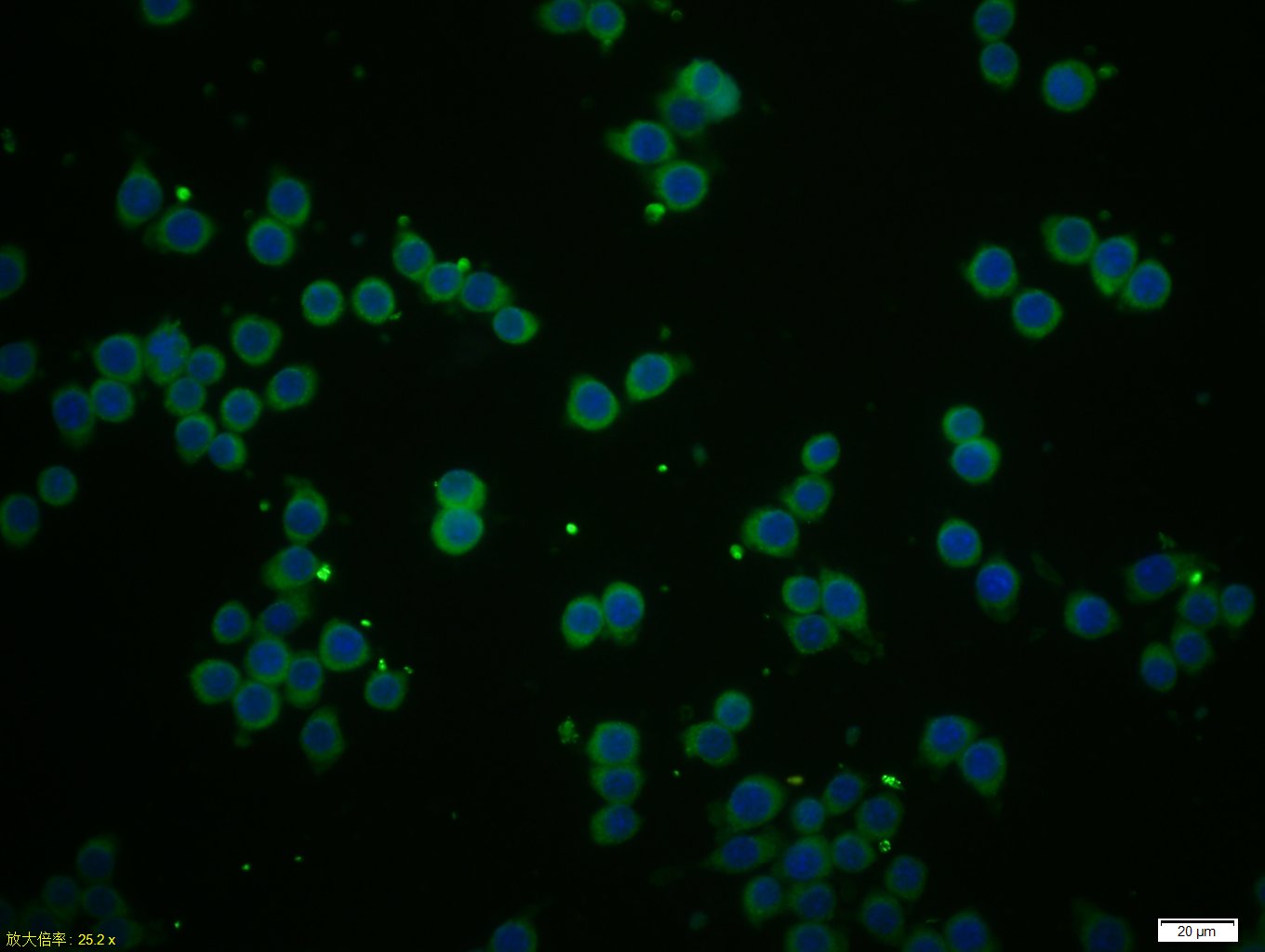 Tissue/cell: BV-2 cell; 4% Paraformaldehyde-fixed; Triton X-100 at room temperature for 20 min; Blocking buffer (normal goat serum, C-0005) at 37°C for 20 min; Antibody incubation with (GFAP) Monoclonal Antibody, Unconjugated (SLM-33065M) 1:50, 90 minutes at 37°C; followed by a conjugated Goat Anti-Mouse IgG antibody (SL0296G-FITC) at 37°C for 90 minutes, DAPI (5ug/ml, blue, C-0033) was used to stain the cell nuclei.
Cartpieces
Totalgoods,subtotals:¥Checkout
References (0)
No References
Bought notes(bought amounts latest0)
No one bought this product
User Comment(Total0User Comment Num)
- No comment


 +86 571 56623320
 +86 18668110335

