
Rabbit Anti-RGC32/C13orf15 antibody
RGC32; Chromosome 13 open reading frame 15; KIAA0564; MGC87338; response gene to complement 32; Response gene to complement 32 protein; RGC 32; RGCC_HUMAN.
View History [Clear]
Details
Product Name RGC32/C13orf15 Chinese Name 补体应答基因32抗体 Alias RGC32; Chromosome 13 open reading frame 15; KIAA0564; MGC87338; response gene to complement 32; Response gene to complement 32 protein; RGC 32; RGCC_HUMAN. literatures Research Area Tumour Cell biology immunology Cyclin Immunogen Species Rabbit Clonality Polyclonal React Species Human, Mouse, Rat, (predicted: Chicken, Dog, Pig, Cow, Horse, Rabbit, Sheep, ) Applications ELISA=1:5000-10000 IHC-P=1:100-500 IHC-F=1:100-500 Flow-Cyt=1ug/test IF=1:50-200 (Paraffin sections need antigen repair)
not yet tested in other applications.
optimal dilutions/concentrations should be determined by the end user.Theoretical molecular weight 15kDa Cellular localization The nucleus cytoplasmic Form Liquid Concentration 1mg/ml immunogen KLH conjugated synthetic peptide derived from human RGC32: 65-137/137 Lsotype IgG Purification affinity purified by Protein A Buffer Solution 0.01M TBS(pH7.4) with 1% BSA, 0.03% Proclin300 and 50% Glycerol. Storage Shipped at 4℃. Store at -20 °C for one year. Avoid repeated freeze/thaw cycles. Attention This product as supplied is intended for research use only, not for use in human, therapeutic or diagnostic applications. PubMed PubMed Product Detail RGC32, also known as C13orf15, is a 137 amino acid protein that localizes to the cytoplasm, as well as to the nucleus and the centrosome. Expressed at high levels in kidney, pancreas and skeletal muscle and at lower levels in brain, heart and placenta, RGC32 functions to modulate the activity of cell cycle-specific kinases, thereby regulating cell cycle progression. Additionally, RGC32 may promote cell cycle arrest at the G2/M phase transition and is thought to inhibit the growth of glioma cells, possibly functioning as a tumor suppressor. Conversely, overexpression of RGC32 may promote cell replication and assist in the pathogenesis of malignancies, suggesting that RGC32 also participates in tumor transformation and progression. RGC32 activity is induced by complement activation and by p53 in response to DNA damage. Multiple isoforms of RGC32 exist as a result of alternative splicing events.
Function:
Modulates the activity of cell cycle-specific kinases. Enhances CDK1 activity. May contribute to the regulation of the cell cycle. May inhibit growth of glioma cells by promoting arrest of mitotic progression at the G2/M transition. Fibrogenic factor contributing to the pathogenesis of renal fibrosis through fibroblast activation.
Subunit:
Interacts with SMAD3. Interacts with CDK1 and PLK1.
Subcellular Location:
Cytoplasm. Nucleus. Cytoplasm ?cytoskeleton ?centrosome. Note: Cytoplasmic in unstimulated cells. Nuclear after activation by complement. Associated with the centrosome during prometaphase and metaphase.
Tissue Specificity:
Detected in brain, heart and liver (at protein level). Highly expressed in liver, skeletal muscle, kidney and pancreas. Detected at lower levels in heart, brain and placenta. Detected in aorta endothelial cells. Overexpressed in colon, breast, prostate, bladder, lung, and ovarian cancer tissues.
SWISS:
Q9H4X1
Gene ID:
28984
Database links:Entrez Gene: 28984 Human
Omim: 610077 Human
SwissProt: Q9H4X1 Human
Product Picture 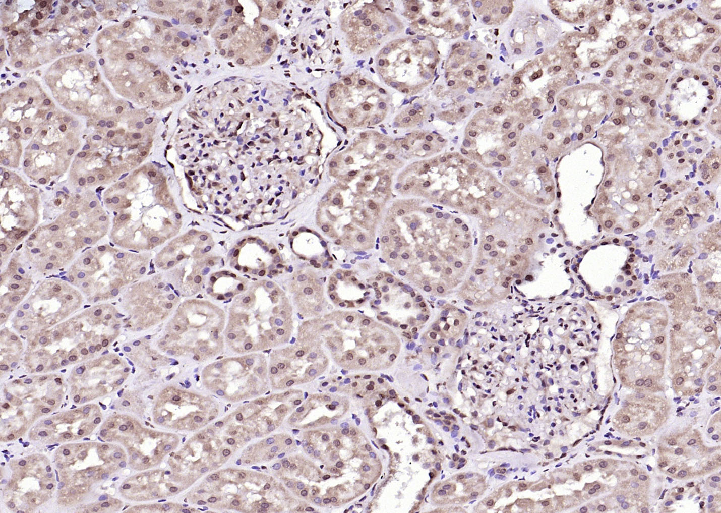 Paraformaldehyde-fixed, paraffin embedded (Human kidney); Antigen retrieval by boiling in sodium citrate buffer (pH6.0) for 15min; Block endogenous peroxidase by 3% hydrogen peroxide for 20 minutes; Blocking buffer (normal goat serum) at 37°C for 30min; Antibody incubation with (RGC32) Polyclonal Antibody, Unconjugated (SL9079R) at 1:200 overnight at 4°C, followed by operating according to SP Kit(Rabbit) (sp-0023) instructionsand DAB staining.
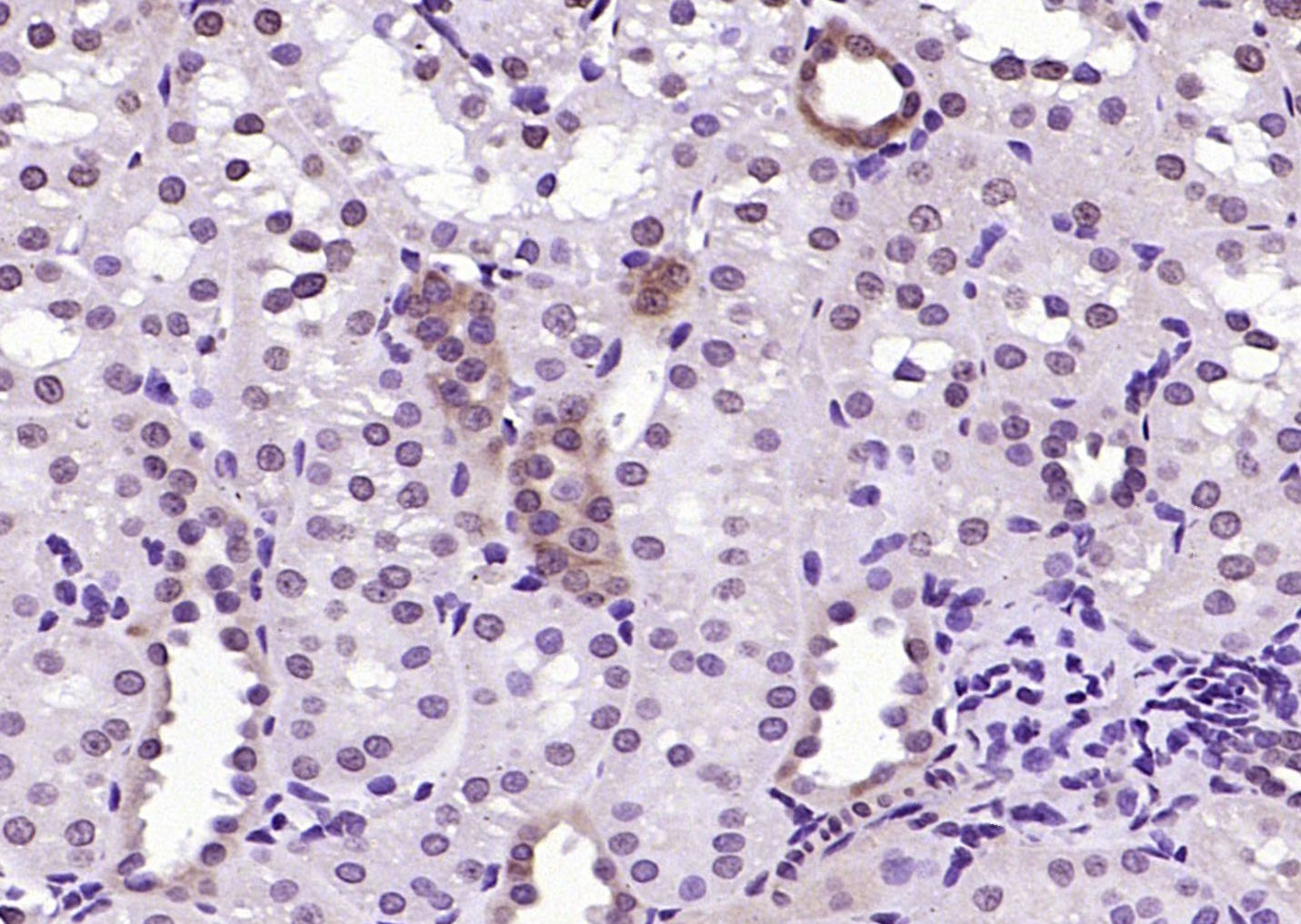 Paraformaldehyde-fixed, paraffin embedded (rat kidney); Antigen retrieval by boiling in sodium citrate buffer (pH6.0) for 15min; Block endogenous peroxidase by 3% hydrogen peroxide for 20 minutes; Blocking buffer (normal goat serum) at 37°C for 30min; Antibody incubation with (RGC32) Polyclonal Antibody, Unconjugated (SL9079R) at 1:200 overnight at 4°C, followed by operating according to SP Kit(Rabbit) (sp-0023) instructionsand DAB staining.
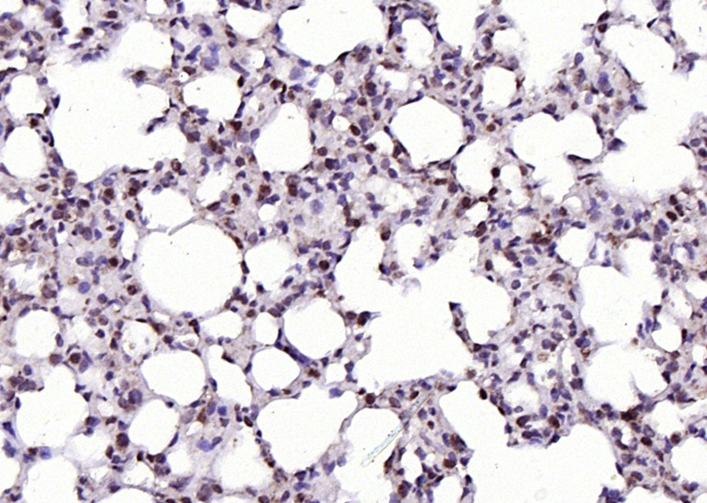 Paraformaldehyde-fixed, paraffin embedded (rat lung); Antigen retrieval by boiling in sodium citrate buffer (pH6.0) for 15min; Block endogenous peroxidase by 3% hydrogen peroxide for 20 minutes; Blocking buffer (normal goat serum) at 37°C for 30min; Antibody incubation with (RGC32) Polyclonal Antibody, Unconjugated (SL9079R) at 1:200 overnight at 4°C, followed by operating according to SP Kit(Rabbit) (sp-0023) instructionsand DAB staining.
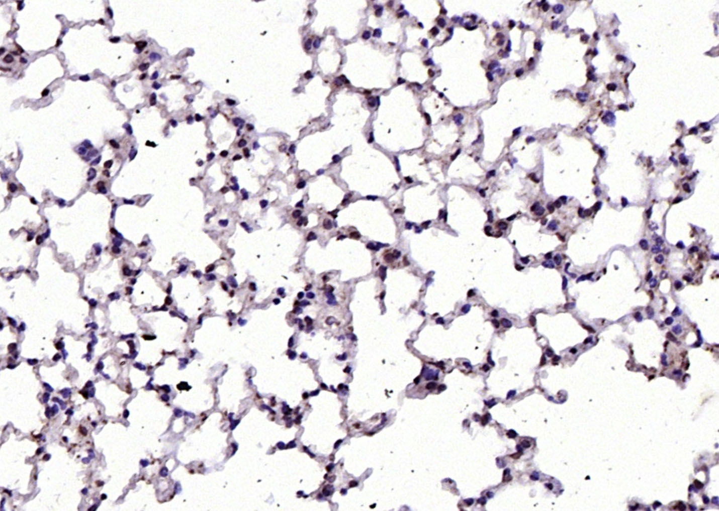 Paraformaldehyde-fixed, paraffin embedded (mouse lung); Antigen retrieval by boiling in sodium citrate buffer (pH6.0) for 15min; Block endogenous peroxidase by 3% hydrogen peroxide for 20 minutes; Blocking buffer (normal goat serum) at 37°C for 30min; Antibody incubation with (RGC32) Polyclonal Antibody, Unconjugated (SL9079R) at 1:200 overnight at 4°C, followed by operating according to SP Kit(Rabbit) (sp-0023) instructionsand DAB staining.
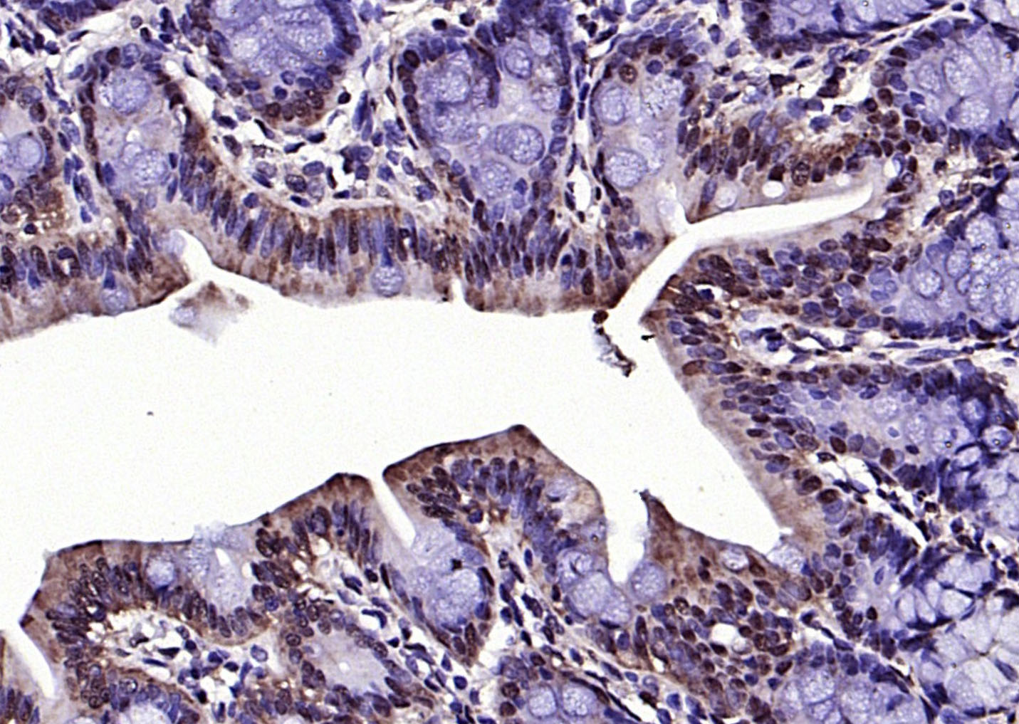 Paraformaldehyde-fixed, paraffin embedded (rat colon); Antigen retrieval by boiling in sodium citrate buffer (pH6.0) for 15min; Block endogenous peroxidase by 3% hydrogen peroxide for 20 minutes; Blocking buffer (normal goat serum) at 37°C for 30min; Antibody incubation with (RGC32) Polyclonal Antibody, Unconjugated (SL9079R) at 1:200 overnight at 4°C, followed by operating according to SP Kit(Rabbit) (sp-0023) instructionsand DAB staining.
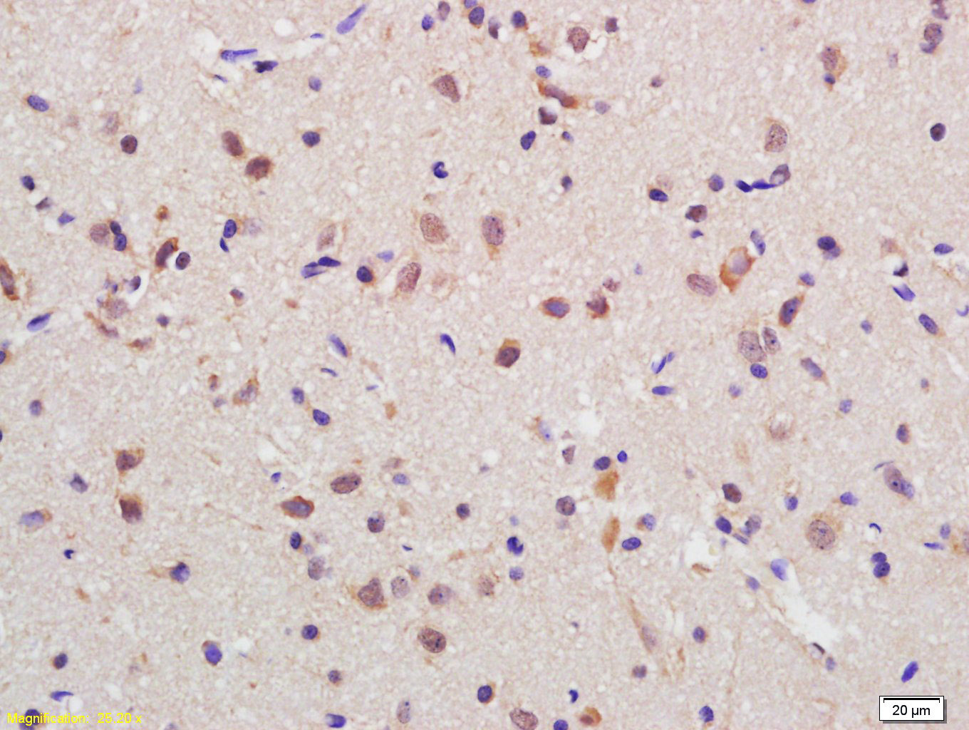 Tissue/cell: rat brain tissue; 4% Paraformaldehyde-fixed and paraffin-embedded;
Antigen retrieval: citrate buffer ( 0.01M, pH 6.0 ), Boiling bathing for 15min; Block endogenous peroxidase by 3% Hydrogen peroxide for 30min; Blocking buffer (normal goat serum,C-0005) at 37℃ for 20 min;
Incubation: Anti-C13orf15 Polyclonal Antibody, Unconjugated(SL9079R) 1:200, overnight at 4°C, followed by conjugation to the secondary antibody(SP-0023) and DAB(C-0010) staining
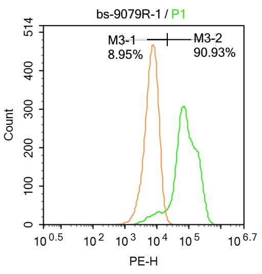 Blank control:Molt-4.
Primary Antibody (green line): Rabbit Anti-C13orf15 antibody (SL9079R)
Dilution: 1μg /10^6 cells;
Isotype Control Antibody (orange line): Rabbit IgG .
Secondary Antibody : Goat anti-rabbit IgG-AF647
Dilution: 1μg /test.
Protocol
The cells were fixed with 4% PFA (10min at room temperature)and then permeabilized with 90% ice-cold methanol for 20 min at-20℃. The cells were then incubated in 5%BSA to block non-specific protein-protein interactions for 30 min at at room temperature .Cells stained with Primary Antibody for 30 min at room temperature. The secondary antibody used for 40 min at room temperature. Acquisition of 20,000 events was performed.
Cartpieces
Totalgoods,subtotals:¥Checkout
References (0)
No References
Bought notes(bought amounts latest0)
No one bought this product
User Comment(Total0User Comment Num)
- No comment


 +86 571 56623320
 +86 18668110335

