
Rabbit Anti-IRE1 antibody
IRE1a; Endoplasmic reticulum (ER) to nucleus signalling 1; Endoplasmic reticulum to nucleus signaling 1; Endoplasmic reticulum-to-nucleus signaling 1; Endoribonuclease; ER to nucleus signaling 1; ERN 1; ERN1; ERN1_HUMAN; hIRE 1p; hIRE1p; Inositol requirin
View History [Clear]
Details
Product Name IRE1 Chinese Name 内质网核Signal transduction蛋白a1抗体 Alias IRE1a; Endoplasmic reticulum (ER) to nucleus signalling 1; Endoplasmic reticulum to nucleus signaling 1; Endoplasmic reticulum-to-nucleus signaling 1; Endoribonuclease; ER to nucleus signaling 1; ERN 1; ERN1; ERN1_HUMAN; hIRE 1p; hIRE1p; Inositol requiring 1; Inositol requiring protein 1; Inositol-requiring protein 1; IRE-1; IRE 1; IRE 1a; IRE 1P; Ire1 alpha; Ire1-alpha; Ire1alpha; IRE1P; MGC163277; Protein kinase/endoribonuclease; Serine/threonine protein kinase/endoribonuclease IRE1. literatures Research Area Cell biology Signal transduction Kinases and Phosphatases Immunogen Species Rabbit Clonality Polyclonal React Species Human, Rat, (predicted: Mouse, Dog, Pig, Cow, Horse, Rabbit, ) Applications WB=1:500-2000 ELISA=1:5000-10000 IHC-P=1:100-500 IHC-F=1:100-500 Flow-Cyt=1ug/test ICC=1:100-500 IF=1:100-500 (Paraffin sections need antigen repair)
not yet tested in other applications.
optimal dilutions/concentrations should be determined by the end user.Theoretical molecular weight 105kDa Cellular localization cytoplasmic The cell membrane Form Liquid Concentration 1mg/ml immunogen KLH conjugated synthetic peptide derived from human IRE1a: 252-260/977 Lsotype IgG Purification affinity purified by Protein A Buffer Solution 0.01M TBS(pH7.4) with 1% BSA, 0.03% Proclin300 and 50% Glycerol. Storage Shipped at 4℃. Store at -20 °C for one year. Avoid repeated freeze/thaw cycles. Attention This product as supplied is intended for research use only, not for use in human, therapeutic or diagnostic applications. PubMed PubMed Product Detail The protein encoded by this gene is the ER to nucleus signalling 1 protein, a human homologue of the yeast Ire1 gene product. This protein possesses intrinsic kinase activity and an endoribonuclease activity and it is important in altering gene expression as a response to endoplasmic reticulum-based stress signals. [provided by RefSeq, Jul 2008]
Function:
Senses unfolded proteins in the lumen of the endoplasmic reticulum via its N-terminal domain which leads to enzyme auto-activation. The active endoribonuclease domain splices XBP1 mRNA to generate a new C-terminus, converting it into a potent unfolded-protein response transcriptional activator and triggering growth arrest and apoptosis.
Subunit:
Homodimer; disulfide-linked. Dimer formation is driven by hydrophobic interactions within the N-terminal luminal domains and stabilized by disulfide bridges. Also binds HSPA5, a negative regulator of the unfolded protein response. This interaction may disrupt homodimerization and prevent activation of ERN1. Interacts with TAOK3 and TRAF2.
Subcellular Location:
Endoplasmic reticulum membrane.
Tissue Specificity:
Ubiquitously expressed. High levels observed in pancreatic tissue.
Post-translational modifications:
Autophosphorylated.
Similarity:
Belongs to the protein kinase superfamily. Ser/Thr protein kinase family.
Contains 1 KEN domain.
Contains 1 protein kinase domain.
SWISS:
O75460
Gene ID:
2081
Database links:Entrez Gene: 2081 Human
Entrez Gene: 78943 Mouse
Omim: 604033 Human
SwissProt: O75460 Human
SwissProt: Q9EQY0 Mouse
Unigene: 133982 Human
Unigene: 592041 Human
Unigene: 700027 Human
Unigene: 20452 Mouse
Unigene: 340943 Mouse
Unigene: 226435 Rat
Product Picture 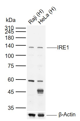 Sample:
Lane 1: Human Raji cell lysates
Lane 2: Human HeLa cell lysates
Primary: Anti-IRE1 (SL8680R) at 1/1000 dilution
Secondary: IRDye800CW Goat Anti-Rabbit IgG at 1/20000 dilution
Predicted band size: 105 kDa
Observed band size: 135 kDa
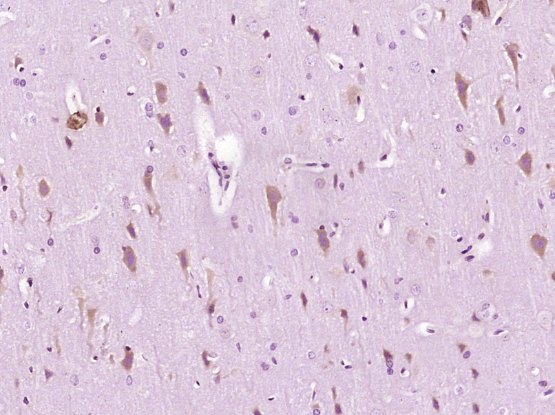 Paraformaldehyde-fixed, paraffin embedded (Rat brain); Antigen retrieval by boiling in sodium citrate buffer (pH6.0) for 15min; Block endogenous peroxidase by 3% hydrogen peroxide for 20 minutes; Blocking buffer (normal goat serum) at 37°C for 30min; Antibody incubation with (IRE1) Polyclonal Antibody, Unconjugated (SL8680R) at 1:400 overnight at 4°C, followed by operating according to SP Kit(Rabbit) (sp-0023) instructionsand DAB staining.
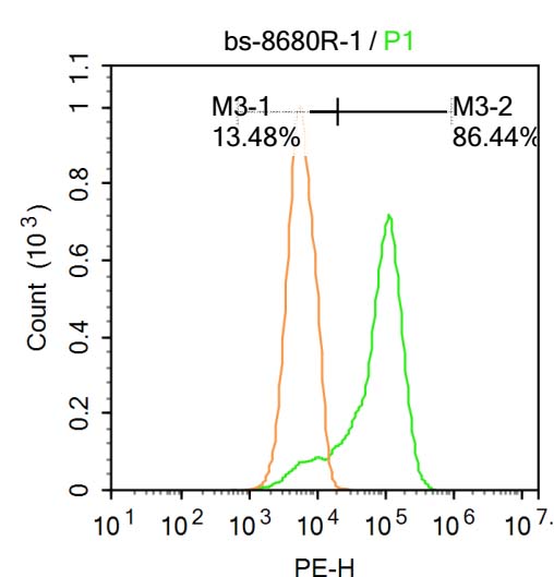 Blank control: Raji.
Primary Antibody (green line): Rabbit Anti-IRE1 antibody (SL8680R )
Dilution: 1μg /10^6 cells;
Isotype Control Antibody (orange line): Rabbit IgG .
Secondary Antibody : Goat anti-rabbit IgG-PE
Dilution: 1μg /test.
Protocol
The cells were fixed with 4% PFA (10min at room temperature)and then permeabilized with PBST for 20 min at room temperature. The cells were then incubated in 5%BSA to block non-specific protein-protein interactions for 30 min at at room temperature .Cells stained with Primary Antibody for 30 min at room temperature. The secondary antibody used for 40 min at room temperature. Acquisition of 20,000 events was performed.
Cartpieces
Totalgoods,subtotals:¥Checkout
References (0)
No References
Bought notes(bought amounts latest0)
No one bought this product
User Comment(Total0User Comment Num)
- No comment


 +86 571 56623320
 +86 18668110335

