
Rabbit Anti-ATG18 antibody
ATG 18; ATG18; Atg18 protein homolog; ATG18A; WD repeat domain phosphoinositide interacting 1; WD repeat domain phosphoinositide interacting protein 1; WD40 repeat protein interacting with phosphoInositides of 49kDa; WIPI 1 alpha; WIPI 1; WIPI 49; WIPI 49
View History [Clear]
Details
Product Name ATG18 Chinese Name 自噬相关蛋白18抗体 Alias ATG 18; ATG18; Atg18 protein homolog; ATG18A; WD repeat domain phosphoinositide interacting 1; WD repeat domain phosphoinositide interacting protein 1; WD40 repeat protein interacting with phosphoInositides of 49kDa; WIPI 1 alpha; WIPI 1; WIPI 49; WIPI 49 kDa ; WIPI49. literatures Research Area Tumour Cell biology immunology Apoptosis transcriptional regulatory factor Immunogen Species Rabbit Clonality Polyclonal React Species Human, Rat, (predicted: Mouse, Pig, ) Applications ELISA=1:5000-10000 IHC-P=1:100-500 IHC-F=1:100-500 Flow-Cyt=1ug/Test IF=1:100-500 (Paraffin sections need antigen repair)
not yet tested in other applications.
optimal dilutions/concentrations should be determined by the end user.Theoretical molecular weight 49kDa Cellular localization cytoplasmic Form Liquid Concentration 1mg/ml immunogen KLH conjugated synthetic peptide derived from mouse WIPI1: 361-446/446 Lsotype IgG Purification affinity purified by Protein A Buffer Solution 0.01M TBS(pH7.4) with 1% BSA, 0.03% Proclin300 and 50% Glycerol. Storage Shipped at 4℃. Store at -20 °C for one year. Avoid repeated freeze/thaw cycles. Attention This product as supplied is intended for research use only, not for use in human, therapeutic or diagnostic applications. PubMed PubMed Product Detail This gene encodes a WD40 repeat protein. Members of the WD40 repeat family are key components of many essential biologic functions. They regulate the assembly of multiprotein complexes by presenting a beta-propeller platform for simultaneous and reversible protein-protein interactions. Members of the WIPI subfamily of WD40 repeat proteins have a 7-bladed propeller structure and contain a conserved motif for interaction with phospholipids. Alternative splicing results in multiple transcript variants. [provided by RefSeq, Mar 2016]
Function:
Plays an important role in autophagy and in particular starvation- and calcium-mediated autophagy, as well as in mitophagy. Functions upstream of the ATG12-ATG5-ATG16L1 complex and LC3, and downstream of the ULK1 and PI3-kinase complexes. Involved in xenophagy of Staphylococcus aureus. Invading S.aureus cells become entrapped in autophagosome-like WIPI1 positive vesicles targeted for lysosomal degradation. Plays also a distinct role in controlling the transcription of melanogenic enzymes and melanosome maturation, a process that is distinct from starvation-induced autophagy. May also regulate the trafficking of proteins involved in the mannose-6-phosphate receptor (MPR) recycling pathway.
Subunit:
Interacts with androgen receptor (AR) and the estrogen receptors ESR1 and ESR2. Binds PtdIns3P and to a lesser extent, PtdIns3,5P2 and PtdIns5P in vitro. Interaction with PtdIns3P is required for recruitment to membranes.
Subcellular Location:
Golgi apparatus, trans-Golgi network. Endosome. Cytoplasmic vesicle, clathrin-coated vesicle. Preautophagosomal structure membrane; Peripheral membrane protein. Cytoplasm, cytoskeleton. Note=Trans elements of the Golgi and peripheral endosomes. Dynamically cycles through these compartments and is susceptible to conditions that modulate membrane flux. Enriched in clathrin-coated vesicles. Upon starvation-induced autophagy, accumulates at subcellular structures in the cytoplasm: enlarged vesicular and lasso-like structures, and large cup-shaped structures predominantly around the nucleus. Recruitment to autophagic membranes is controlled by MTMR14. Labile microtubules specifically recruit markers of autophagosome formation like WIPI1, whereas mature autophagosomes may bind to stable microtubules.
Tissue Specificity:
Ubiquitously expressed. Highly expressed in skeletal muscle, heart, testis, pancreas and placenta. Highly expressed in G361, Sk-mel-28, Sk-mel-13, WM852 and WM451 cells. Up-regulated in a variety of tumor tissues.
Similarity:
Belongs to the WD repeat SVP1 family.
Contains 7 WD repeats.
SWISS:
Q5MNZ9
Gene ID:
52639
Database links:Entrez Gene: 55062 Human
Entrez Gene: 52639 Mouse
Omim: 609224 Human
SwissProt: Q5MNZ9 Human
SwissProt: Q8R3E3 Mouse
Unigene: 463964 Human
Unigene: 35817 Mouse
Product Picture 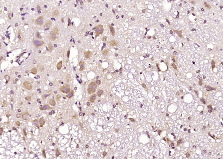 Paraformaldehyde-fixed, paraffin embedded (rat brain); Antigen retrieval by boiling in sodium citrate buffer (pH6.0) for 15min; Block endogenous peroxidase by 3% hydrogen peroxide for 20 minutes; Blocking buffer (normal goat serum) at 37°C for 30min; Antibody incubation with (ATG18) Polyclonal Antibody, Unconjugated (SL3871R) at 1:200 overnight at 4°C, followed by operating according to SP Kit(Rabbit) (sp-0023) instructionsand DAB staining.
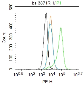 Blank control:U87MG.
Primary Antibody (green line): Rabbit Anti-ATG18 antibody (SL3871R)
Dilution: 1μg /10^6 cells;
Isotype Control Antibody (orange line): Rabbit IgG .
Secondary Antibody : Goat anti-rabbit IgG-PE
Dilution: 1μg /test.
Protocol
The cells were fixed with 4% PFA (10min at room temperature)and then permeabilized with 0.1%PBST for 20 min at room temperature.The cells were then incubated in 5%BSA to block non-specific protein-protein interactions for 30 min at room temperature .Cells stained with Primary Antibody for 30 min at room temperature. The secondary antibody used for 40 min at room temperature. Acquisition of 20,000 events was performed.Blank control:U87MG.
Primary Antibody (green line): Rabbit Anti-ATG18 antibody (SL3871R)
Dilution: 1μg /10^6 cells;
Isotype Control Antibody (orange line): Rabbit IgG .
Secondary Antibody : Goat anti-rabbit IgG-PE
Dilution: 1μg /test.
Protocol
The cells were fixed with 4% PFA (10min at room temperature)and then permeabilized with 0.1%PBST for 20 min at room temperature.The cells were then incubated in 5%BSA to block non-specific protein-protein interactions for 30 min at room temperature .Cells stained with Primary Antibody for 30 min at room temperature. The secondary antibody used for 40 min at room temperature. Acquisition of 20,000 events was performed.
Cartpieces
Totalgoods,subtotals:¥Checkout
Bought notes(bought amounts latest0)
No one bought this product
User Comment(Total0User Comment Num)
- No comment


 +86 571 56623320


