
Rabbit Anti-phospho-LLGL1 + LLGL2 (Ser650+Ser654)antibody
LLGL1 + LLGL2 (phospho S650 + S654); LLGL1 + LLGL2 (phospho Ser650 + Ser654); p-LLGL1 + p-LLGL2 (S650 + S654); p-LLGL1/LLGL2 (Ser650 + Ser654); p-LLGL1/LLGL2 (S650 + S654); p-LLGL1/LLGL2 (Ser650/654); DLG4; HGL; HUGL; HUGL1; lethal giant larvae homolog 1;
View History [Clear]
Details
Product Name phospho-LLGL1 + LLGL2 (Ser650+Ser654) Chinese Name 磷酸化相似Tumour抑制基因HUGL抗体 Alias LLGL1 + LLGL2 (phospho S650 + S654); LLGL1 + LLGL2 (phospho Ser650 + Ser654); p-LLGL1 + p-LLGL2 (S650 + S654); p-LLGL1/LLGL2 (Ser650 + Ser654); p-LLGL1/LLGL2 (S650 + S654); p-LLGL1/LLGL2 (Ser650/654); DLG4; HGL; HUGL; HUGL1; lethal giant larvae homolog 1; lethal giant larvae homolog 2; Lethal(2) giant larvae protein homolog 1; Lethal(2) giant larvae protein homolog 2; LGL2; LLGL; L2GL1_HUMAN; L2GL2_HUMAN. Product Type Phosphorylated anti Research Area Tumour Cell biology Signal transduction Cyclin Cytoskeleton Immunogen Species Rabbit Clonality Polyclonal React Species Rat, (predicted: Human, Mouse, Chicken, Dog, Pig, Cow, Horse, Guinea Pig, ) Applications ELISA=1:5000-10000
not yet tested in other applications.
optimal dilutions/concentrations should be determined by the end user.Theoretical molecular weight 117kDa Cellular localization cytoplasmic The cell membrane Form Liquid Concentration 1mg/ml immunogen KLH conjugated synthesised phosphopeptide derived from mouse LLGL1 around the phosphorylation site of Ser650+Ser654: PL(p-S)RVK(p-S) Lsotype IgG Purification affinity purified by Protein A Buffer Solution 0.01M TBS(pH7.4) with 1% BSA, 0.03% Proclin300 and 50% Glycerol. Storage Shipped at 4℃. Store at -20 °C for one year. Avoid repeated freeze/thaw cycles. Attention This product as supplied is intended for research use only, not for use in human, therapeutic or diagnostic applications. PubMed PubMed Product Detail LLGL1 is a protein that is similar to a tumor suppressor in Drosophila. The protein is part of a cytoskeletal network and is associated with nonmuscle myosin II heavy chain and a kinase that specifically phosphorylates this protein at serine residues. The gene for LLGL1 is located within the Smith-Magenis syndrome region on chromosome 17. LLGL2 is a protein similar to lethal giant larvae of Drosophila. In fly, the protein's ability to localize cell fate determinants is regulated by the atypical protein kinase C (aPKC). In human, this protein interacts with aPKC-containing complexes and is cortically localized in mitotic cells.
Function:
Cortical cytoskeleton protein found in a complex involved in maintaining cell polarity and epithelial integrity. Involved in the regulation of mitotic spindle orientation, proliferation, differentiation and tissue organization of neuroepithelial cells.
Subunit:
Associated with nonmuscle myosin II heavy chain. Interacts with PRKCI/aPKC, PARD6B/Par-6 and PARD6A. Interacts with STX4A.
Subcellular Location:
Cytoplasm, cytoskeleton. Note=Localized to the lateral membrane during the polarization and formation cell-cell contacts.
Tissue Specificity:
Expressed in brain, kidney, and muscle but is barely seen in heart and placenta. Down-regulated or lost in all cell lines and in most of the tumor samples analyzed. Loss was associated with advanced stage of the disease.
Post-translational modifications:
Phosphorylated upon DNA damage, probably by ATM or ATR. Phosphorylated at least at Ser-663 by PRKCI.
Similarity:
Belongs to the WD repeat L(2)GL family.
Contains 14 WD repeats.
SWISS:
Q15334
Gene ID:
3996
Database links:Entrez Gene: 3996 Human
Omim: 600966 Human
SwissProt: Q15334 Human
Unigene: 513983 Human
Product Picture 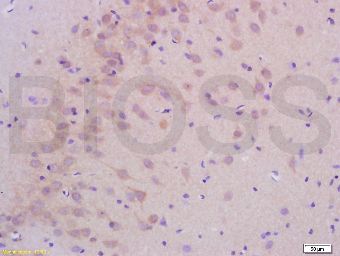 Tissue/cell: rat brain tissue; 4% Paraformaldehyde-fixed and paraffin-embedded;
Antigen retrieval: citrate buffer ( 0.01M, pH 6.0 ), Boiling bathing for 15min; Block endogenous peroxidase by 3% Hydrogen peroxide for 30min; Blocking buffer (normal goat serum,C-0005) at 37℃ for 20 min;
Incubation: Anti-phospho-LLGL1+LLGL2(Ser650+Ser654) Polyclonal Antibody, Unconjugated (SL6070R) 1:200, overnight at 4°C, followed by conjugation to the secondary antibody(SP-0023) and DAB(C-0010) staining
Cartpieces
Totalgoods,subtotals:¥Checkout
References (0)
No References
Bought notes(bought amounts latest0)
No one bought this product
User Comment(Total0User Comment Num)
- No comment


 +86 571 56623320
 +86 18668110335

